INTRODUCTION
Larval development is one of the most important periods of the decapod life cycle; recruitment does not occur unless the larval period is completed. Larval data can be useful in evaluating species diversity in a region and in specifying the reproduction time of brachyuran species. Morphological features of larvae are complementary characters for crustacean taxonomy and phylogeny. The study of larvae is important for the problem of introduced species arriving in ballast waters or on fouled ships. However, insufficient attention has been paid to decapod larval development in the Russian Far East Seas. Many keys exist for the identification of brachyuran larvae in different regions of the World Ocean (Ingle, Reference Ingle1992; Paula, Reference Paula1996; Báez, Reference Báez1997; Pessani et al., Reference Pessani, Burri and Salton1998; Anosov, Reference Anosov2000; Puls, Reference Puls and Shanks2001; Santos & González-Gordillo, Reference dos Santos and González-Gordillo2004; Rice & Tsukimura, Reference Rice and Tsukimura2007), but only one taxonomic guide is known for the Sea of Japan (Konishi, Reference Konishi, Chihara and Murano1997). There is no comprehensive key for identification of the brachyuran larvae in Russian waters of the Sea of Japan; however, the species list of this region considerably differs from that of coastal waters of Japan. The aim of this work is to provide a key for the identification of brachyuran larvae in Peter the Great Bay.
MATERIALS AND METHODS
The key is based on the accounts previously published by various authors (Table 1) and new original descriptions of brachyuran larvae both taken from plankton samples and reared from ovigerous crab females in the laboratory.
List of species and sources of descriptions of brachyuran zoeae included in the identification key.

Zooplankton was sampled in Vostok Bay (inner bay of Peter the Great Bay, Sea of Japan) between May and November 2002 using a Norpac net with a ring diameter of 40 cm and a filtering cone made of a 168 mm mesh, and in Amursky and Ussurijsky Bays (inner bays of Peter the Great Bay, Sea of Japan) between April and October 2007 using a Juday net with a ring diameter of 38 cm and a filtering cone made of a 168 mm mesh (Figure 1).
Map showing the sampling area.

It is very difficult to identify the larvae (especially the larvae of congeneric species) using drawings and descriptions of different authors in more or less detail. So, zoeae of most considered species were obtained under the laboratory conditions. This material was used for the construction of the key and the original figures.
Ovigerous crab females were maintained in an aerated seawater aquarium until larvae hatched. After hatching, larvae were concentrated at the edge of the aquarium using a point-light source and transferred to 1-l glass vessels with filtered and UV-sterilized seawater and reared to the megalopal stage. The density of larvae was about 100 specimens l−1. The water in the vessels was changed daily. The larvae were fed with newly hatched nauplii of Artemia salina. Very small zoea of varunid and pinnotherid crabs were reared using nauplii of the rhizocephalan crustacean, Polyascus polygenea, as a food. Earlier, this method was described in detail (Kornienko & Korn, Reference Kornienko and Korn2005a).
All larvae were fixed in 4% formaldehyde for light microscopic studies.
The dichotomous identification key is based mainly on external morphological characters, which are easy to observe under a stereomicroscope MBS-10 without specimen dissection. When these features are insufficient, the morphology and setation of appendages has been included. Moreover, new original figures were provided to make identification easier. The outlines of the larvae were drawn using a camera lucida attached to a binocular Ergaval microscope (Carl Zeiss Jena).
The key was constructed for zoea I, but the characters used in the key do not change or change slightly through the successive zoeal stages, with the exception of peculiar cases. Main characters of brachyuran zoea used for the identification are represented in Figure 2.
Main characters used for the identification of brachyuran zoea.

RESULTS
Key for the identification of brachyuran zoea I (interspecific distinctions)
1a. Carapace with rostral, dorsal and lateral spines (Figure 3A)………2
1b. Carapace with rostral spine only (Figure 3B) or with rostral and dorsal spines (Figure 3C), lateral spines absent………11
2a. Abdominal somites 3–5 with well developed posterolateral spines; furcal rami with lateral and dorsal spines (Figure 4A)………3
2b. Abdominal somites 3–5 with poorly developed posterolateral spines (Figure 4B, C); furcal rami without lateral and dorsal spines (Figure 4B) or with lateral spines only (Figure 4C)………7
3a. Furcal rami longer than proximal part of telson; dorsal and lateral furcal spines short (Figure 5A)………4
3b. Furcal rami shorter than proximal part of telson; lateral furcal spines long (Figure 5B, C)………6
4a. All carapace spines spinulated; dorsal spine straight; posterolateral spines longer than half abdominal somite; each furcal ramus with three spines………Chionoecetes opilio (Figure 11A)
[Lateral spines long, slightly shorter dorsal and rostral spines. Antenna biramous; protopod spinulated, approximately equal rostral spine; endopod with three terminal setae of unequal length. Lateral knobs on abdominal somites 2–3 long, those of third one reach end of the same somite. Larvae found from April to July.]
4b. All carapace spines smooth; dorsal spine slightly curved; posterolateral spines shorter than half abdominal somite; each furcal ramus with two spines………5
5a. Abdominal somite 2 with a pair of lateral knobs; outermost pair of inner setae on posterior telson margin dentated on inside surface (Figure 6A, A′); posterolateral spines very short in zoea I and considerably lengthen in successive stages.………Cancer amphioetus (Figure 11B)
[Antenna biramous; protopod with two rows of spinules, nearly half of rostral spine; exopod with two terminal setae of unequal length. Larvae found from June to September.]
5b. Abdominal somites 2 and 3 with a pair of lateral knobs; all inner setae on posterior telson margin plumodenticulate; posterolateral spines prominent in all zoeal stages (Figure 6B)………Charybdis japonica (Figure 11C)
[Antenna biramous; protopod with two rows of spinules, nearly half of rostral spine; exopod with two terminal setae of unequal length. Larvae not found yet.]
6a. Abdominal somite 2 with a pair of lateral knobs; posterolateral spines of somite 4 shorter somite 5; lateral telsonal spine shorter than half furcal ramus; each furcal ramus with three inner setae (Figure 5B)………Telmessus cheiragonus (Figure 11D)
[Dorsal spine straight; rostral and dorsal spines spinulated in distal part. Antennal protopod spinulated, shorter than rostral spine; exopod with two nearly equal setae. Abdominal somites 3–5 with long posterolateral spines. Each furcal ramus with three spines: one long lateral spine and two short dorsal spines. Larvae found in April and May.]
6b. Abdominal somites 2 and 3 with a pair of lateral knobs; posterolateral spines of somite 4 shorter than somite 5; lateral telsonal spine longer than half furcal ramus; each furcal ramus with four inner setae (Figure 5C)………Erimacrus isenbeckii (Figure 11E)
[Dorsal spine straight; rostral and dorsal spines spinulated in distal part. Antennal protopod spinulated, shorter than rostral spine; exopod with two nearly equal setae. Abdominal somites 3–5 with long posterolateral spines. Each furcal ramus with three spines: long lateral spine and two short dorsal spines. Larvae found in April and May.]
7a. Lateral carapace spines arranged as in typical brachyuran larvae; all abdominal somites cylindrical; telson triangular; furcal rami without spines (Figure 7A, B)………8
7b. Lateral carapace spines arranged more ventrally than in typical brachyuran zoea; abdominal somites 4–5 not cylindrical but laterally expanded; telson subrectangular; furcal rami without spines or with minute lateral spines (Figure 7D–F)………10
8a. Dorsal and rostral spines longer than carapace; dorsal carapace spine straight; antenna uniramous (Figure 8A)………Tritodynamia rathbuni (Figure 11F)
[Antennal protopod with two rows of spinules and with short seta near base. Posterolateral spines on abdominal somites 3–5 highly reduced. Larvae found from June to September.]
8b. Dorsal and rostral spines slightly shorter than carapace; dorsal carapace spine slightly curved; antenna biramous (Figure 8B)………9
9a. Abdominal somites 2–4 with a pair of lateral knobs (lateral knobs on somite 4 disappear in successive stages); antennule with 2 aesthetascs; antennal exopod nearly half protopod (Figure 9A)………Eriocheir japonicus (Figure 11G)
[Antennal exopod as tapering spine with two small unequal subterminal setae. Larvae found from June to September.]
9b. Abdominal somites 2–3 with a pair of lateral knobs; antennule with 3 aesthetascs; antennal exopod nearly 2/3 protopod (Figure 9B)………Hemigrapsus sanguineus, H. penicillatus, H. longitarsis (Figure 11H)
[Antennal exopod as tapering spine with small unequal subterminal setae. Larvae found from June to September.]
10a. Abdominal somites 4–5 expanded; furcal rami shorter than proximal part of telson, with minute lateral spines (Figure 7D)………Pinnaxodes mutuensis (Figure 11I)
[Antenna uniramous; protopod with two rows of spinules. Lateral telsonal margins slightly convex. Larvae found in July.]
10b. Abdominal somite 5 as a horseshoe; furcal rami longer than proximal part of telson, without lateral spines………Pinnixa rathbuni (Figure 11J)
[Rostral and dorsal carapace spines straight. Antenna uniramous; protopod with two rows of spinules and with short seta near base. Furcal rami without spines (Figure 7E). Larvae found from May to November.]
11a. Carapace with rostral spine only………Sakaina yokoyai (Figure 11K)
[Antenna uniramous; protopod with two rows of spinules. Abdominal somites 4–5 as a horseshoe. Telson nearly rectangular; furcal rami shorter than proximal part of telson, without spines; median notch virtually absent (Figure 7F). Larvae found in June–August.]
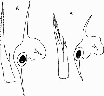
11b. Carapace with rostral and dorsal spines (Figure 10)………12
12a. Rostral and dorsal spines three times longer than carapace; dorsal spine straight………Paradorippe granulata (Figure 11L)
[Dorsal spine with blunt spinules, rostral spine with acute spinules. Antenna biramous; protopod and exopod sub-equal. Abdominal somite 2 with pair of lateral knobs. Telson narrow and long, with a pair of lateral spines; furcal rami twice exceeding proximal part of telson. Posterior telsonal margin with two inner setae only ( Figure 10A, A′). Larvae found in July and August.]
12b. Rostral and dorsal spine shorter than carapace, dorsal spine slightly curved (Figure 10B)………Pugettiaquadridens, Pisoides bidentatus (Figure 11M)
[Rostral spine nearly one-third antenna; dorsal spine with sparse blunt spinules. Antenna biramous; protopod and exopod sub-equal. Abdominal somites 2 with a pair of lateral knobs. Furcal rami with lateral spines (Figure 4C). Larvae found from June to September.]
Carapace of zoea in Eriocheir japonicus (A), Sakaina yokojai (B) and Pisoides bidentatus (C).

Abdomen and telson of zoea in Chionoecetes opilio (A), Tritodynamia rathbuni (B) and Pisoides bidentatus (C).

Telson of zoea in Chionoecetes opilio (A), Telmessus cheiragonus (B) and Erimacrus isenbeckii (C).

Abdomen and telson of zoea in Cancer amphioetus (A, A′) and Charybdis japonica (B) (after Yatsuzuka et al., Reference Yatsuzuka, Sakai and Del Rio Roman1984).

Carapace of zoea in Tritodynamia rathbuni (A) and Pinnaxodes mutuensis (C); telson of zoea in Tritodynamia rathbuni (B); Pinnaxodes mutuensis (D), Pinnixa rathbuni (E) and Sakaina yokoyai (F).

Antenna and carapace of zoea in Tritodynamia rathbuni (A) and Eriocheir japonicus (B).

Antenna, antennule and abdomen of zoea in Eriocheir japonicus (A) and Hemigrapsus sanguineus (B).

Carapace (A) and telson (A′) of zoea in Paradorippe granulata; carapace of zoea in Pugettia quadridens (B).

Lateral view of zoea in Chionoecetes opilio (A), Cancer amphioetus (B), Charybdis japonica (C), Telmessus cheiragonus (D), Erimacrus isenbeckii (E), Tritodynamia rathbuni (F), Eriocheir japonicus (G), Hemigrapsus sanguineus (H), Pinnaxodes mutuensis (I), Pinnixa rathbuni (J), Sakaina yokoyai (K), Paradorippe granulata (L), and Pisoides bidentatus (M).

Most Brachyura from Peter the Great Bay pass through five zoeal stages. Majiod crabs (Pisoides bidentatus, Pugettia quadridens, and Chionoecetes opilio) have an abbreviated development including only two zoeal stages. Two of three pinnotherid crabs also undergo an abbreviated development: Sakaina yokoyai passes through probably three zoeal stages; Pinnaxodes mutuensis, through four zoeal stages. Four zoeal stages are known also for Paradorippe granulata.
Different zoeal stages of brachyuran crabs (age distinctions) are easily determined using the number of natatory setae on the exopods of maxillipeds, the number of setae along the posterior telsonal margin, the presence of uniramous or biramous pleopod buds and some other morphological features. The same stages in the species with abbreviated development are more advanced in the number of characters over those of the species with longer development.
Identification of different zoeal stages of brachyuran crabs (age distinctions)
Zoea I. Eyes sessile; exopod of maxilliped each with 4 natatory setae; abdomen consists of 5 somites and telson; posterior telsonal margin with 3+3 setae (in Paradorippe granulata, posterior telsonal margin with two setae only in all zoeal stages).
Zoea II. Eyes stalked; exopod of maxilliped each with 6 natatory setae; in most species abdomen consists of 5 somites and telson; pleopod buds absent; posterior telsonal margin with 3+3 setae. In Pisoides bidentatus, Pugettia quadridens, and Chionoecetes opilio, zoea II is the last stage; hence, the sixth somite is delineated, somites 2–6 with biramous pleopod buds. In Sakaina yokoyai, pleopod buds uniramous. In Charybdis japonica and Chionoecetes opilio, posterior telsonal margin with 4+4 setae.
Zoea III. Exopod of maxilliped each with 8 natatory setae; the sixth somite delineated, with the exception of Pinnixa rathbuni which has 5 somites in all zoeal stages. In Cancer amphioetus, Trithodynamia rathbuni, Erimacrus isenbeckii, Telmessus cheiragonus, Eriocheir japonicus and Hemigrapsus species, posterior telsonal margin with 4+4 setae; in Charybdis japonica and Pinnaxodes mutuensis, with additional small unpaired setae. In Sakaina yokoyai, pleopod buds biramous.
Zoea IV. Exopod of maxilliped each with 10 natatory setae; somites 2–6 with uniramous pleopod buds. In Cancer amphioetus and Tritodynamia rathbuni, posterior telsonal margin with 5+5 setae.
Zoea V. Exopod of maxilliped each with 12 natatory setae; pleopod buds biramous. In Eriocheir japonicus and Hemigrapsus species, posterior telsonal margin with 5+5 setae.
DISCUSSION
According to Adrianov & Kussakin (Reference Adrianov and Kussakin1998), 20 brachyuran species belonging to 8 families and 16 genera inhabit Peter the Great Bay (Russian waters of the Sea of Japan). We found larvae of only 16 species from 9 families and 14 genera occurring in the plankton of Peter the Great Bay (Table 1). To date, we have not found both adults and larvae of Goetice depressus (de Haan, 1835), Helice tridens de Haan, 1833, and Paradromia japonica (Henderson, 1888). According to Vassilenko (Reference Vassilenko1990), adult specimens of Pinnixa tumida Stimpson, 1858 are found only in Possyet Bay (eastern Peter the Great Bay). Brachyuran larvae occur in Peter the Great Bay from April to November.
Zoea of each species represented in this key has been previously described. Only the larvae of Sakaina yokoyai are not known yet; however, the morphology of closely related species, S. japonica, has been described (Konishi, Reference Konishi1981a). Both adults and larvae of S. yokoyai are rarely found in Peter the Great Bay, so we failed to obtain the total series of their larval stages. Nevertheless, we believe that S. yokoyai, like S. japonica, passes through three zoeal stages.
Frequently, the distinction between larvae of congeneric species is based on slight differences. The larvae of congeneric species, described on the basis of specimens hatched in the laboratory from ovigerous females, can be identified only to the generic level when collected in nature. In our case, the specific identification of three Hemigrapsus species (H. sanguineus, H. penicillatus and H. longitarsis) is very difficult. Zoea I and zoea II of these species are nearly identical. The larvae are distinguishable only from zoea III. Zoea III–V of Hemigrapsus species differ in the number of dorsomedial setae on the abdominal somite I and in the number of setae on the posterodorsal arch. It is pertinent to note that among the three Hemigrapsus species, zoea and megalopa of H. penicillatus differ greatly (Kornienko et al., Reference Kornienko, Korn and Kashenko2008). Despite the great similarity of larvae in varunid crabs of the genera Hemigrapsus and Eriocheir, the latter possess a number of distinctive features in all developmental stages (Kornienko & Korn, Reference Kornienko and Korn2005a; Kornienko et al., Reference Kornienko, Korn and Kashenko2008).
It was recently shown that zoea of Pisoides bidentatus and Pugettia quadridens belonging to different families are also nearly identical, with the exception of a more intensive coloration of the latter. Some differences appear only in the megalopal stage. Based on the larval similarity (Kornienko & Korn, Reference Kornienko and Korn2007) and high genetic identity of adults (Zaslavskaya et al., Reference Zaslavskaya, Kornienko and Korn2007) these two species should be assigned to one genus.
The larvae of the other ten species belonging to different families and genera are easy to identify in the plankton. The features used in the key were chosen to enable identification of zoea to the species level and do not reflect any systematic arrangement of decapod families.
ACKNOWLEDGEMENTS
The project was supported by the Far East Branch of the Russian Academy of Sciences (grant no. 06-III-A-06-164) and by the Russian Foundation for Fundamental Researches (grant no. 08-04-00929).