Homografts are widely used in congenital cardiac surgery to create a valved conduit between the right ventricle and the pulmonary arteries,Reference Perron, Moran, Gauvreau, del Nido, Mayer and Jonas1, Reference Powell, Lock, Keane and Perry2 for example, in patients with pulmonary atresia, tetralogy of Fallot, truncus arteriosus, and valvular aortic stenosis. Although the short-term results are excellent, the longevity of the homograft remains a problem, mostly because of the development of stenosis time.Reference Tatebe, Nagakura, Boyle and Duncan3, Reference Oosterhof, Meijboom and Vliegen4
The optimal approach to this problem is still unclear.Reference Hanley5, Reference Yuan, Mishaly, Shinfeld and Raanani6 Currently, there are several treatment options, such as balloon valvuloplasty, stenting of the homograft, percutaneous pulmonary valvular implantation, and surgical valve replacement. Balloon dilatation has been used to dilate the stenosed homograft, but it is unclear whether this catheter intervention influences the longevity of the homograft significantly.Reference Tatebe, Nagakura, Boyle and Duncan3 The reported series of homograft valvuloplasty are small, ranging from only 3 to a maximum of 12 patients. The results are not favourable: one study describes that catheter intervention did not prolong the longevity of the homograft;Reference Tatebe, Nagakura, Boyle and Duncan3 two others report that they have abandoned this technique after unfavourable results.Reference Oosterhof, Meijboom and Vliegen4, Reference Bohm7
However, postponing percutaneous pulmonary valve implantation or surgical valve replacement could be beneficial, in order to reduce the number of valve replacements that patients with a homograft may need in their life. Especially in children, the effect of balloon dilatation could be beneficial if the stenosis is the result of degeneration or calcification and not caused by outgrowth of the homograft, even if the necessary intervention or surgery is postponed only by 1 or 2 years. During this time, the child can grow and it might become possible to either implant a larger size homograft surgically or perform percutaneous pulmonary valve implantation.Reference Zeevi, Keane, Perry and Lock8, Reference Nordmeyer, Coats and Bonhoeffer9
Therefore, we retrospectively studied balloon dilatation of homograft stenosis performed in two university medical centres in the Netherlands. The aim of our study was to evaluate the results of this treatment and to identify factors that might influence or are able to predict the effect of the homograft dilatation. To our knowledge, this study describes the largest series of balloon dilatations of homograft stenosis.
Materials and methods
A retrospective study was performed in two university medical centres in the Netherlands. All patients who were diagnosed with stenosis of a homograft in pulmonary position and were treated by balloon dilatation between 1998 and 2007 were included. A cardiac catheterisation was performed in those patients who had a peak systolic pressure gradient over the stenotic homograft on echocardiography of more than 50 millimetres of mercury. All procedures were performed under general anaesthesia. Right ventricle and pulmonary artery pressures were measured and a right ventricle angiogram was perfomed simultaneously in antero-posterior and left-lateral projections. In general, a balloon dilatation was performed if the right ventricle pressure was more than half of the systemic pressure during cardiac catheterisation.
Methods
In order to determine the effect of a percutaneous balloon dilatation, we used two criteria: one concerning the direct effect of the dilatation and one describing the lasting of the effect during follow up, as the direct effect can be of short duration. These were the decrease of the peak systolic pressure gradient in the homograft during catheterisation for the first criterion and the time until re-intervention – second balloon dilatation – or replacement of the homograft for the second criterion. In general, the indications for re-intervention or replacement of the homograft were the same as the indications for the balloon dilatation, meaning a peak systolic pressure gradient over the stenotic homograft on echocardiography of more than 50 millimetres of mercury. In this study, we designated a dilatation as successful, if the peak systolic pressure gradient was reduced by more than 33% and re-intervention or replacement was postponed by more than 1 year and 6 months.
In search of factors that might be able to predict or influence the success of dilatation, we compared the successful with the unsuccessful dilatations. Data collected and analysed included: age, gender, date of primary surgical correction, date of implantation of the homograft, size and type of homograft, systemic systolic blood pressure; echocardiographic peak systolic pressure gradient in the stenotic homograft – in millimetres of mercury – and tricuspid regurgitation gradient before and after the balloon dilatation – in millimetres of mercury; right ventricle and pulmonary artery pressure measured before and after the balloon dilatation – in millimetres of mercury; the location of the stenosis; presence of calcification; balloon type, size, and pressure; and time to re-intervention or valve replacement of the homograft. The location of the stenosis at angiography – left lateral projection – was categorised as: proximal, near the site of the proximal anastomosis of the homograft; valvular, at the level of the valve; distal, near the site of the distal anastomosis of the homograft; tubular, if a larger part of the homograft was stenotic.
Statistical analysis
Statistical calculations were made by the use of SPSS 17.0 (IBM Company, Chicago, Illinois, United States of America) statistical program. For creating and calculating survival curves, Kaplan–Meier survival curves and Log-Rank (Mantel–Cox) tests were used. Means for scale numeric data were compared with the independent sample T-test, equality of variance was tested by the Levene's test. Categorical data were compared in cross tables and tested with the Pearson chi-square test.
Results
A total of 41 balloon dilatations were performed in 36 patients. Among them, one patient was lost to follow-up because the family had moved abroad. There were two patients who still met the criteria for homograft replacement after balloon dilatation of the homograft, but owing to social-developmental comorbidity conservative treatment was preferred. Therefore, they were included in the study and the mean time between balloon dilatation and homograft replacement was set at 0 months. Eventually, 40 procedures of balloon dilatations in 34 patients were included in the study. In one patient, the homograft was dilated again 3 years after the first dilatation. In five other patients, the homograft was replaced after the first unsuccessful dilatation and later on this new homograft was dilated.
The mean age of the patients at the time of the procedure was 13.6 years – standard deviation plus or minus 7.3 years, range 3–29 years, 40 procedures; 16 procedures were done in patients younger than 10 years of age. The origin of the homograft was aortic in 18 procedures, pulmonary in 21 procedures, and unknown in one procedure. The mean size of the homograft was 18 millimetres – standard deviation plus or minus 3.8 millimetres, range 12–27 millimetres. The mean time between initial homograft implantation and balloon dilatation was 6.9 years – standard deviation plus or minus 4.3 years, range 1–18 years, 40 procedures.
Indication for balloon dilatation
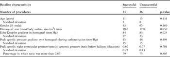
In all except two procedures, the peak systolic echo-Doppler gradient in the homograft was more than 50 millimetres of mercury before dilatation. In one procedure, the gradient could not be obtained, but the right ventricular pressure – based on tricuspid regurgitation gradient on echocardiography – was 88 millimetres of mercury. In another procedure, the echo-Doppler gradient was 47 millimetres of mercury, but the patient had a clearly reduced exercise tolerance at that time. The mean echo-Doppler gradient in the homograft was 83 millimetres of mercury – standard deviation plus or minus 24 millimetres of mercury, range 47–154 millimetres of mercury, 39 procedures. During cardiac catheterisation, the right ventricular peak systolic pressure was more than half the systemic pressure in all procedures, except for two in which the right ventricular pressure was 43% and, respectively, 47% of the systemic pressure. In two other procedures, the systemic blood pressure was not documented adequately. The mean peak systolic right ventricular pressure was 78% of the systemic pressure – standard deviation plus or minus 17%, range 43–110%, 38 procedures. Table 1 summarises the patient characteristics in the two groups of successful and unsuccessful procedures.
Characteristics of successful and unsuccessful procedures before balloon dilatation.

Procedures
Most dilatations were performed using a Z-med (26 procedures) or Mullins (7 procedures) high-pressure balloon (Numed, Incorporated, Hopkinton, New York, United States of America). In one case, a Mansfield (Boston Scientific, Corporation, Maple Grove, Minnesota, United States of America) was used, and in another case an Osypka high-pressure balloon (Osypka, Rheinfelden, Germany). The pressure applied varied between 2 and 16 atmosphere. In a number of patients, a Tyshak balloon (Numed, Incorporated, Hopkinton, New York, United States of America) was used to test whether or not the stenosis would rebound after dilatation, because this balloon can be introduced through a sheath of smaller size. In five cases, the stenosis could be dilated, but did rebound, and therefore the procedure was terminated. In all other cases, the final dilatation was performed using a high-pressure balloon.
Effect of balloon dilatation
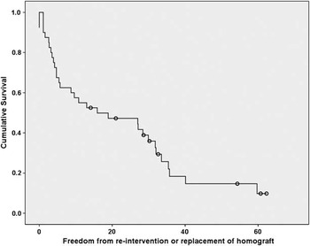
The mean reduction of the peak systolic pressure gradient over the homograft was 30% – standard deviation plus or minus 22%, range 0–70%, 40 procedures. Re-intervention or homograft replacement was postponed by a mean of 1 year and 7 months – standard deviation plus or minus 18 months, range 0 months to 5 years and 2 months, 40 procedures. The survival curve depicting freedom from replacement of the homograft or re-intervention after balloon dilatation is shown in Figure 1.
Freedom from re-intervention or replacement of the homograft after balloon dilatation in months (40 procedures). Cases that did not reach the end-point (re-intervention or replacement of the homograft) before the end of the study period are depicted in circles (censored cases).

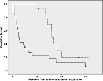
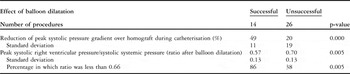
In all, 14 balloon dilatations (35%) were successful according to our definition. In these procedures, the mean reduction of the peak systolic pressure gradient was 49% – standard deviation plus or minus 11%, range 33–70%, 14 procedures. Re-intervention or replacement of the homograft was postponed by a mean of 2 years and 10 months – standard deviation plus or minus 12 months, range 19 months to 5 years and 2 months, 14 procedures. A total of 26 balloon dilatations were unsuccessful, with a mean reduction of the peak systolic pressure gradient over the homograft of 20% – standard deviation plus or minus 19%, range 0–68%, 26 procedures. Re-intervention or replacement of the homograft was postponed by a mean of 1 year and 1 month – standard deviation plus or minus 17 months, range 0 months to 5 years and 1 month, 26 procedures. Homograft replacement was postponed by more than 1.5 years in five procedures in this group, because the right ventricular systolic pressure during catheterisation was reduced to less than half of the systolic systemic pressure, making immediate replacement of the homograft unnecessary. However, these five procedures did not meet the second criterion of a more than 33% peak systolic pressure gradient reduction, and therefore they were not included in the group of successful dilatations. The freedom from re-intervention or replacement of the homograft in the group of successful and unsuccessful procedures is presented in Figure 2. The effect of the balloon dilatation in both groups is summarised in Table 2.
Freedom from re-intervention or replacement of the homograft after balloon dilatation in months, for the group of procedures that were successful (dotted line) and the group of procedures that were unsuccessful (continuous line) (p-value of 0.004, 40 procedures). Cases that did not reach the end-point (re-intervention or replacement of the homograft) before the end of the study period are depicted in boxes or circles (censored cases) in the two different groups.

Effect of balloon dilatation in successful and unsuccessful procedures.

Factors influencing longevity of homograft
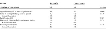
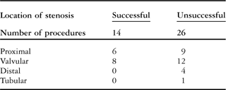
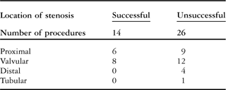
The two groups of procedures were compared for factors that might be able to predict or influence the success of dilatation. The number of procedures in which a pulmonary homograft was implanted versus the number of aortic homografts was the same in both groups. In addition, the location of the stenosis did not seem to differ in the two groups, but the subgroups were too small to test significancy. On the other hand, in the group of successful procedures, the time that the homografts had been in situ tended to be longer, the presence of calcification tended to be higher, the homograft/balloon ratio tended to be lower, and the pressure applied during balloon dilatation tended to be higher; however, none of these factors was statistically significant (see Tables 3 and 4).
Factors influencing longevity of homograft in successful and unsuccessful procedures.

Location of stenosis in homograft in successful and unsuccessful procedures.
Complications
Balloon dilatation in this series of procedures did not result in any mortality, during the procedure or at follow-up. There were two major complications. In one patient, the Tyshak balloon ruptured and had to be surgically removed out of the femoral vein, without further sequelae. In another patient, the homograft (13 millimetres) ruptured after dilatation using an oversized Mullins balloon (15 millimetres). The patient underwent surgical valve replacement the next day and his outcome was favourable. In four other cases, an oversized balloon (homograft/balloon ratio of 0.8) was used without any complications. In all other cases, the balloon size was equal or smaller than the size of the homograft. During follow-up of the patients in this study, no patient needed treatment for haemodynamically important pulmonary regurgitation.
Discussion
In this study, we examined the effect of balloon dilatation of homograft stenosis in pulmonary position. The mean reduction of the peak systolic pressure gradient over the homograft during cardiac catheterisation in all procedures was 30%. Re-intervention or replacement of the homograft was postponed by a mean of 1 year and 7 months. We found that 14 (35%) out of 40 dilatations were successful according to our definition: a clear reduction of the peak systolic pressure gradient over the homograft of more than 33% and postponement of re-intervention or replacement of the homograft by more than of 1 year and 6 months. Whether or not a balloon dilatation can be designated successfully is arbitrary, and to our knowledge there are no internationally accepted criteria. In the short term, success depends on the immediate and significant reduction in right ventricular pressure and the peak systolic pressure gradient over the homograft after intervention. In the long run, success depends on the increase in longevity of the homograft. Owing to the fact that 35% of the patients in this study had a successful procedure, we tried to find factors that might predict the chance of success, and therefore might be of use in patient selection, and factors that might improve the success of dilatation.
Factors influencing longevity of the homograft
In this series of procedures, there was a trend towards a better result in procedures with a homograft that had been in situ for a longer period of time and in which there was calcification of the homograft. Larger series of procedures are needed to evaluate whether this trend is significant. The cause of this phenomenon is not clear, but homografts tend to calcify over time and a limited rupture of the calcified homograft could be an integral part of the success of the dilatation. This finding contrasts with the report of Powell et al,Reference Powell, Lock, Keane and Perry2 who state that “angiographic improvement in the antero-posterior view was most strongly associated with the absence of calcification and younger conduit age in a multivariate model”. However, the outcome variable in this study was an angiographic improvement in the antero-posterior view and is difficult to compare with our outcome variable: reduction of the peak systolic pressure gradient and postponement of surgery.
Balloon pressure and balloon size compared with initial homograft size might be factors that can influence the effect of balloon dilatation. Although these factors seem to correlate with the effect, in this study no significant correlation was found between the pressure applied in the balloon and the success of the dilatation. We did use high-pressure balloons to dilate the stenosis in the patients described in this study. Sohn et alReference Sohn, Kashani and Rothman10 report that “high-pressure balloons may result in better gradient relief by eliminating the balloon waist in a similar manner as that reported for angioplasty of branch pulmonary artery stenosis”. Furthermore, it is not clear whether balloon size might influence the outcome of dilatation. In cases of porcine bioprosthetic valves in pulmonary position, it is described that a balloon should be used with the same diameter as the valve annulus.Reference Waldman, Schoen, Kirkpatrick, Mathewson, George and Lamberti11 On the other hand, Zeevi et alReference Zeevi, Keane, Perry and Lock8 illustrate that there was no significant difference in balloon/valve ratio in the successful and unsuccessful cases of balloon dilatation. In our series of procedures, we could not find a significant difference related to the homograft diameter/balloon diameter ratio. A larger prospective study would be necessary to analyse the effect of balloon size and the pressure applied in the balloons.
The location of the stenosis did not correlate with the outcome in this study. Sanatani et alReference Sanatani, Potts, Human, Sandor, Patterson and Gordon Culham12 confirm this finding and report that the location of the obstruction was not predictive of the response to balloon dilatation. On the other hand, other authors describe that balloon dilatation was most successful in patients with a valvular stenosis and not in those who had a proximal stenosis.Reference Zeevi, Keane, Perry and Lock8, Reference Sohn, Kashani and Rothman10, Reference Lloyd, Marvin, Mahoney and Lauer13
Approach to homograft stenosis
The best treatment option for homograft stenosis is still unclear.Reference Hanley5, Reference Yuan, Mishaly, Shinfeld and Raanani6 As reported, we treated patients with balloon dilatation and not by implanting stents in the homograft because of the risk of severe regurgitation. Homograft regurgitation can also be a complication of balloon dilatation.Reference Sreeram, Hutter and Silove14 However, during follow-up of the patients in this study, no patient needed treatment for haemodynamically important regurgitation, and several authors confirm that there is no increase in the severity of regurgitation after balloon dilatation alone.Reference Waldman, Schoen, Kirkpatrick, Mathewson, George and Lamberti11, Reference Sanatani, Potts, Human, Sandor, Patterson and Gordon Culham12 The use of stents to reduce the stenosis has been reported to be successful.Reference Powell, Lock, Keane and Perry2, Reference Aggarwal, Garekar, Forbes and Turner15, Reference Peng, McElhinney and Nugent16 However, these reports also mention an increase in homograft regurgitation when the stent is placed over the valve, therefore creating an increased volume load to the right ventricle. This may increase susceptibility to arrhythmias and right ventricular dysfunction, which can be irreversible in some cases.Reference Aggarwal, Garekar, Forbes and Turner15
In 2000, a new technique was introduced by Bonhoeffer, creating a method for percutaneous implantation of a bovine pulmonary valve in a stent in pulmonary position. It is now an accepted treatment option for right ventricular to pulmonary (graft) obstruction in adolescents and adults with good medium-term results.Reference Zeevi, Keane, Perry and Lock8, Reference Nordmeyer, Coats and Lurz17, Reference Lurz, Bonhoeffer and Taylor18 Unfortunately, at this moment the technique is not suitable for younger children under 25 kilograms, because of the delivery system of more than 7 millimetres.Reference Bokenkamp, Hazekamp, Schalij, Clur, Ottenkamp and Blom19 Research is done to expand the use to the younger age group.Reference Boudjemline, Laborde and Pineau20, Reference Clur, Baan, Lurz, Bonhoeffer and Ottenkamp21 However, in the long run, these bovine grafts can be expected to degenerate, as is experienced in the surgical implanted bovine grafts, and therefore long-term follow-up studies are in progress.
Balloon dilatation seems to be a safe and valuable way to postpone surgical pulmonary valve replacement or percutaneous pulmonary valve implantation for a subgroup of patients, according to the data presented in this study. Especially in young children, balloon dilatation might be of clinical significance if the homograft stenosis is caused by degeneration or calcification and is not the result of the child outgrowing the homograft. In these cases, balloon dilatation will delay surgical pulmonary valve replacement or gain enough time to consider percutaneous pulmonary valve implantation.
Limitations
In literature, no uniform criteria are present to define failure of the homograft in the case of stenosis and the need for catheter intervention or surgical homograft replacement. Some authors describe that a peak systolic pressure gradient in excess of 50 millimetres of mercury is used as an indication for intervention.Reference Tatebe, Nagakura, Boyle and Duncan3 Others use a ratio of the right ventricle to systemic blood pressure of more than 0.65 as an indication for intervention.Reference Aggarwal, Garekar, Forbes and Turner15 As our study was retrospective in design, we cannot exclude small changes in indications for balloon dilatations, re-interventions or replacements of homografts over the years. This problem has been reported in comparable studies,Reference Powell, Lock, Keane and Perry2 and they even report that “the threshold for surgical intervention might have been influenced by the prior intervention”. We cannot exclude that this happened in our study too.
This study was performed retrospectively and the echocardiographic data of the pulmonary regurgitation after balloon dilatation was not sufficiently documented to draw more specific statistical conclusions on the change of regurgitation. Unfortunately, the same applies to the right ventricular function, which is also known to be difficult to measure reliably,Reference Helbing, Bosch and Maliepaard22 and to tricuspid insufficiency.
Although this is the largest series reported in children, adolescents, and young adults, the numbers are still too small to draw conclusions on factors influencing longevity after balloon dilatation and on subgroups of patients. Larger series are needed. Goals for further study include refining patient selection for the different techniques, optimising timing of the proper intervention and performing long-term follow-up.
Conclusion
In this series of balloon dilatations of stenotic homografts, peak systolic pressure gradients over the homograft were reduced by a mean of 30% and re-intervention or replacement of the homograft was postponed by a mean of 1 year and 7 months. According to our definition, it was successful in 35%, with a mean reduction of the peak systolic pressure gradient over the homograft of 49% and a mean postponement of homograft replacement of 2 years and 10 months. This study indicates that balloon dilatation alone is a safe procedure and can be of clinical significance in a subgroup of patients, postponing either percutaneous valve implantation or surgical pulmonary valve replacement.