

Introduction
The family Diplostomidae is well represented worldwide (Behrmann-Godel, Reference Behrmann-Godel2013; Georgieva et al., Reference Georgieva, Soldánová, Pérez-del-Olmo, Dangel, Sitko, Sures and Kostadinova2013; Locke et al., Reference Locke, Al-Nasiri and Caffara2015; Gordy & Hanington, Reference Gordy and Hanington2019; Hoogendoorn et al., Reference Hoogendoorn, Smit and Kudlai2020). In Argentina, Drago & Lunaschi (Reference Drago and Lunaschi2015) have reported the adults of the following eight genera (number of species in parenthesis): Austrodiplostomum Szidat & Nani, 1951 (2); Diplostomum von Nordmann, 1832 (1); Dolichorchis Dubois, Reference Dubois1961 (2); Hysteromorpha Lutz, 1931 (1); Neodiplostomum Railliet, 1919 (1); Posthodiplostomum Dubois, 1936 (6); Sphincterodiplostomum Dubois, 1936 (1); and Tylodelphys Diesing, 1850 (3). The type-genus Diplostomum Von Nordmann, 1832 has a complex systematic and taxonomic history. It was divided into three subgenera, Diplostomum, Tylodephys and Dolichorchis (Dubois, Reference Dubois1961), which were subsequently raised to the genus level (Niewiadomska & Laskowski, Reference Niewiadomska and Laskowski2002). The genus Dolichorchis shows a combination of traits of the other genera, such as having an asymmetrical anterior testis and a symmetrical and bilobed posterior testis with elongated lobes (as in Diplostomum), and a copulatory bursa enclosing the genital cone (as in Tylodelphys) (Niewiadomska & Laskowski, Reference Niewiadomska and Laskowski2002).
The genus Dolichorchis is distributed in South America (Dolichorchis bonariensis Ostrowski de Nuñez, 1970; Dolichorchis lacombeensis Lunaschi & Drago, 2006), Africa (Dolichorchis tregenna Nazmi Gohar, 1932; Dolichorchis marahoueense Baer, 1957), Asia (Dolichorchis buteii Vidyarthi, 1937; Dolichorchis ketupanense ketupanense Vidyarthi, 1937; Dolichorchis heronei Srivastava, 1954; Dolichorchis ketupanense vietnamiae Odening, 1963) and Oceania (Dolichorchis auriculosum Dubois et Pearson, 1967 and Dolichorchis galaxiae Smith et Hickman, 1983). Sudarikov (Reference Sudarikov and Skrjabin1971) suggested that Dolichorchis mashonense may belong to the genus Tylodelphys and this was further supported by molecular evidence (Chibwana et al., Reference Chibwana, Blasco-Costa, Georgieva, Hosea, Nkwengulila, Scholz and Kostadinova2013). So far, the only known metacercariae for this genus are D. tregenna from Africa (Khalil, Reference Khalil1963), D. galaxiae from Oceania (Smith & Hickman, 1983) and D. heronei from Asia (Srivastava, 1954).
The difficulty in identifying diplostomid metacercariae to the species level has been attributed to their morphological variation depending on host species, size and age, and on infection density, among other factors (Pelegrini et al., Reference Pelegrini, Gião, Vieira, Müller, da Silva, de León, Kozlowiski de Azevedo and Abdallah2019). In addition, the recovery of adult flukes from the definitive hosts and the experimental completion of the life cycle are hard tasks to accomplish. Such limitations can be overcome by applying an integrative approach that combines morphological and molecular data (Laskowski & Niewiadomska, Reference Laskowski and Niewiadomska2002; Chibwana & Nkwengulila, Reference Chibwana and Nkwengulila2010; Chibwana et al., Reference Chibwana, Blasco-Costa, Georgieva, Hosea, Nkwengulila, Scholz and Kostadinova2013; Otachi et al., Reference Otachi, Locke, Jirsa, Fellner-Frank and Marcogliese2014; García-Varela et al., Reference García-Varela, Sereno-Uribe, Pinacho-Pinacho, Domínguez-Domínguez and Pérez-Ponce de León2016a, Reference García-Varela, Sereno-Uribe, Pinacho-Pinacho, Hernández-Cruz and Pérez-Ponce de León2016b; Blasco-Costa & Locke, Reference Blasco-Costa and Locke2017). Until now, life-cycle studies of Argentinean diplostomids have exclusively focused on characterizing the developmental stages recovered from experimentally infected intermediate (e.g. snails and fishes) and definitive hosts (e.g. chickens and rats) (Ostrowski de Núñez, Reference Ostrowski de Núñez1973, Reference Ostrowski de Núñez2017; Ritossa et al., Reference Ritossa, Flores and Viozzi2013, among others). On the other hand, sequences of diplostomid metacercariae from Argentina have been only published for Austrodisplostomum mordax (Sereno-Uribe et al., Reference Sereno-Uribe, Andrade-Gómez, de León and García-Varela2019b), Tylodelphys jenynsiae (Locke et al., Reference Locke, Al-Nasiri and Caffara2015) and Diplostomum spp. (Locke et al., Reference Locke, Drago, Núñez, Souza and Takemoto2020).
During a parasitological survey of fishes from Iguazú National Park, Misiones, Argentina, we detected diplostomid metacercariae in the brain of Erythrinus cf. erythrinus (Bloch & Schneider, 1801). The objective of this work was to determine the identity and phylogenetic affiliation of this parasite based on morphological and molecular analyses.
Materials and methods
Sample collection
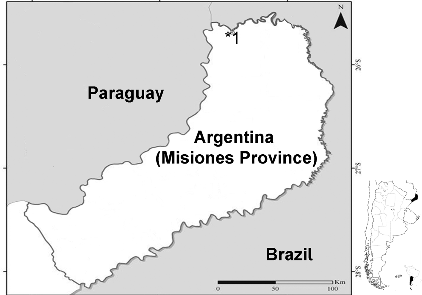
In autumn and spring 2016, fish of the genus E. cf. erythrinus were collected from an unnamed stream upstream from the waterfalls in the basin of the Iguazú River (fig. 1) using crab traps. Fish were transported alive to the field laboratory, where they were cold-anaesthetized and euthanized by cervical dissection. After measuring body weight, total length and standard length, animals were necropsied. Digeneans (metacercariae) found in the brain were removed, heat-killed in water and fixed in 10% formalin. For molecular analysis, live specimens were preserved in cold 96% ethanol and stored until DNA extraction.
Sampling site of Erythrinus cf. erythrinus with Dolichorchis lacombeensis metacercaria in the brain. The asterisk indicates the location of the unnamed stream from Iguazú National Park (Argentina).

DNA extraction, amplification and sequencing
Total genomic DNA was extracted from two individual specimens using Wizard® Genomic DNA Purification Kit (Promega) according to the manufacturer's protocol. The partial fragment of each gene was amplified for each specimen using polymerase chain reaction (PCR) on an Eppendorf Mastercycler thermal cycler (Hamburg, Germany). Amplification of the partial sequence of the 28S rDNA gene (28S) was performed with the forward primer LSU-5 (5′-TAGGTCGACCCGCTGAAYTTAAGCA-3′) (Littlewood & Olson, Reference Littlewood, Olson, Littlewood and Bray2001) and the reverse primer 1500R (5′ -GCTATCCTGAGGGAAACTTCG-3′) (Tkach et al., Reference Tkach, Littlewood, Olson, Kinsella and Swidersk2003). The fragment of the internal transcribed spacer (ITS)1-5.8S-ITS2 gene was amplified using the forward primer MITFF (5′-CGTAACAAGGTTTCCGTAG-3′) (Tkach & Curran, Reference Tkach and Curran2015). A fragment of the partial cytochrome c oxidase subunit I (COI) were amplified using the forward primer DICE 1 F (5′-ATTAACCCTCACTAAATT WCNTTRGATCATAAG- 3′) and the reverse primer DICE 14R (5′ –TAATACGAC TCACTATACCHACMRTAAACATATGATG- 3′), as proposed by Van Steenkiste et al. (Reference Van Steenkiste, Locke, Castelin, Marcogliese and Abbott2015). The PCRs were made with GoTAQ Master Mix (Promega) according to the manufacturer's protocol. Amplification was done under conditions based on the protocols described for 28S and ITS by Tkach et al. (Reference Tkach, Littlewood, Olson, Kinsella and Swidersk2003), and for COI by Montes et al. (Reference Montes, Barneche, Pagano, Ferrari, Martorelli and de León2021b). Sequencing was performed on an ABI 3730XL sequencer by Macrogen Inc. (Korea).
Sequence comparison and phylogenetic analysis
Sequences obtained in this study were assembled using the platform Geneious 5.0.4 (Drummond et al., Reference Drummond, Ashton, Buxton, Cheung, Cooper, Duran and Wilson2016). COI sequences were checked for the presence of pseudogenes using the translated amino acid sequences based on the invertebrate mitochondrial genetic code, in Geneious 5.0.4. All the resulting sequences of 28S, ITS1 and COI (table 1) were used to search for homologous sequences in GenBank with Basic Local Alignment Search Tool (BLAST). Sequences were aligned using the online version of MAFFT 7 (Katoh & Standley, Reference Katoh and Standley2013), yielding three matrices, one for each gene. Ambiguously aligned, hypervariable regions in the 28S rDNA and ITS1-5.8S-ITS2 rDNA dataset were removed with Gblocks online version 0.91b (Castresana, Reference Castresana2000; Talavera & Castresana, Reference Talavera and Castresana2007), according to a secondary structure model, with the parameter settings of a less stringent selection (allowing smaller final blocks, gap position within the final blocks and less strict flanking positions). Two species of Clinostomum were used as outgroup (table 1). The best partitioning scheme and substitution model for the DNA partition was chosen under the Bayesian information criterion (Schwarz, Reference Schwarz1978), using JModelTest 2.1.4 (Darriba et al., Reference Darriba, Taboada, Doallo and Posada2012). The appropriate nucleotide substitution models resulting after the Gblock program were TVM + I + G for the 28S matrix and TPM2uf + G for the ITS matrix. The COI dataset was partitioned into first-, second-, and third- codon positions with the appropriate nucleotide substitution model implemented for each codon position (TPM1uf + I + G for the first, HKY + G for the second and TIM3 + I + G for the third codon position).
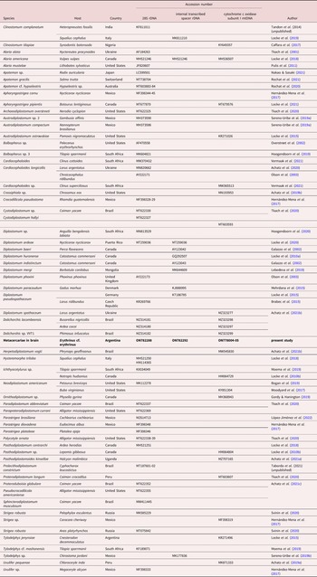
Collection data and GenBank accession numbers for the digeneans analysed in this study.

New sequences in boldface type.
Phylogenetic reconstruction was carried out using Bayesian inference (BI) through MrBayes 3.2.3 (Ronquist et al., Reference Ronquist, Teslenkovan and van der Mark2012). Phylogenetic trees were constructed using two parallel analyses of metropolis-coupled Markov chain Monte Carlo) for 20 million generations each, to estimate the posterior probability (PP) distribution. Topologies were sampled every 1000 generations and the average standard deviation (SD) of split frequencies was observed to be less than 0.01, as suggested by Ronquist et al. (Reference Ronquist, Teslenkovan and van der Mark2012). The robustness of the clades was assessed using Bayesian PP, where PP > 0.90 was considered strongly supported. A majority consensus tree with branch lengths was reconstructed for each run after discarding the first 25% of sampled trees.
Additionally, the proportion (p) of absolute nucleotide sites (p-distance) (Nei & Kumar, Reference Nei and Kumar2000) was obtained (including gaps in pairwise comparison) to compare the genetic distance among lineages. The P-value matrix was obtained using MEGA X with the bootstrap method (1000 replicates) and with nucleotide substitution (transition + transversions) uniform rate, and gaps/missing data treatment as complete deletion (Kumar et al., Reference Kumar, Stecher, Li, Knyaz and Tamura2018). Newly generated sequences were submitted to the National Center for Biotechnology Information GenBank database (http:// www.ncbi.nlm).
Morphological analysis
Specimens preserved in 10% formalin solution were whole-mounted, stained with hydrochloric carmine and processed according to standard parasitological techniques (Pritchard & Kruse, Reference Pritchard and Kruse1982). Digital images of the specimens were taken with an Olympus Bx51 microscope equipped with an AmScope MU 1000 10 MP digital camera. Measurements were made using ImageJ software (Schneider et al., Reference Schneider, Rasband and Eliceiri2012). Drawings were made with the aid of a drawing tube.
Ten metacercariae were photographed and the structures measured using the program ImageJ. The mean followed by minimum and maximum values in parentheses are given in micrometres (μm). The 95% Bayesian credible intervals (95%BCIs), the mean, SD, and the 2.5th and 97.5th percentiles of the distribution were calculated for each character. The software WinBUGS 1.4.3 (Lunn et al., Reference Lunn, Thomas, Best and Spiegelhalter2000) was used to generate 100,000 samples (with normal and noninformative prior distribution) from the posterior distributions for each of the analyses after discarding the initial 10,000 samples as ‘burn in’. The Bayesian approach is based on the posterior distribution where the parameters are random quantities. Therefore, in the BCIs, the mean of a posterior distribution is the estimate of the parameter and its percentiles provide the credible intervals (Stokes et al., Reference Stokes, Chen and Gunes2014).
Results
Molecular analyses
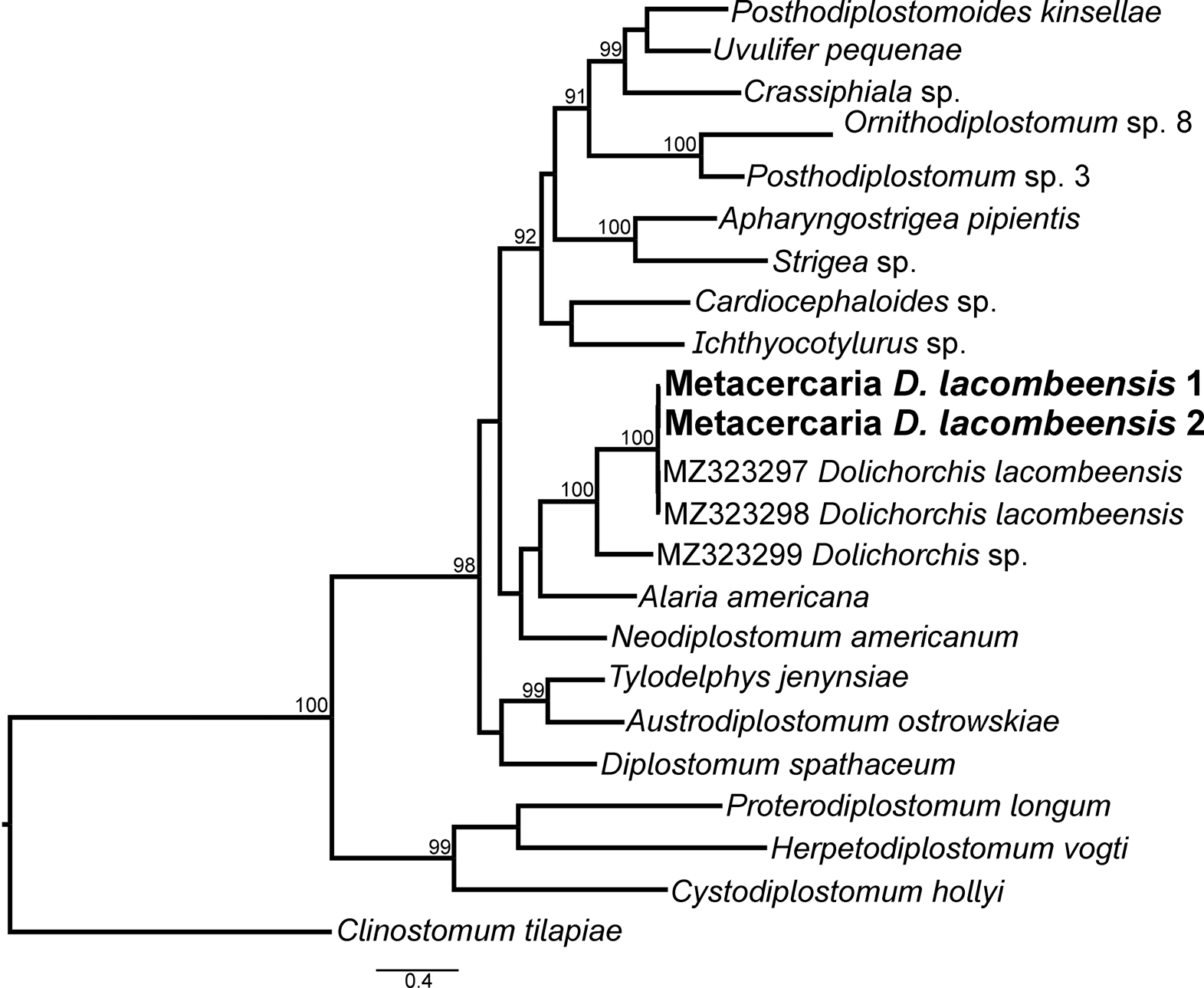
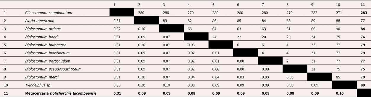
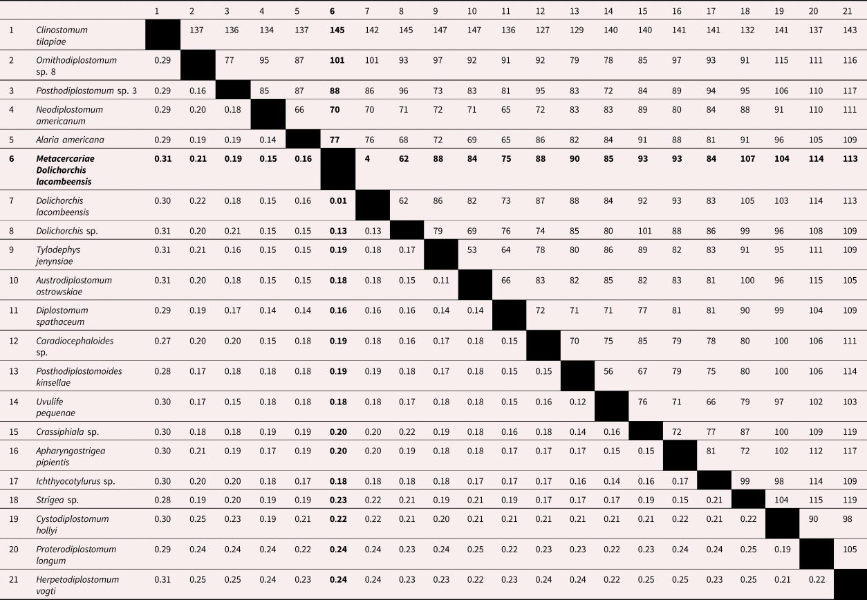
We obtained four sequences, one of 28S, one of ITS and two of COI. The sequence of 28S was 1223 base pairs (bp) long and the matrix was constructed with 50 taxa and 1263 bp. The ITS sequence was 1075 bp long and the matrix had 11 taxa and 1182 bp. The COI sequences were 651–657 bp long and the matrices were generated with 23 taxa and 812 bp.
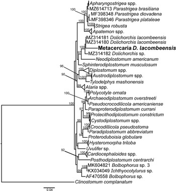
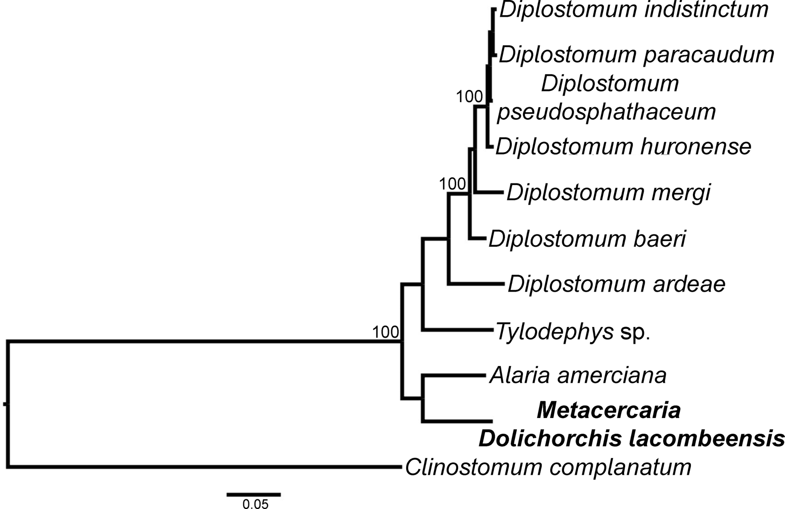
In the 28S phylogram (fig. 2), the metacercaria from E. cf. erythrinus is clustered with Dolichorchis sp., with a p-distance of 0.00 between them (table 2). In the ITS phylogram (fig. 3) this metacercaria is clustered with Alaria americana Hall & Wigdor, 1918; the genetic distances between this metacercaria with A. americana, Diplostomum spp. and Tylodelphys sp. are 0.09, 0.08–0.09 and 0.10, respectively (table 3). In the COI phylogram (fig. 4), the metacercariae are clustered with D. lacombeensis and a p-distance of 0.01 (table 4). The COI sequences corroborated those metacercariae as D. lacombeensis.
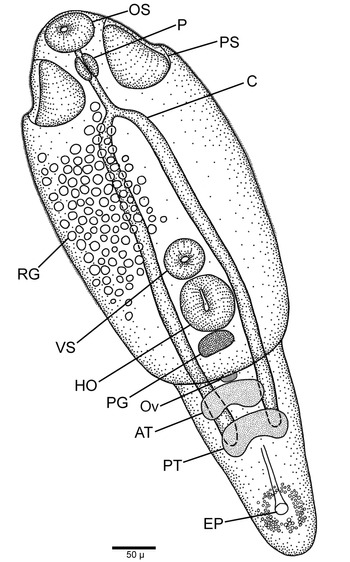
Line drawing of metacercaria of Dolichorchis lacombeensis. Whole specimen in ventral view. Abbreviations: AT, anterior testis; C, ceca; EP, excretory pore; HO, holdfast organ; OS, oral sucker; Ov. ovary, P, pharynx; PG, proteolytic glands; PS, pseudosucker; PT, posterior testis; RG, refringent granules; and VS, ventral sucker.

Phylogram from Bayesian inference of partial 28S rDNA gene sequences of Dolichorchis lacombeensis metacercaria found in the brain of Erythrinus cf. erythrinus from Iguazú National Park (Argentina).

Phylogram from Bayesian inference of partial internal transcribed spacer ITS gene sequences of Dolichorchis lacombeensis metacercaria found in the brain of Erythrinus cf. erythrinus from Iguazú National Park (Argentina).

Species of family Diplostomidae analysed for the 28S rDNA gene.

Genetic divergence (uncorrected p-distance) and number of nucleotide difference are below and above the diagonal line, respectively. New sequences in boldface type.
Species of family Diplostomidae analysed for the internal transcribed spacer rDNA gene.

Genetic divergence (uncorrected p-distance) and number of nucleotide difference are below and above the diagonal line, respectively. New sequences in boldface type.
Species of family Diplostomidae analysed for the cytochrome c oxidase subunit I mtDNA gene.

Genetic divergence (uncorrected p-distance) and number of nucleotide difference are below and above the diagonal line, respectively. New sequences in boldface type.
Morphological analysis
(fig. 5)
Phylogram resulting from Bayesian inference of partial cytochrome c oxidase subunit I mtDNA gene sequences of Dolichorchis lacombeensis metacercariae found in the brain of Erythrinus cf. erythrinus from Iguazú National Park (Argentina).

Description based on 10 whole-mounted specimens: Body fusiform, bipartite, 495–696 (595) long. Forebody spatulate, 425–471 (448) long and 206–250 (228) wide, slightly concave ventrally; hind-body conical, 181–229 (205) long and 129–149 (139) wide. Forebody/hind-body length ratio 1:1.97–2.46 (1:2.22). Anterior part of forebody to ventral sucker or holdfast organ covered with fine spines. Oral sucker 47–53 (50) long and 58–64 (61) wide, sub-terminal, nearly same size as ventral sucker. Ventral sucker 42–46 (44) long × 46–50 (48) wide, located in middle third of forebody. Pseudosuckers 52–62 (68) long × 28–36 (32) wide. Oral sucker/pseudosuckers length ratio 1:1.16–1.27 (1:1.22). Body/pseudosuckers length ratio 1:9.63–11.26 (1:10.44). Forebody/pseudosuckers length ratio 1:6.53–8.05 (1:7.30). Distance between oral sucker and ventral sucker 214–240 (227). Distance between suckers/forebody length ratio 0.5–0.51. Suckers’ width ratio 1:1.03–1.19 (1:1.11). Holdfast organ oval, 65–77 (71) long × 57–72 (64) wide, immediately posterior to ventral suck. Distance between ventral sucker and holdfast 5–9 (7); proteolytic gland below holdfast organ. Body/holdfast organ length ratio 1:7.23–9.25 (1:8.24). Forebody/holdfast organ length ratio 1:5–6.46 (1:5.74). Prepharynx short; pharynx 41–54 (48) long × 23–26 (24) wide. Oesophagus 25–30 (27), intestinal ceca reach to middle of posterior testis. Oral sucker/pharynx length ratio 1:0.93–1.19 (1:1.06). Pseudosuckers/pharynx length ratio 1:1.21–1.43 (1:1.32). Primordia of testes in tandem, relatively large, extended transversally occupying hind-body. Anterior and posterior testes symmetrical, curved, bilobed, not always visible. Ovary median, ellipsoidal, pretesticular at junction of forebody and hind-body, not always visible. Excretory pore terminal.
Taxonomic summary
Metacercaria host: Erythrinus cf. erythrinus.
Type host: Ardea cocoi (L) (Argentina).
Other hosts from Brazil: Ardea cocoi (L) and Busarellus nigricollis (Latham).
Site of infection: brain.
Type locality and collection date: unnamed stream in Iguazú National Park (25° 40′ 49″ S 54° 26′ 55″ W). March and September 2016.
Prevalence of infestation: 100% (n = 30 fish).
Mean intensity and abundance: over 200 metacercariae per infected host.
Voucher material: MLP-He 7768; deposited in the Museo de Ciencias Naturales, La Plata, Buenos Aires, Argentina.
Sequences of adult Dolichorchis lacombeensis from Brazil: the 28S rDNA sequences MZ314180–81. The COI mtDNA sequences MZ323297–98.
New sequences of Dolichorchis lacombeensis metacercariae: the 28S rDNA sequence ON782288, the ITS rDNA sequence ON782292 and the COI mtDNA sequences ON778004–05.
Remarks
The DNA analysis indicated that the metacercariae from E. cf. erythrinus belonged to D. lacombeensis. This species only were reported for Lunaschi and Drago (Reference Lunaschi and Drago2006) when describing the species and for Achatz et al. (Reference Achatz, Martens and Kostadinova2021b) reporting the 28S and COI sequences for the species. Therefore, below we offer a morphological comparison between the adult described by Lunaschi and Drago (Reference Lunaschi and Drago2006) and the metacercaria under study.
The length of the metacercaria is almost half that of the adult. The forebody/hind-body length ratio differs between stages: the forebody of the metacercaria is almost twice as long as its hind-body (1:1.97–2.46), while the ratio becomes smaller in the adult (1:1.25–1.53) due to the enlargement of the hind-body by the development of the genital complex. The similarity in the oral sucker/pseudosuckers length ratio between stages (1.16–1.27 and 1.10–1.20 for the metacercaria and adult, respectively), indicates that their size remains almost unchanged in the adult. The ratios of body and forebody length to pseudosuckers’ length are smaller in the metacercaria because the adult is longer (total body length: 495–696 vs. 1027–1123; forebody length: 425–471 vs. 619–720). The suckers’ width ratio is almost the same in the metacercaria (1.03–1.19) and the adult (0.90–1.00), suggesting that their size remains unchanged between these stages.
The body/holdfast length ratio (7.23–9.25 and 8.00–8.90 for the metacercaria and adult, respectively) and the forebody/holdfast length ratio (5.00–6.46 and 4.90–5.70 for the metacercaria and adult, respectively) appear not to vary with growth.
The oral sucker/pharynx length ratio is smaller for the metacercaria (0.93–1.19) than for the adult (1.40–1.50). Considering that pharynx length does not vary with growth (41–54 × 23–26 in the metacercaria and 46–51 × 23–31 in the adult), this difference seems to be due to the smaller oral sucker of the metacercaria (47–53 × 58–64 vs. 67–78 × 74–78).
The pseudosuckers/pharynx length ratio is similar in both stages (1.21–1.43 in the metacercaria and 1.2–1.4 in the adult) despite the larger size of the adult.
It is worthwhile to note that although the distance between the oral and ventral suckers is larger for the adult (269–361 vs. 214–240), the relationship of this distance to forebody length is slightly greater for the metacercaria (0.50–0.51 vs. 0.43–0.50).
The distance between ventral sucker and holdfast organ is smaller in the metacercaria (5–7 vs.14–29), indicating that these structures become more separated with growth. This would be related to the site of infection: the larger distance between suckers in the adult may allow better attachment to overcome the strong peristaltic movement in the intestine of the bird host.
The oesophagus length remains almost similar in both stages (25–30 in the metacercariae and 19–34 in the adult).
Discussion
When we collected the metacercariae from E. cf. erythrinus there were few experimental studies on the life cycles of Diplostomidae from Argentina, thus precluding their identification to the genus level. In the present work, the use of molecular methods provided valuable insight into the phylogeny of this family. In the ITS tree, the metacercariae were clustered with A. americana, but the fact that this gene was poorly represented in the GenBank database made it less reliable for the molecular identification of Diplostomidae genera. The analysis of the 28S gene sequence searched using the BLAST tool of GenBank indicated that the metacercariae belonged to Diplostomidae and that they were closely related to Dolichorchis. Moreover, they were located similarly in the COI and the 28S trees and would belong to D. lacombeensis, based on genetic divergence and number of nucleotide differences.
The comparison of the morphology of the metacercaria from E. cf. erythrinus and the adult of Dolichorchis sp. revealed that some structures (e.g., pharynx, oesophagus and pseudosuckers) reach their final size at the metacercarial stage. The oral sucker, ventral sucker and holdfast organ, as well as the distance between oral and ventral suckers, are larger in the adult but they increase in proportion to features such as body length, forebody and hind-body. On the other hand, hind-body length and width are larger in the adult as a consequence of the development of the genital complex. In addition, the adult shows a larger distance between the ventral sucker and the holdfast organ.
Further studies using an integrative approach that combines morphological and molecular characterizations will help confirm the affiliation of other species to the genus Dolichorchis. For example, using morphological measurements, Chibwana & Nkwengulila (Reference Chibwana and Nkwengulila2010) described three types of metacercariae coexisting in the cranial cavity of Clarias gariepinus (Burchell): Tylodelphys types 1 and 2; and Diplostomum type 3 (=Diplostomum mashonense (Beverley-Burton, 1963)). Later, Chibwana et al. (Reference Chibwana, Blasco-Costa, Georgieva, Hosea, Nkwengulila, Scholz and Kostadinova2013) recognized Dolichorchis mashonense as Tylodelphys mashonense based on the molecular evidence. Interestingly, Chibwana & Nkwengulila (Reference Chibwana and Nkwengulila2016) stated that the adults of T. mashonense and Dolichorchis sp. shared the presence of a genital cone and an asymmetrical anterior testis. These features (symmetrical/asymmetrical anterior testis and genital cone) are used to discriminate among the genera. The genus Dolichorchis shows a combination of traits of the other genera, such as having an asymmetrical anterior testis and a symmetrical and bilobed posterior testis with elongated lobes (as in Diplostomum), and a copulatory bursa enclosing the genital cone (as in Tylodelphys) (Niewiadomska & Laskowski, Reference Niewiadomska and Laskowski2002). If the results obtained by Chibwana & Nkwengulila (Reference Chibwana and Nkwengulila2016) and Chibwana et al. (Reference Chibwana, Blasco-Costa, Georgieva, Hosea, Nkwengulila, Scholz and Kostadinova2013) were not due to sample contamination and the sequence actually belonged to Di. mashoense or to a type of Tylodephys associated with a Dolichorchis morphology, then the characters selected to separate the genera Tylodelphys, Diplostomum and Dolichorchis may not be supported by molecular evidence. Another possibility to consider is that Dolichorchis from different continents are actually cryptic genera.
Acknowledgement
We specially thank Maria Marcia Montes for making the drawings.
Financial support
We are grateful to CONICET, FONCYT and UNLP for financial support, to CEPAVE for providing facilities and equipment. This research was supported by Fondo para la Investigación Científica y Tecnológica (FONCYT) to M.M. Montes (PICT 2016-4153), Universidad Nacional de la Plata to M.M. Montes (PPID N039) and CEPAVE (PUE 3334/16).
Conflicts of interest
None.
Ethical approval
The authors assert that all procedures contributing to this work comply with the ethical standards of the relevant national and institutional guides on the care and use of laboratory animals.