Introduction
Basal cell carcinoma (BCC) is the most frequently encountered non-melanoma skin cancer of the body, with the head and neck region being the most common site of occurrence.Reference Koplin and Zarem1–Reference Manstein, Manstein and Beidas3 The worldwide incidence of BCC varies widely according to geographical location. The highest incidence of BCC has been reported in Australia at approximately 726 per 100 000 person-years, followed by the USA and Europe.Reference Veronese, Farinelli, Zavattaro, Zuccoli, Bonvini and Leigheb4 In China, however, the incidence of BCC is relatively low, at about 1.1 per 100 000 inhabitants each year.Reference Ding and Lin5
The most common aetiological factor involved in the pathogenesis of BCC is ultraviolet radiation, and this is the reason why most of these tumours occur in the head and neck region. The clinical course of small BCC is generally characterised by slow growth, minimal local invasiveness, lower morbidity and a high cure rate. On the other hand, giant BCC, in which the tumour measures 5 cm or greater in diameter,Reference Page, Fleming and Fritz6 is a rare and biologically aggressive variant, which usually carries a higher risk of complications and mortality.
While metastasis of BCC is rare, with an incidence as low as 0.03 per cent,Reference Snow, Sahl, Lo, Mohs, Warner and Dekkinga7 giant BCCs have been associated with a higher incidence of metastasis. Sahl et al. reported that systematic metastasis is prevalent in patients with a giant BCC lesion that exceeds 100 cm2 in area or 25 cm in diameter.Reference Sahl, Snow and Levine8
It is well known that patient neglect or denial may contribute to these large tumours, which have usually been present for an extended period of time. Giant BCC of the head and neck will have a significant influence on patients’ physical functioning, psychological functioning and social interaction because of the exposed nature of these regions. In addition, these large cancers tend to ulcerate and necrose over a long period, creating significant hygienic and aesthetic problems.
Although BCC therapy is relatively straightforward and mortality rates are extremely low,Reference Kiiski, de Vries, Flohil, Bijl, Hofman and Stricker9 giant lesions, as presented in this study, require extensive resection, and reconstruction with a local or free flap, creating surgical challenges and placing a major burden on the limited healthcare resources in many developing countries.
Given the rare nature of giant BCCs, few studies have reported on their incidence, resection and reconstruction, and no studies have been conducted in China. The main purpose of this study, therefore, was to retrospectively analyse the reconstruction methods and surgical outcomes of patients with giant head and neck BCC treated at the Department of Oral and Maxillofacial Surgery of the First Affiliated Hospital of Xinjiang Medical University (one of the main referral hospitals for the treatment of patients with head and neck cancers) over the past 13 years.
Materials and methods
At our hospital, from January 2000 to December 2012, giant BCCs in the head and neck region of 17 patients were surgically excised with subsequent reconstruction. We retrospectively reviewed and analysed the medical charts of these patients to abstract the pertinent information, including: demographic data (e.g. age and sex), symptom duration, lesion size at the time of presentation, lesion anatomical sites (e.g. scalp-forehead, eyelid, temporo-auricular, nasal-cheek or lips-chin-cervical regions), histological type (nodular, superficial or infiltrative), treatment pattern, reconstruction method and treatment outcome. Tumour size was classified as 5–6 cm, 7–8 cm, 9–10 cm, or 11 cm or greater according to the largest diameter of the lesion.
Because of the dimensions of the lesions, all patients consented to wide resection with adequate margins, followed by immediate reconstruction with a local or free flap depending upon the extent of the tumour.
All patients were followed up for at least two years on a regular basis. Follow-up information was obtained from out-patient records, interviews with family members and relevant medical examinations.
The study was approved by the institutional review board of the Division of Medical Ethics of the First Affiliated Hospital of Xinjiang Medical University.
Results
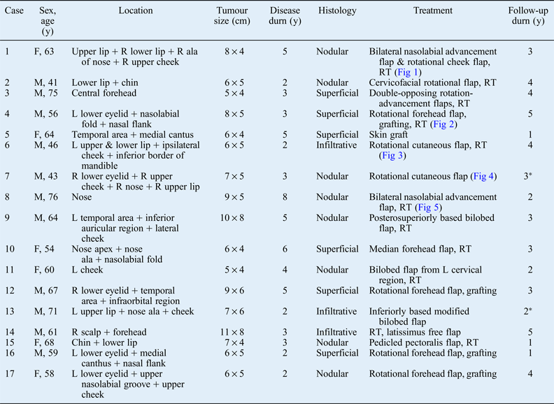
A total of 17 giant BCCs of the head and neck were treated at our hospital during the period from January 2000 to December 2012 (Table I and Figures 1–5). There were 11 (64.7 per cent) men and 6 (35.3 per cent) women in our study, with a male to female ratio of 1.83:1. Median age at presentation was 61 years (range, 41–76 years).
(a) A 63-year-old woman with an 8 × 4 cm nodular basal cell carcinoma of the entire upper lip, extending into the right ala and upper cheek. (b) Intra-operative soft tissue defect after resection of this giant lesion. (c) Reconstruction afterwards with bilateral nasolabial advancement flap and rotational cheek flap. (d) Two-week post-operative view showed reasonable morphology of the upper lip with insignificant scarring.

(a) A 56-year-old man who developed a giant superficial basal cell carcinoma over the left lower eyelid, nasolabial fold and nasal flank during a 3-year period. (b) A forehead free flap measuring 8 × 5 cm was planned; the extent of lesion resection was marked intra-operatively. (c) The lesion was resected with a surgical margin of more than 1 cm and the pedicled forehead flap was raised. (d) The forehead flap and abdominal skin graft were transposed to the defect. (e) View after suturing the flaps. (f) One month post-operatively, the patient presented an acceptable facial appearance given the size of defect being constructed.
(a) A 46-year-old man with giant nodular basal cell carcinoma of the left upper and lower lip, with extension into the ipsilateral cheek and inferior border of the mandibular body. (b) Surgical field after resection of the lesion. (c & d) Primary and final closure of the surgical defects.
(a) A 43-year-old man with large basal cell carcinoma of the right lower eyelid, right upper cheek, nose and right upper lip. (b) The extent of tumour resection was marked and a rotational cheek flap was designed. (c) Giant soft tissue defects of the right half of the face. (d & e) The rotational cheek flap was transposed to the defect and the defect was primarily closed. (f) Intra-operative view after complete closure of the defect.
(a) A 76-year-old man with giant nodular basal cell carcinoma of the nose. (b) The lesion was resected and reconstructed using a bilateral nasolabial advancement flap in a single-stage procedure. (c) The patient presented moderate nasal deformity with surgical scarring at the two-year follow up.
Summary of patients with giant head and neck BCC
*Presented with local recurrence. BCC = basal cell carcinoma; y = years; durn = duration; F = female; R = right; RT = radiotherapy; Fig = Figure; M = male; L = left
The youngest patient was a 41-year-old man with a 6 × 5 cm BCC in the lips-chin-cervical region, while the oldest was a 76-year-old man with a 9 × 5 cm lesion on the nose. Regarding anatomical site, the lesion was located on the scalp and forehead in three cases (17.7 per cent), the inferior orbital region in four cases (23.5 per cent), the temporo-auricular region in two cases (11.8 per cent), the nasal-cheek region in four cases (23.5 per cent), and the lips-chin-cervical region in four cases (23.5 per cent). The largest diameter of the lesions ranged from 5 to 11 cm, with 5–6 cm being the most common size at the time of presentation (Figure 6). The main complaints of the patients were pain, visual disturbance, drooling, discomfort during eating and speaking, odour from the lesion, and severe social interference. Six of the 17 patients reported that they had had their lesions resected at a local hospital and the lesion had become very large thereafter. The time interval between discovery of the lesion to diagnosis ranged from two to five years. None of the patients had evidence of local or distant metastasis at the time of diagnosis. Post-operative histopathological examination was performed for all patients; nodular type was seen most frequently (52.9 per cent), followed by superficial (35.3 per cent) and infiltrative (11.8 per cent) types.
Distribution of tumours according to the largest diameter of each tumour (in centimetres).
Because of the large size of the tumours, all patients underwent wide excision with adequate margins, followed by immediate reconstruction with a local or free flap depending upon the extent of the tumour. All lesions were resected with more than 1 cm surgical margins to obtain clear free margins as validated by intra-operative frozen sections. Three patients presented positive surgical margins (two infiltrative and one solid type) after primary resection; these patients subsequently underwent secondary procedures to confirm a free margin prior to immediate reconstruction.
A rotational forehead flap was used in four patients with giant lower eyelid BCC that extended into the adjacent structures without invading the orbital contents. One patient underwent subtotal rhinectomy and another underwent total upper lip cheilectomy that necessitated extensive resection and reconstruction with a bilateral nasolabial advancement flap. Only one latissimus free flap was used, to reconstruct a huge BCC of the scalp and forehead. Post-operative external radiotherapy was also administered to 11 patients with higher risk tumours.
The most common early complications occurred within 5 days after the operation, and were resolved with conservative treatment. These complications included difficulty with opening and closing the eyelid in two patients, flap inflammation in three patients, and difficulty with breathing in the patient with nasal carcinoma. All patients were followed up for at least one year on a regular basis, with follow-up duration ranging from one to five years. During the follow-up period, two patients had late complications of lower eyelid ectropion, one patient had a moderate nasal deformity, and one patient had difficulty in saliva control due to the lack of lip support.
One 43-year-old man with extensive BCC of the right pre-orbital and cheek region, and another 71-year-old man with infiltrative BCC of the paranasal-lip region, presented with local recurrence at 3 and 2 years follow up, respectively. No tumour metastases have been identified in any of our patients thus far. Patient and family satisfaction has been reasonable given the extensive nature of the surgery, with alleviation of pain, and improved appearance and hygiene.
Discussion
Although small-sized BCC is considered to be the most common cutaneous malignancy in many countries, giant BCC, in which the tumour measures 5 cm or greater in diameter, is a very rare skin malignancy that accounts for less than 1 per cent of all basal cell tumours.Reference Zoccali, Pajand, Papa, Orsini, Lomartire and Giuliani10 Few clinical studies have reported on giant BCC since Eckhoff first described the giant, recurrent, multiple and metastasising BCC in 1951.Reference Eckhoff11 By September 2011, only 91 cases of giant BCC had been reported in the English literature,Reference Zoccali, Pajand, Papa, Orsini, Lomartire and Giuliani10 and tumours located in the head and neck region are even rarer.
While ultraviolet radiation is the primary cause of BCC, the risk factors for development of giant BCC have not yet been clearly defined.Reference Archontaki, Stavrianos, Korkolis, Arnogiannaki, Vassiliadis and Liapakis12 It is generally accepted that giant BCC mainly occurs in patients who have an aggressive histological type (morpheaform, micronodular or metatypical), and in cases where there is a long period of time from discovery to presentation. Some authors have reported patients’ negligence as the leading factor for continuous growth of the tumour over a long period of time. However, Randle et al. considered that inadequate previous treatment of smaller tumours and the resulting local recurrence are the most important variables in the development of such large tumours.Reference Randle, Roenigk and Brodland13 Nevertheless, few scientific studies have endorsed the association between increased growth rate and a prolonged period between discovery and presentation with a large sized tumour.
The large size of lesions in our study is probably a result of either the lengthy period between discovery and presentation or patients’ poor economic status: most of the patients were farmers who came from remote, rural areas of the country, where public health awareness, accessibility of health services, and realisation of diagnosis and therapy are quite low. This study further demonstrates the pivotal role of patient neglect in the development of sizeable tumours, especially given the conspicuity of large facial lesions.
Archontaki et al. claimed that giant BCC correlated with chronic alcoholism, because compulsive and uncontrolled consumption of alcoholic beverages will generate an immune response deficiency in patients.Reference Archontaki, Stavrianos, Korkolis, Arnogiannaki, Vassiliadis and Liapakis12
Our study consisted of 17 cases of head and neck giant BCC, predominantly occurring in the eyelid and paranasal region of the face. The reasons why most of the tumours frequently emerge in the middle-third of the face and why tumours in this region generally run a more aggressive course are still a matter of controversy.Reference Zoccali, Pajand, Papa, Orsini, Lomartire and Giuliani10 Previous studies have attributed this phenomenon to the close proximity of the medial canthus and nasolabial fold to underlying bone and cartilage.Reference Roenigk, Ratz, Bailin and Wheeland14
The histological subtype of the tumour is another key factor for the development of aggressive and giant BCC, and has been closely associated with the clinical course of the tumour. According to the various histological types of BCC, giant BCC can also be divided into non-aggressive and aggressive groups.Reference LeBoit, Burg, Weedon and Sarasin15 Nodular and superficial BCC are tumour subtypes known for less aggressive tendencies, whereas morpheaform and infiltrating varieties carry a greater risk of progressing to giant BCC. Randle et al. reported that the majority of giant BCCs were either micronodular or infiltrative.Reference Randle, Roenigk and Brodland13 Our study also demonstrated the relatively high incidence of the infiltrative subtype in patients with a larger tumour size, but these patients had a shorter disease duration.
Metastatic spread from giant BCC is extremely rare, even in patients with recurrent tumours or aggressive histological subtypes. The metastatic rates for BCC depend largely on the interval between initial discovery and treatment, and the size of the tumour. An overall metastatic rate of 0.03 per cent was reported recently, but the rate increases dramatically to 1.9 per cent when tumour size reaches more than 3.0 cm in diameter.Reference Snow, Sahl, Lo, Mohs, Warner and Dekkinga7 Snow et al. further stated that, in tumours larger than 10 cm in diameter, the incidence of metastasis and/or fatal outcome was 45 per cent.Reference Snow, Sahl, Lo, Mohs, Warner and Dekkinga7 Lymph nodes are the most frequent site of distal spread (40–83 per cent), followed by the lungs (35–53 per cent), bone (20–28 per cent), skin (10–17 per cent) and liver (9 per cent).Reference Martin, Edwards, Cawte, Sewell and McMasters16, Reference Talmi, Gal, Finkelstein, Nobel, Shvilli and Zohar17 Metastases occur in men more than women, at a 2:1 ratio, with a median survival range (after metastasis) of 8–14 months.Reference Ting, Kasper and Arlette18 Metastases will have a significant impact on patients’ quality of life, and survival diminishes remarkably if they have either local or distant metastasis. Fortunately, none of our patients had any metastasis before the operation or during follow up.
• This study presents 17 cases of giant basal cell carcinoma (BCC) of the head and neck
• The lesions had grown over a mean period of four years mainly due to patient neglect and inappropriate earlier treatment
• Aggressive and giant BCCs pose severe cosmetic, functional and social problems
• Treatment of these large tumours is surgically challenging
• All patients underwent resection and reconstruction using a single-stage procedure, with acceptable outcomes and no severe post-operative complications
• Head and neck giant BCC can be successfully treated with a local flap or skin graft in a single-stage approach
The treatment pattern is of paramount importance, especially when aiming to achieve a low local recurrence rate and longer survival. Surgical resection is the treatment of choice for giant BCC, with cure rates averaging 90–91 per cent.Reference Dubin and Kopf19 Given the high residual tumour rate, a wide surgical excision with adequate free margins (more than 1 cm) is extremely important for reducing post-operative recurrence and, in turn, for long-term survival. However, extensive resection of giant BCC of the face may not only result in extirpation of important organs such as the eye, ear, nose and lips, but may also create large complex soft tissue and bone defects, with exenteration of the orbit and brain exposure. Loss of these parts of the human body will have a significant negative influence on patients’ cosmetic appearance, physical and psychological functioning, and social interaction. Histological margin control, therefore, plays a major role in reducing potential recurrences, while sparing normal and unaffected tissue from unnecessary surgical resection. In our series, all resected lesions were subjected to intra-operative frozen section testing to obtain a clear margin.
The reconstruction of mixed bony and soft tissue defects following surgical resection of giant BCC of the head and neck region is a great challenge to most oral, maxillofacial and plastic surgeons in the developing world. This is especially true with lesions in aesthetically or functionally sensitive areas, whereby we may opt for composite flaps.Reference Zoccali, Pajand, Papa, Orsini, Lomartire and Giuliani10
The reconstructive options are determined both by the anatomical site of the lesion and the extent of the defect. The mean size of the lesions in our study was 7.2 cm, and none of the defects were too deep or too large. We therefore preferred to use local cutaneous flaps or skin grafts. This is in contrast to previous studies, which reconstructed giant BCC defects with: only skin grafts,Reference Manstein, Gottlieb, Manstein and Manstein20 exclusively with free flapsReference McCombe, Donato, Hofer and Morrison21 or with a combination of techniques. The selection of an appropriate flap largely depends on tumour location, donor site availability and the patient's general health. Even though most of our patients were treated with a local flap, the post-operative cosmetic and functional outcomes were acceptable given the size of the defect being constructed, and none of the patients experienced flap loss or severe post-operative complications.