Introduction
Adding sub-therapeutic doses of antibiotics (AB) to feed has been widely used in the pig industry to enhance production efficiency (Cromwell, Reference Cromwell2002). Concerns about potential risks for human health due to the use and misuse of AB in animal feeds (Dewey et al., Reference Dewey, Cox, Straw, Bush and Hurd1997) have led to their ban as growth-promoters throughout Europe since 1 January 2006 (Regulation (EC) No. 1831/2003). Risks to human health include the possibility of AB residues in meat, unapparent carriage of anti-microbial drug-resistant bacteria, and exchange of plasmids from AB-resistant bacteria of swine to human pathogens making them resistant to AB (Dewey et al., Reference Dewey, Cox, Straw, Bush and Hurd1997; Anadon and Martinez-Larranaga, Reference Anadon and Martinez-Larranaga1999; Pugh, Reference Pugh2002). In-feed AB were used not only to improve growth but also to control enteric infections during critical periods such as weaning. Initial experience of an in-feed AB ban in Sweden and Denmark indicated that there was a reduced performance and increased morbidity in nursery pigs, which emphasised the therapeutic use of AB in farms (Stein, Reference Stein2002). This underlines the need to develop alternative strategies.
In pigs, the post-weaning period is characterised by an immediate, but transient, drop in feed intake, resulting in alterations in gut architecture and function, making piglets highly sensitive to digestive diseases (Pluske et al., Reference Pluske, Hampson and Williams1997; Lallès et al., Reference Lallès, Boudry, Favier, Le Floc’h, Luron, Montagne, Oswald, Pié, Piel and Sève2004). To help piglets to cope with this transition, various nutritional approaches have been proposed (Lallès et al., Reference Lallès, Bosi, Smidt and Stokes2007), including supplementing the diet with substances that increase appetence or have anti-microbial and/or immunostimulating properties. Amongst the alternatives to in-feed AB, strategies aiming at boosting natural host defences (e.g. immunomodulators), as opposed to those that act directly on the microflora, are attracting a greater level of attention. Indeed, the correct functional development of the gastro-intestinal tract is of crucial importance in controlling potential pathogens during the neonatal and post-weaning period. The development of both innate and adaptive immunity at the mucosal intestinal surface is critical in determining the outcome of the large exposure to new antigens at these times. Paradoxically, the gut is required to be sufficiently permeable to allow the absorption of nutrients, whilst at the same time it must prevent the harmful effects associated with the replication and absorption of potential pathogens (or their products). If for the adult the main aim is the maintenance of gut homoeostasis, for the young animal it is to establish a favourable steady state. Weaning affects the ontogeny of immune functions, largely as a consequence of the withdrawal of milk, which has important implications for passively modulating immune responses through both suppressive and enhancing pathways. The aim of immunomodulating substances is to help piglets develop ‘appropriate’ active responses from both innate and acquired immunity.
Immunomodulating substances used as in-feed additives have to fulfil a variety of properties. From a technical point of view, they have to be resistant to the processes used in feed manufacturing. From a regulatory viewpoint, they have to be safe for farm animals and must not affect food safety. Those products should also share a positive image towards the public, who are increasingly sensitive to ethical considerations and whose opinion influences legislation (Florkowski et al., Reference Florkowski, Elnagheeb and Huang1998). Thus, products from natural sources will probably be easily accepted by the public. Finally, these alternatives must be effective in their purpose, namely to act as growth-promoters and provide health benefits to piglets, which will be the main focus of the present review.
Following a brief description of the development of the piglet mucosal immune system, this paper will focus on groups of in-feed alternatives that have been suggested to show immunomodulatory properties and that have been the purpose of in vivo investigations in pigs. These include yeast derivates, plant extracts and animal by-products.
Piglet immunity: implications for the oral application of immunomodulators
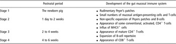
The immune response to oral delivery of antigens is orchestrated by a well-developed local intestinal mucosal immune system (Figure 1). On the one hand it must respond with vigour to potential pathogens, whilst on the other inappropriate responses to dietary antigens and commensal bacteria may lead to harmful allergic or damaging inflammatory conditions. In adult animals, these potentially harmful responses are downregulated by a process known as oral tolerance, but at birth the piglet’s mucosal immune system is poorly developed and it has been hypothesised that this may lead to inappropriate responses to food and microbial antigens in the post-weaning period. The kinetics of development of the gut immune system, which will be described in the following (summarised in Table 1), may therefore play a critical role in determining the outcome of the response to feed antigens.
Stages in the development of the gut mucosal immune system of the neonatal piglet

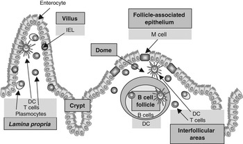
Schematic representation of the distribution of ‘immune cells’ in the small intestinal tract. Abbreviations: IEL = intraepithelial lymphocyte; DC = dendritic cell.

The development of the gut mucosal immune system
Organised lymphoid tissues
The porcine small intestinal wall contains Peyer’s patches (PP) in the jejunum and ileum (Figure 1). Whereas jejunal PP are several centimetres in length and located at the anti-mesenteric site of the gut, the ileal PP is a continuous band of up to 2 m in length (Rothkötter and Pabst, Reference Rothkötter and Pabst1989). The postnatal development of these two types of PP is different. For a short period of time, the ileal PP resembles the characteristics of the ileal PP in lambs with tall, elongated follicles and a small dome area (Barman et al., Reference Barman, Bianchi, Zwart, Pabst and Rothkötter1997). Jejunal PPs have rounded follicles and a larger dome area. Microfold cells (M cells) are present in the follicle-associated epithelium of both types of PP (Gebert et al., Reference Gebert, Rothkötter and Pabst1994), and play a specialised role in sampling antigen from the gut lumen. Their microfolded apical cell membrane enables endocytotic antigen uptake, and with lymphocytes located in pockets of their basolateral cell membrane, a close interaction of antigens and lymphocytes is possible (Gebert et al., Reference Gebert, Rothkötter and Pabst1996). Similarly, cellular processes of dendritic cells (DC) are also in close to the basolateral membrane of M cells (Bimczok et al., Reference Bimczok, Post, Tschernig and Rothkötter2006). Following antigen uptake and presentation in the PP, studies from various species have demonstrated that PP B-cells migrate via the mesenteric lymph prior to homing back to the lamina propria crypts where they differentiate into immunoglobulin (Ig)-producing plasma cells (Bienenstock et al., Reference Bienenstock, Befus, McDermott, Mirski and Rosenthal1983). The rapid migration of IgA+ immunoblasts from intestinal lymph into the intestinal mucosa has also been demonstrated in lymph duct cannulation experiments in pigs (Rothkötter et al., Reference Rothkötter, Hriesik, Barman and Pabst1999b).
Lamina propria lymphocytes
The lamina propria of adult pig is well supplied with leucocytes (Figure 1), and in contrast to many other species the immunological organisation of the lamina propria in the pig intestine shows a high level of organisation (Bailey et al., Reference Bailey, Haverson and Stokes1996; Haverson et al., Reference Haverson, Bailey and Stokes1999). Within the villous lamina propria the tissue deep to the capillary plexus contains predominately CD4+ T-cells whilst CD8+ cells occur luminally and in the epithelium. Antigen-presenting cells are present in large numbers in the lamina propria of many species including the pig (Haverson et al., Reference Haverson, Singha, Stokes and Bailey2000). The lamina propria around the intestinal crypts contains cells staining for Igs (predominantly IgA, presumably plasma cells), with few T-cells or DC, but with myeloid cells with the characteristics of macrophages and granulocytes (Rothkötter et al., Reference Rothkötter, Ulbrich and Pabst1991).
At birth, only small numbers of leucocytes are found in the lamina propria and in pigs it becomes populated according to a clearly staged time course. DC that are strongly major histocompatibility complex class II (MHC II+) and co-express CD45 and CD16 appear within the first week (Haverson et al., Reference Haverson, Singha, Stokes and Bailey2000). In contrast, T-cells appear more slowly and undergo a phased pattern of appearance. An unusual cell type, characterised by the expression of CD2, but lacking CD4 and CD8, has been shown to co-express CD3 and can therefore be classified as CD4−CD8− T-cells (Rothkötter et al., Reference Rothkötter, Ulbrich and Pabst1991; Haverson et al., Reference Haverson, Bailey and Stokes1999). Together with a second T-cell population, characterised as CD2+CD3+CD4−CD8α/α+, they form the dominant T-cells migrating into the jejunal tissue during the first week to 10 days of life. Interestingly, while conventional CD4+ and CD8α/β+ T-cells in this site in adult animals express low levels of CD45RC, consistent with advanced memory status, a significant proportion of these unusual CD2+CD3+CD4−CD8α/α+ T-cells express moderate to high levels of CD45RC, suggesting that they may be less antigen-experienced (Bailey et al., Reference Bailey, Plunkett, Clarke, Sturgess, Haverson and Stokes1998; Haverson et al., Reference Haverson, Bailey and Stokes1999). During the second and third weeks of life, increasing numbers of CD4+ T-cells appear and CD8+ cytotoxic T-cells only appear in significant numbers after the third week of life (Rothkötter et al., Reference Rothkötter, Ulbrich and Pabst1991; Haverson et al., Reference Haverson, Bailey and Stokes1999). Similarly, IgA+ plasma cells have been reported to appear in significant numbers as late as 3 to 6 weeks of life. The final architecture containing large numbers of DC and CD4+ T-cells of resting, advanced memory phenotype, transcribing interleukin-4 (IL-4) but being unable to secrete IL-2 and responding to further activation by apoptosis is not achieved until the pig is approximately 6 weeks old (Bailey et al., Reference Bailey, Haverson, Inman, Harris, Jones, Corfield, Miller and Stokes2005).
Intraepithelial lymphocytes
The intraepithelial lymphocytes (IEL) are a large population of cells with up to 2 × 10⁷ cells present per gram of porcine jejunal mucosa (Figure 1). They are mostly CD8+ T-cells with a significant proportion expressing the CD8αα homo-dimer (Rothkötter et al., Reference Rothkötter, Mollhoff and Pabst1999a). In the postnatal period, the frequency of IEL increases with age and this can be influenced by the luminal contents. For example, their number is markedly reduced with total parenteral nutrition (Kansagra et al., Reference Kansagra, Stoll, Rognerud, Niinikoski, Ou, Harvey and Burrin2003).
The induction of immune responses in the postnatal period
In order to generate an immune response, the ingested antigens must pass the epithelial barrier by either transcellular (vesicular transport) or paracellular routes. The uptake of antigens occur via either fluid-phase or receptor-mediated transport, followed by intracellular processing or simply exocytosis.
Enterocytes are non-phagocytic cells that, in health, prevent the passage of macromolecules through the epithelium (Shah and Walker, Reference Shah and Walker2002; Oswald, Reference Oswald2006). Additionally, access to the enterocyte surface is restricted by local secretions like IgA and mucins, closely packed villi, and a thick glycocalyx. However, the epithelial barrier cannot completely prevent antigen uptake (Macdonald and Monteleone, Reference Macdonald and Monteleone2005). Although the paracellular antigen entry by diffusion from the lumen is avoided by tight junctions at the apical poles, antigens can cross through breaks in tight junctions (Neutra et al., Reference Neutra, Mantis and Kraehenbuhl2001). Additionally, the tightness of tight junctions can be affected by host mediators and soluble environmental factors (Bouhet et al., Reference Bouhet, Hourcade, Loiseau, Fikry, Martinez, Roselli, Galtier, Mengheri and Oswald2004). For example, in cases of immaturity and diseases like viral gastroenteritis, bacterial infection and inflammatory bowel disease, the well-integrated function of the intestinal epithelium is affected, and antigens of every kind can enter. In health, only penetrating pathogens (Niedergang and Kweon, Reference Niedergang and Kweon2005) or invading bacteria (Cossart and Sansonetti, Reference Cossart and Sansonetti2004) find a way into the epithelial cell.
DC are located throughout the intestinal immune system and as sentinel cells they perform important immunosurveillance functions (Stockwin et al., Reference Stockwin, McGonagle, Martin and Blair2000). They are found in the villous lamina propria, in the sub-epithelial dome and the interfollicular regions of the PP (Figure 1), as well as in the mesenteric lymph nodes (MLN). Although it is feasible that DC residing in the MLN take up free antigens or pathogens derived from afferent lymph, antigen uptake is primarily performed by the immature peripheral DC located in the villous lamina propria and the sub-epithelial dome of PP. Interestingly, even non-replicating particulate antigens can generate specific immune responses, although they cannot normally cross the intestinal barrier. One important mechanism responsible for this may be antigen uptake by lamina propria DC that extend cytoplasmic processes into the gut lumen between the enterocytes (Niess and Reinecker, Reference Niess and Reinecker2005). These cytoplasmic processes also exist in the pig, but they are relatively rare (Bimczok et al., Reference Bimczok, Post, Tschernig and Rothkötter2006). After antigen uptake, the cells alter their chemokine receptor pattern, which allows them to migrate to T-cell regions of organised lymphoid tissue (i.e. interfollicular regions of the PP or MLN), where antigen presentation takes place (Fleeton et al., Reference Fleeton, Contractor, Leon, Wetzel, Dermody and Kelsall2004). Cannulation studies of pseudo-afferent intestinal lymph in rats and in pigs have shown that DC are constantly migrating from the gut wall to the MLN, even in the absence of overt antigenic stimulation (Macpherson and Uhr, Reference Macpherson and Uhr2004). This would suggest that these cells play an important role in the maintenance of tolerance. Indeed, a significant population of these cells contained particles of apoptotic cells (Huang et al., Reference Huang, Platt, Wykes, Major, Powell, Jenkins and MacPherson2000). Phenotypic analyses of migrating DC in porcine pseudo-afferent intestinal lymph suggest that migration routes are mainly from the villous lamina propria to MLN, and from the sub-epithelial domes to the interfollicular regions of the PP (Bimczok et al., Reference Bimczok, Sowa, Faber-Zuschratter, Pabst and Rothkötter2005).
The postnatal period is critical in terms of the development of an effective mucosal immune system that is capable of the adaptation to commensal bacteria, tolerance towards nutritional components and defence against pathogens in the gut lumen (Pié et al., Reference Pié, Lallès, Blazy, Laffitte, Seve and Oswald2004 and Reference Pié, Awati, Vida, Falluel, Williams and Oswald2007; Stokes et al., Reference Stokes, Bailey, Haverson, Harris, Jones, Inman, Pié, Oswald, Williams, Akkermans, Sowa, Rothkötter and Miller2004; Rothkötter et al., Reference Rothkötter, Sowa and Pabst2005). Weaning is a period during which there is considerable change in the magnitude and diversity of exposure to environmental antigens derived from food and potentially pathogenic organisms. Under ‘natural conditions’ weaning is a gradual process, and in piglets it is not complete until 10 to 12 weeks of age. This is however not normal husbandry practice and piglets are more commonly weaned abruptly at 3 to 5 weeks. As a consequence, neonatal pigs experience a transient immune hypersensitivity to dietary antigens when weaned at 3 weeks of age, often associated with clinical symptoms, e.g. diarrhoea, weight loss and enteritis (Bailey et al., Reference Bailey, Miller, Telemo, Stokes and Bourne1993; Stokes et al., Reference Stokes, Bailey, Haverson, Harris, Jones, Inman, Pié, Oswald, Williams, Akkermans, Sowa, Rothkötter and Miller2004). In contrast, if piglets are allowed to suckle their dams, until they voluntarily wean themselves at approximately 12 weeks, they develop oral tolerance to the dietary antigens without clinical symptoms (Bailey et al., Reference Bailey, Miller, Telemo, Stokes and Bourne1994). The initial and characteristic lesions of post-weaning diarrhoea are a crypt hyperplasia and a severe villous atrophy in the small intestine, which is associated with a fall in brush border enzymes (disaccharidases, sucrase and lactase) and malabsorption. These changes occur within 3 to 4 days of weaning and a partial recovery occurs after 7 to 10 days. They are partly related to a transient hypersensitivity reaction to antigens in the post-weaning diet as comparable effects are observed in the complete absence of bacteria. However, the weaning effects are most commonly associated with an increase in the proliferation of gut bacteria and the hypersecretory effects of bacterial enterotoxins (Sahin et al., Reference Sahin, Ro, el-Naggar, Wilson, Teague, Blick and Ayala1991).
Natural alternatives to in-feed antibiotics
In the following, the capacity of selected plant extracts and other natural substances to improve the immune status of the pig is reviewed. For each type or class of substances, effort has been made to synthesise in tables major elements concerning experimental designs, and immune parameters which have been studied by differentiating the local intestinal immune response and the systemic immunity. Indeed, in most of the experiments, the peripheral blood immune cells are more easily studied than mucosal immune cells. However, only two percent of all lymphocytes are in the peripheral blood (Blum and Pabst, Reference Blum and Pabst2007) and their half life in the blood is about 30 min. Thus, the observed reactions reflect the responses of cells that may have their origin in the PP, in the MLN or in the tonsils – all compartments of the mucosal immune system. Alternatively, the peripheral blood lymphocytes may be spleen-derived or come from peripheral lymph nodes. Thus, immune reactions after an in-feed application of natural immunomodulators cannot directly be linked to a defined site of origin. So far, it is often difficult to explain whether their reaction pattern depends on mucosal or systemic immune responses.
Yeast derivates
Interest in the impact of polysaccharides on immunity has increased rapidly over the past decade and this is reflected in the large number of reviews dealing with this topic (Bohn and BeMiller, Reference Bohn and BeMiller1995; Williams, Reference Williams1997; Tzianabos, Reference Tzianabos2000; Brown and Gordon, Reference Brown and Gordon2003). A variety of polysaccharides from different natural sources have the ability to modulate the immune system. Among those, β-d-glucans and the carbohydrate portion of mannoproteins, the α-d-mannans, have been recognised to be responsible for modulating the immune responses in mammals through specific interactions with different immunocompetent cells (Kogan and Kocher, Reference Kogan and Kocher2007). A direct interaction of β-d-glucans with macrophages and polymorphonuclear cells confers to these products their immunomodulatory properties (Tzianabos, Reference Tzianabos2000; Brown and Gordon, Reference Brown and Gordon2003). Mannans alter the immune response by specific links with mannose receptors, present on many cells of the immune system (Lee, Reference Lee1988), such as macrophages (Tzianabos, Reference Tzianabos2000).
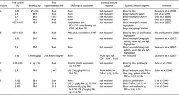
Glucans and mannans are already proposed as potential immunomodulatory agents for prophylaxis and therapy of infections in human (Masihi, Reference Masihi2000) and in farm animals including pigs (Sohn et al., Reference Sohn, Kim, Kim and Han2000; Kogan and Kocher, Reference Kogan and Kocher2007). Numerous commercial preparations derived from yeast cell walls, particularly rich in glucans and mannans, are available as in-feed supplements for pigs (Table 3). However, the composition and the purity of these products often remain unknown. This could partly explain the high variation of incorporation levels (from a factor 1 to 500 for glucans, and a factor 1 to 30 for mannans) of these commercial preparations in feed (Table 2). Moreover, the literature dealing with diet incorporation of yeast cell wall extracts often refers to only one of those two compounds, but yeast derivates usually contain both polysaccharides.
Immune parameters measured in in vivo experiments where pigs are dietary supplied with β-glucans

PW = post-weaning; d = day; wk = week; PRRS = porcine reproductive and respiratory syndrome; Ig = immunoglobulin; LC = lymphocyte; LPS = lipopolysaccharide; TNF-α = tumour necrosis factor-α; IL = interleukin; IL-1Ra = interleukin1-receptor antagonist.
A: Macrogard-S™ (Biotec-Mackzymal A/S, Tromso, Norway; or Provesta Corp., Bartlesville, OK, USA; or Jeil Vet. Chem. Co., Seoul, Korea); B: β1,3/1,6-glucans (Antaferm MG, Dr Eckel GmbH, Niederzissen, Germany) – insoluble fraction containing 25% of β-d-glucans, and 10% of mannans; C: Glucagen (Enbiotec Company, Seoul, Korea); D: Energy Plus (Natural Chem Industries, Ltd, Houston, TX, USA); E: From baker’s yeast, manual manufacturing – should contain 86% of β-glucans.
aStarting 1 wk post-weaning; bTreatment also administered in suckling piglets by gavage; cPRRSV (Ingelvac®PRRS MLV, Boehringer, Ingelheim, Germany); dPfizer Co., Seoul, Korea.
Glucans
β-glucans extracted from the cell wall of Saccharomyces cerevisiae is the most common source used for animal in-feed complements. It is a β-1,3-glucan with long β-1,6-glucan branches, whose structure is different from the β-glucans extracted from bacteria (linear β-1,3-glucan), cereals (β-1,3/1,4-glucan) and fungi (short β-1,6-glucan branched β-1,3-glucan). Their different chemical structures would be expected to be reflected in their different bioactivities (Bohn and BeMiller, Reference Bohn and BeMiller1995). Furthermore, depending on the extraction procedure, soluble and insoluble fractions that have different activities can be present together or separately (Tzianabos, Reference Tzianabos2000). Thus, the form under which glucans are included in diet will probably influence their biological properties.
Literature dealing with effects of glucans on local intestinal immune response in pigs is very scarce (Table 2). In finishing pigs, the ileal contents of IgM, IgA, or CD4+ and CD8+ T-cells were not modified by glucans supplementation (Sauerwein et al., Reference Sauerwein, Schmitz and Hiss2007). A higher incorporation dose of β-glucans (2.5%) led to increased intestinal tumour necrosis factor-α (TNF) and IL-1β mRNA but also in intestinal IL1-receptor antagonist (IL-1Ra) mRNA in pigs challenged 4 h earlier with lipopolysaccharide (LPS) (Eicher et al., Reference Eicher, McKee, Carroll and Pajor2006). These changes were not associated with alterations in blood TNF-α within hours following the inflammatory challenge. Biological significance of these results is difficult to assess as those cytokines are implicated in both pro-inflammatory (TNF-α and IL-1β) and anti-inflammatory (IL-1Ra) processes.
Much interest has been paid to the effects of glucans on systemic immune responses, particularly innate immunity. β-glucans have been shown to have anti-inflammatory properties. In vitro, IL-6 and TNF-α production by lymphocytes isolated from peripheral blood of weaned piglets supplemented with β-glucan and stimulated with LPS was decreased, whereas IL-10 production was enhanced (Li et al., Reference Li, Xing, Li, Wang, Zhao, Lv and Huang2005). Those cytokine patterns were confirmed in vivo in piglets fed diets supplemented with glucans, and challenged intraperitoneally with LPS (Li et al., Reference Li, Xing, Li, Wang, Zhao, Lv and Huang2005 and Reference Li, Li, Xing, Cheng and Lai2006). Taken together, these in vitro and in vivo analyses show that dietary β-glucans prevent the elevation in pro-inflammatory cytokines whilst enhancing the production of anti-inflammatory cytokines in response to an inflammatory challenge. Consistently, glucans have been shown to modulate the acute phase response, whose regulation is known to be orchestrated by pro-inflammatory cytokines like IL-1, IL-6 or TNF (Baumann and Gauldie, Reference Baumann and Gauldie1994; Niewold, Reference Niewold2007). In piglets, blood haptoglobin concentrations increase during the 2 weeks following an early weaning (18 days), then remain stable (Dritz et al., Reference Dritz, Shi, Kielian, Goodband, Nelssen, Tokach, Chengappa, Smith and Blecha1995). A supplementation with 0.025% or 0.05% of β-glucans partly suppresses this increase (Dritz et al., Reference Dritz, Shi, Kielian, Goodband, Nelssen, Tokach, Chengappa, Smith and Blecha1995). These results were not confirmed in piglets weaned at 28 days (Hiss and Sauerwein, Reference Hiss and Sauerwein2003; Sauerwein et al., Reference Sauerwein, Schmitz and Hiss2007). However, it should be noted that in these later experiments, haptoglobin concentration increased only during 1 week post-weaning, which could have limited the impact of glucans on this response.
As a known target for glucans, the function of neutrophils and macrophages has been widely investigated (Brown and Gordon, Reference Brown and Gordon2003). Dietary β-glucans had no consistent effects on the ability of peripheral blood neutrophils to generate reactive oxygen intermediates, neutrophil-mediated antibody-dependent cellular cytotoxicity and on neutrophil phagocytic activity (Dritz et al., Reference Dritz, Shi, Kielian, Goodband, Nelssen, Tokach, Chengappa, Smith and Blecha1995; Sauerwein et al., Reference Sauerwein, Schmitz and Hiss2007). Similarly, lung macrophage production of superoxide anion and bactericidal activity, as well as the expression of CD14, were not influenced by the inclusion of β-glucans in the diet (Dritz et al., Reference Dritz, Shi, Kielian, Goodband, Nelssen, Tokach, Chengappa, Smith and Blecha1995). Thus, the predicted modulation of neutrophil and macrophage functions by glucans was not confirmed in in vivo experiments, which may partly be explained by the limiting ability of glucans to be delivered to those cells after an oral supplementation. Whereas the ability of lymphocytes to proliferate is also insensitive to a feed supplementation with glucans (Hiss and Sauerwein, Reference Hiss and Sauerwein2003), the production of the different classes of Igs is influenced in a dose-dependent manner with lower and higher doses favouring IgA and IgG responses, respectively (Sauerwein et al., Reference Sauerwein, Schmitz and Hiss2007). This could be partly correlated to the different pattern of lymphocyte subclasses (helper and cytotoxic T-cells) observed in response to a supplementation with β-glucans (Hahn et al., Reference Hahn, Lohakare, Lee, Moon and Chae2006). However, those variations are inconstantly reported, and appear to be related to the period of supplementation (Kim et al., Reference Kim, Hyun, Sohn, Woo, Kim and Han2000; Hahn et al., Reference Hahn, Lohakare, Lee, Moon and Chae2006).
The effect of dietary supplementation with β-glucans on the response to systemic immunisation has produced contrasting results. For example, whereas β-glucan-supplemented piglets vaccinated with atrophic rhinitis vaccine produced a transiently lower antibody response (Hahn et al., Reference Hahn, Lohakare, Lee, Moon and Chae2006), pigs injected with ovalbumin and receiving β-glucans at a dose of 0.005% mounted a transiently higher antibody response (Li et al., Reference Li, Xing, Li, Wang, Zhao, Lv and Huang2005). β-glucans did not enhance the efficiency of a vaccination with porcine reproductive and respiratory syndrome (PRRS) virus (Hiss and Sauerwein, Reference Hiss and Sauerwein2003), or different EnteroToxinogenic Escherichia coli (ETEC) antigens (Decuypere et al., Reference Decuypere, Dierick and Boddez1998).
The effect of β-glucans on the passive transfer of immunity has also been studied by immunisation of sows with strains of E. coli implicated in neonatal diarrhoea (Decuypere et al., Reference Decuypere, Dierick and Boddez1998). Whereas the titre of antibodies specific to K88ab and K99 was not enhanced in colostrum of glucan-fed sows, it was elevated in their milk as compared to control sows. The authors noted that the health score (mainly due to neonatal diarrhoea) of piglets whose mother was submitted to a β-glucan regimen was severely impaired during 14 days post partum, but that the aetiological agent was not one of the ETEC strains against which sows were immunised.
In conclusion, the effects of glucans on immunity are not predictable and their ability to act as growth-promoters is also not reliable (Table 2). It should be highlighted that many of the studies have been performed in ‘clean’ environments, where morbidity rates are low, and that beneficial effects on health are much more difficult to detect in such environments.
Mannans
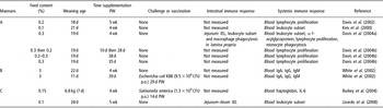
The potential protective activities of mannans may include their ability to ‘adsorb’ enteric pathogens and to modulate immune functions (Sohn et al., Reference Sohn, Kim, Kim and Han2000). To our knowledge, only two publications have investigated the effects of dietary mannan supplementation on local intestinal immune response in swine (Table 3). Even if mannans do not have any effect on the number of macrophages in intestinal lamina propria, their function seems to be enhanced. Indeed, the phagocytosis of sheep red blood cells by macrophages isolated from jejunal lamina propria was increased by inclusion of 0.3% mannans in the basal diet (Davis et al., Reference Davis, Maxwell, Erf, Brown and Wistuba2004a). The recruitment of lymphocytes into the small intestinal lamina propria is reduced in piglets fed mannans (Lizardo et al., Reference Lizardo, Nofrarias, Guinvarch, Justin, Auclair and Brufau2008) and their profile is also influenced. Indeed, the ratio of CD3+ CD4+/CD3+ CD8+ T-cells was lowered 21 days post-weaning, indicating a greater proportion of CD8+ T-cells in mannan-supplemented pigs (Davis et al., Reference Davis, Maxwell, Erf, Brown and Wistuba2004a). After birth, infiltration of lamina propria with CD8+ T-cells is usually delayed (4 weeks after birth) as compared to infiltration with CD4+ cells, which can begin as early as 2 weeks of age (Bailey et al., Reference Bailey, Plunkett, Rothkötter, Vega-Lopez, Haverson and Stokes2001). This observation could be an indication that mannan supplementation would enhance the establishment of a mature T-cell repertoire within the gastro-intestinal tract. However, further work is still needed to confirm this hypothesis.
Immune parameters measured in in vivo experiments where pigs are dietary supplied with mannans

PW = post-weaning; d = day; wk = week; IEL = intraepithelial lymphocytes; Ig = immunoglobulin; CFU = colony forming unit; IL = interleukin.
A: Bio-Mos® (Alltech Inc., Nicholasville, KY, USA); B: From brewer’s yeast (Brewtech, International Ingredient Corp., St Louis, MO, USA) – mannan content was found to be 5.2%; C: SAF-Mannan® (Lesaffre Feed Additives, Marquette-Lez-Lille, France).
Systemic immune response to dietary mannans has been studied in more detail. For example under normal breeding conditions, serum α-1-acid glycoprotein concentration was not altered by inclusion of dietary mannans (Davis et al., Reference Davis, Maxwell, Erf, Brown and Wistuba2004a), whereas piglets fed with 0.3% of phosphorylated mannans exhibited a decreased blood neutrophils : lymphocytes ratio, suggesting that mannan supplementation could alleviate the inflammatory response associated with the stress of weaning (Davis et al., Reference Davis, Maxwell, Erf, Brown and Wistuba2004a). The increased blood lymphocyte population among leucocytes in mannan-fed animals could be linked more to B than to T-cells. Indeed, 3% of brewer’s yeast (which corresponds to a final level of 0.16% of mannan oligosaccharide) only tended to increase the piglet serum level of IgG when used alone, but substantially increased this level when fed with the acidifier, citric acid (White et al., Reference White, Newman, Cromwell and Lindemann2002). Conversely, blood proportions of CD4+ or CD8+ lymphocytes are insensitive to mannan supplementation (Kim et al., Reference Kim, Hyun, Sohn, Woo, Kim and Han2000). The proliferative response of peripheral blood lymphocytes was generally found not to be affected by the inclusion of mannans in the diet of nursery piglets (Davis et al., Reference Davis, Maxwell, Brown, de Rodas, Johnson, Kegley, Hellwig and Dvorak2002, Reference Davis, Maxwell, Erf, Brown and Wistuba2004a and Reference Davis, Brown, Maxwell, Johnson, Kegley and Dvorak2004b), but when continuously incorporated at a dose rate of 0.3% the ability of lymphocytes to proliferate after a stimulation with a pokeweed mitogen (non-specific proliferation of B- and T-cells) or with phytohemaglutinin (proliferation of primarily T-cells) was reduced (Davis et al., Reference Davis, Brown, Maxwell, Johnson, Kegley and Dvorak2004b). The authors suggested that this altered immune function may explain the beneficial effects of mannans for piglet growth. However, there are no data concerning morbidity or mortality to assess the health effects of such an immune depression.
In piglets challenged with Salmonella enterica serotype Typhimurium, serum haptoglobin concentrations were increased in 0.15% mannan-fed piglets 6 and 13 days post-infection as compared to piglets fed the basal diet either alone or enriched with AB (Burkey et al., Reference Burkey, Dritz, Nietfeld, Johnson and Minton2004). Serum IL-6 was not altered by mannan supplementation, but this could be explained by the absence of a response on this cytokine following challenge with this pathogen (Burkey et al., Reference Burkey, Dritz, Nietfeld, Johnson and Minton2004). Contrary to carbadox, mannans failed to reduce the length of the period of hyperthermia observed after infection with S. enterica and did not promote growth (Burkey et al., Reference Burkey, Dritz, Nietfeld, Johnson and Minton2004). Total levels of serum IgA, IgM and IgG were not modified by dietary mannan, in piglets challenged or not with an enterotoxigenic strain of E. coli K88 (White et al., Reference White, Newman, Cromwell and Lindemann2002).
As for glucans, the influence of mannans on immunity is not always reliable, as well as their effects on piglet performances. In piglets challenged with enteric pathogens (E. coli K88, S. enterica), health benefits of dietary mannans are not consistent.
Plant extracts
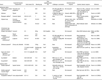
Empirical evidence suggests that plant extracts may offer benefits in boosting the immune system; thus, preventing disease in production animals (Wenk, Reference Wenk2003) and plant-derived products have gained increasing interest as possible feed additives for non-ruminant species (Windisch et al., Reference Windisch, Schedle, Plitzner and Kroismayr2008). Plants, and where they have been identified their bioactive components, are very diverse and their potential to enhance pig health and immunity has only been scarcely evaluated in vivo (see Table 4). Moreover, most studies have used a mixture of compounds, which does not allow the investigation of the immune properties of a specific bioactive component.
Immune parameters measured in in vivo experiments where pigs are dietary supplied with plant extracts

PW = post-weaning; d = day; wk = week; IL = interleukin; TNF-α = tumour necrosis factor-α; Ig = immunoglobulin; CFU = colony forming unit; IEL = intraepithelial lymphocytes; PP = Peyer’s patches; MLN = mesenteric lymph nodes; LPS = lipopolysaccharide; PRRS = porcine reproductive and respiratory syndrome; NK = natural killer; IFN-γ = interferon-γ; Ab = antibody; AGP=alpha-1-acid glycoprotein.
aEqual amount of powder extracted from eight herbal medicines purchased from a drug market in Taiwan; bCommercial phytogenic additive containing 53% inulin, 8% essential oil mix, 3% tannins – analysis revealed a 6% carvacrol and 0.14% thymol content of this additive; cPlant extract combination (Pancosma, S.A., Geneva, Switzerland) containing 5% carvacrol (Origanum spp.), 3% cinnamaldehyde (Cinnamonum spp.) and 2% capsicum oleoresin (Capsicum annum); dOregpig®, Oregpig GmbH, Pecs, Hungary; eHongSheng Herbal Drug Store, Beijing, China, manual manufacturing; fBeijing, People’s Republic of China, manual manufacturing; gEchinacea pressed juice from aerial part of the plant, commercial product; hShin Mitsui Sugar Co., Ltd, Japan; iCSI Metabolics, Newport Beach, CA, USA; jWiley Organics, Coshocton, OH, USA; kDesert King International, Chula Vista, CA, USA; lAcros Organics, Geel, Belgium; mAxiss France SAS, Bellegarde-sur-Valserine, France; nNo information; oExcept the 1st wk: 0.075%; pSupplementation only during wk 1–3 and 7–9; qThree consecutive d/wk; rPorcilis Ery ad us. Vet., Intervet, Unterschleißheim, Germany.
Herbaceous plants
Herbs and spice extracts have been used extensively in different parts of the World to treat a variety of human diseases (Stein and Kil, Reference Stein and Kil2006). Gastro-intestinal disorders have been treated with a number of different plant extracts including garlic, peppermint, chamomile or aloe (Amagase et al., Reference Amagase, Petesch, Matsuura, Kasuga and Itakura2001; Akerreta et al., Reference Akerreta, Cavero, Lopez and Calvo2007; Rodriguez-Fragoso et al., Reference Rodriguez-Fragoso, Reyes-Esparza, Burchiel, Herrera-Ruiz and Torres2008). Some herbs like Echinacea, liquorice, cat’s claw and garlic are claimed as immuno-enhancers (Craig, Reference Craig1999). The essential oil of the plant is often the biologically active component, but other extracts can also display biological activities.
The Labiatae constitutes a large family of herbs: basil, dill, fennel, marjoram, mint, rosemary, oregano, sage and thyme (Craig, Reference Craig1999). Mixtures of essential oils based on thymol and carvacrol whose major sources are thyme and oregano, respectively (Burt, Reference Burt2004), seem promising due to their anti-microbial (Kim et al., Reference Kim, Marshall and Wei1995; Cosentino et al., Reference Cosentino, Tuberoso, Pisano, Satta, Mascia, Arzedi and Palmas1999; Baydar et al., Reference Baydar, Sagdic, Ozkan and Karadogan2004) and potential immunomodulatory properties (Woollard et al., Reference Woollard, Tatham and Barker2007). A plant extract containing 6% of carvacrol and 0.14% of thymol, incorporated at 0.05% to 0.15% in a pig diet, had no effect on the plasma levels of the acute phase proteins, haptoglobin and C-reactive protein (Muhl and Liebert, Reference Muhl and Liebert2007) and the inclusion of a commercial plant product composed of oregano oil mixed with anis and citrus oils did not improve the health status of piglets (Kommera et al., Reference Kommera, Mateo, Neher and Kim2006). In contrast, an extract of Origanum vulgare, enriched with both thymol and carvacrol in similar proportions, was reported to protect low-weight growing-finishing pigs from disease (Walter and Bilkei, Reference Walter and Bilkei2004). This health benefit was associated with an increased proportion of CD4+, CD8+ and double-positive T-cells in peripheral blood and MLN (Walter and Bilkei, Reference Walter and Bilkei2004). Thymol used alone enhances total IgA and IgM serum levels and exhibits some local anti-inflammatory properties, as indicated by a reduction in TNF-α mRNA in the stomach (Trevisi et al., Reference Trevisi, Merialdi, Mazzoni, Casini, Tittarelli, De Filippi, Minieri, Lalatta-Costerbosa and Bosi2007). In vitro, cinnamaldehyde, the main component of cinnamon essence, also has anti-microbial (Burt, Reference Burt2004) and immunomodulatory (Koh et al., Reference Koh, Yoon, Kwon, Jeong, Nam and Han1998) properties. A plant extract containing 5% of carvacrol (Origanum spp.), 3% of cinnamaldehyde (Cinnamonum spp.) and 2% of capsicum oleoresin (Capsicum annum), included in the feed at a 0.03% level, led to a decreased number of jejunal IEL, and an increased number of lymphocytes in the colonic lamina propria (Manzanilla et al., Reference Manzanilla, Nofrarias, Anguita, Castillo, Perez, Martin-Orue, Kamel and Gasa2006). Conversely, mononuclear cell subsets from ileal PP were not affected by this plant extract combination (Nofrarias et al., Reference Nofrarias, Manzanilla, Pujols, Gibert, Majo, Segales and Gasa2006) and only the percentage of B lymphocytes was reduced in lymph nodes of piglets (Nofrarias et al., Reference Nofrarias, Manzanilla, Pujols, Gibert, Majo, Segales and Gasa2006).
Chinese pharmacopoeia describes the use of numerous herbal formulations to cure a wide variety of diseases. Among those, Bahzen is a medicine composed of eight different herbs (Atractylodes ovata, Codonopsis pilosula, Poria cocos, Glycyrrihiza uralensis, Angelica sinensis, Ligusticum chuanxiong, Paeonia albiflora and Rehmannia glutinosa) whose effects have been tested in vivo in pigs (Lien et al., Reference Lien, Horng and Wu2007). Bahzen increased white blood cell counts, without leukocytosis, and enhanced IL-6 and TNF-α in the serum of pigs challenged with LPS, compared to negative and positive (AB) control animals (Lien et al., Reference Lien, Horng and Wu2007). Serum IgG levels and blood neutrophil activity in Bahzen and AB groups were increased. However, Bahzen did not modulate acquired immunity, as measured by specific antibody responses directed towards different antigens (sheep red blood cells, ovalbumin) (Lien et al., Reference Lien, Horng and Wu2007). Piglets fed with Bahzen also grew faster than control piglets (Lien et al., Reference Lien, Horng and Wu2007). The authors speculated that Bahzen-fed piglets had an increased ability to respond to an inflammatory stress, and that this may be beneficial for piglet health. Equally, it is possible to argue that an increased inflammatory response could be detrimental to piglet health.
An abundance of plants of the Echinacea family in pasture is used as an indicator of ‘good health’. The main bioactive components of Echinacea purpurea are chicory acid and alkamids. When included as juice or cobs in the post-weaning diet or that of finishing pigs, growth performances are not improved, but feed efficiency tends to be increased (Maass et al., Reference Maass, Bauer, Paulicks, Bohmer and Roth-Maier2005). Blood parameters, including cell count and lymphocyte proliferation, were not modified by dietary treatment but the health status of piglets was good throughout the trial. Moreover, the response to immunisation of piglets with a vaccine against Erysipelothrix rhusiopathiae was enhanced by the inclusion of E. purpurea into the diet of finishing pigs (Maass et al., Reference Maass, Bauer, Paulicks, Bohmer and Roth-Maier2005). This could be an indication for the health-promoting properties of Echinacea, but further studies would be required to confirm this.
Whilst the bulk of β-glucans used in feed industry is derived from yeast cell wall (see previous section), β-glucans from the Chinese herb Astragalus membranaceus have also been tested (Mao et al., Reference Mao, Piao, Lai, Li, Xing and Shi2005; Yuan et al., Reference Yuan, Piao, Li, Kim, Lee and Guo2006). Dietary A. membranaceus increased the white blood cell count, mainly through the contribution of CD4+ lymphocytes (Yuan et al., Reference Yuan, Piao, Li, Kim, Lee and Guo2006). The proliferation of T-cells isolated from peripheral blood in weanling pigs was also increased in a dose-dependent manner in β-glucans-fed piglets (Mao et al., Reference Mao, Piao, Lai, Li, Xing and Shi2005). Concomitantly, β-glucans from Astragalus increased blood concentration in IL-2 (Mao et al., Reference Mao, Piao, Lai, Li, Xing and Shi2005; Yuan et al., Reference Yuan, Piao, Li, Kim, Lee and Guo2006) and interferon-γ (IFN-γ) (Yuan et al., Reference Yuan, Piao, Li, Kim, Lee and Guo2006), but IL-4 and IL-10 concentrations remained unchanged (Yuan et al., Reference Yuan, Piao, Li, Kim, Lee and Guo2006). This cytokine profile suggests a Th1 bias, which is in accordance with the enhancement of cellular immunity conferred by glucans. Plant β-glucans do not influence humoral immunity, as their inclusion in diet did not alter specific antibody titres following immunisation with ovalbumin (Yuan et al., Reference Yuan, Piao, Li, Kim, Lee and Guo2006). A 0.05% level of glucans extracted from A. membranaceus counteracted the increased plasma concentrations of IL-1β and prostaglandin E2 induced by an LPS challenge. Surprisingly, this effect was less pronounced when included at a dose of 0.1% in the diet (Mao et al., Reference Mao, Piao, Lai, Li, Xing and Shi2005). Thus, β-glucans from A. membranaceus exhibited anti-inflammatory properties (decrease in IL-1β synthesis) and promoted T-lymphocyte proliferation (which may be partly linked to the enhanced serum IL-2), but had no positive effects on humoral immunity. These immune modulations may be beneficial for the piglets to fight against infections, but the health status of piglets was not stated in these studies.
Sugar cane extracts display a wide range of biological effects, including immunostimulating and growth-promoting properties as shown in chicken (El-Abasy et al., Reference El-Abasy, Motobu, Shimura, Na, Kang, Koge, Onodera and Hirota2002). In pigs, sugar cane extract prepared by chromatographic separation on an ion exchange column could enhance innate immunity, through increased NK (natural killer) cell cytotoxicity and phagocytic function of monocytes and neutrophils (Lo et al., Reference Lo, Chen, Chien, Koge, Hosono, Kaminogawa and Lee2005). As morbidity and mortality rates remained very low in this experiment, it is not possible to speculate whether those immune modulations induced by sugar cane extract would improve the resistance of piglets to infection. Moreover, the potential bioactive component(s) from this particular extract of sugar cane have not been identified.
Genistein and daidzein, two isoflavones found in soybean products, have also been suggested to act as immunomodulators when given orally. After oronasal inoculation of piglets with PRRS virus, dietary daidzein failed to decrease serum titres of virus (Greiner et al., Reference Greiner, Stahly and Stabel2001b), whereas genistein minimised the viremia from day 4 to day 24 post-inoculation, as well as the serum concentration of IFN-γ (Greiner et al., Reference Greiner, Stahly and Stabel2001a). Serum α-1-acid glycoprotein concentration was not modulated by daidzein (Greiner et al., Reference Greiner, Stahly and Stabel2001b), but was increased during periods of high viremia by genistein (Greiner et al., Reference Greiner, Stahly and Stabel2001a). This enhanced α-1-acid glycoprotein response in genistein-fed piglets supports the hypothesis of a greater and more effective immune response, which could explain the lower viremia (Greiner et al., Reference Greiner, Stahly and Stabel2001a). Accordingly, lower serum IFN-γ concentration in genistein-fed animals is in agreement with the greater virus elimination and a quicker return of IFN-γ to basal levels (Greiner et al., Reference Greiner, Stahly and Stabel2001a). Despite the different effects on viremia, both isoflavones were efficient in promoting growth in piglets challenged with PRRS virus, which suggests that their mechanisms of action would differ. It would be of interest in future to test their ability to enhance immune responses and health during a challenge with an intestinal pathogen.
Ligneous plants
An extract of the South American tree Quillaja saponaria is widely used as a vaccine adjuvant (Kensil et al., Reference Kensil, Mo and Truneh2004), and whose active ingredient appears to be the saponin fraction (Milgate and Roberts, Reference Milgate and Roberts1995). As saponins are known to enhance immunity via the intestinal route, their use in animal nutrition has been the subject of investigation (Francis et al., Reference Francis, Kerem, Makkar and Becker2002). Dietary treatment with crude soap bark of Q. saponaria, ranging from 125 to 500 mg/kg, did not counteract the negative effects on feed intake and growth induced transiently by a challenge with Salmonella enterica serovar Typhimurium (Turner et al., Reference Turner, Dritz, Higgins, Herkelman and Minton2002b). Q. saponaria also failed to modulate the rise in serum haptoglobin, α-1-acid glycoprotein and IgM concentrations induced by this challenge 7 and 14 days post-infection (Turner et al., Reference Turner, Dritz, Higgins, Herkelman and Minton2002b). The phagocytic function of peripheral white blood cells tended to be depressed in challenged pigs fed with high doses of Q. saponaria, but not with low doses (Turner et al., Reference Turner, Dritz, Higgins, Herkelman and Minton2002b). It has been suggested that these ‘weak immune modulations’ may be due to the low purity of the extract used (Ilsley et al., Reference Ilsley, Miller and Kamel2005), which could reach a maximal level of 10% of saponins. Moreover, tannins, which could be as high as 15% in a crude extract of Q. saponaria, might have induced a detrimental response, as they are known to have anti-nutritional properties (Singh et al., Reference Singh, Bhat and Singh2003). Thus, Ilsley et al. (Reference Ilsley, Miller and Kamel2005) incorporated a saponin extract from Q. saponaria in the diet, alone or in combination with curcumin, which has been shown to modulate lymphocyte-mediated immune functions in mice (Churchill et al., Reference Churchill, Chadburn, Bilinski and Bertagnolli2000). The combination of both products did not result in adverse or synergistic effects (Ilsley et al., Reference Ilsley, Miller and Kamel2005). Whereas piglet immune responses were not influenced by curcumin, the feed intake and serum IgA, IgG and C-reactive protein concentrations were transiently increased in saponin-fed piglets. The subsequent negative impact of saponin on feed utilisation, assessed by a decreased feed : weight gain ratio, could result from increased dietary requirements to mount an immune response (Ilsley et al., Reference Ilsley, Miller and Kamel2005). However, as the health status of piglets remained very good throughout the experiment, it is difficult to predict whether such a response may have been beneficial or detrimental to piglets during the time of an infection.
Seaweed extracts
Seaweed extracts are also known to have immunomodulatory properties (Yoshizawa et al., Reference Yoshizawa, Enomoto, Todoh, Ametani and Kaminogawa1993). To date, we are only aware of one study that presents data on influence of a seaweed extract on the immune parameters in pig production. This extract was derived from Ascophyllum nodosum, and different levels of incorporation ranging from 0% to 2% were tested in piglets orally challenged with 6 × 10⁹ CFU (colony forming units) of Salmonella enterica serovar Typhimurium (Turner et al., Reference Turner, Dritz, Higgins and Minton2002a). Increasing levels of A. nodosum tended to linearly enhance the feed intake, but decreased the feed efficiency during the 4 weeks following the weaning of piglets. The challenge with Salmonella had only moderate effects on piglets, and dietary A. nodosum had no influence on immune responses irrespective of whether the piglets were infected or not. In vitro, a dose of 10 mg/ml of A. nodosum (greatly exceeding the highest level of feed incorporation level of 2%) was able to activate porcine alveolar macrophages to secrete prostaglandin E2. However, this dose did not alter the secretion of IL-10 by splenocytes (Turner et al., Reference Turner, Dritz, Higgins and Minton2002a). Taken together, the in vitro and in vivo data suggest that A. nodosum extract probably has only little direct effect on gut immune system in pig.
Animal by-products
Spray-dried animal plasma
The biological properties of spray-dried plasma (SDP), a by-product of commercial slaughtering facilities, have been investigated in pig nutrition (Coffey and Cromwell, Reference Coffey and Cromwell2001). Spray-dried animal plasmas are characterised by a rich protein content, whose digestibility and amino acid composition are similar to those of the proteins in sow’s milk (van Dijk et al., Reference van Dijk, Everts, Nabuurs, Margry and Beynen2001), and whose Igs content can represent 24% to 25% (Niewold et al., Reference Niewold, van Dijk, Geenen, Roodink, Margry and van der Meulen2007). Plasma from both bovine and porcine origins seem efficient in improving performances of piglets, and the IgG fraction would appear to be the main component responsible for the growth-promoting properties for these products (Pierce et al., Reference Pierce, Cromwell, Lindemann, Russell and Weaver2005). The Igs may prevent viruses and bacteria from interacting with the gut wall, resulting in an improvement of gut function (Coffey and Cromwell, Reference Coffey and Cromwell2001). One major problem is the stability of Igs during the manufacturing process of SDP. The most critical step would be spray-drying, which can lead to a 30% loss of Igs activity (Niewold et al., Reference Niewold, van Dijk, Geenen, Roodink, Margry and van der Meulen2007). Further processing to pellets would not lead to appreciable losses. It is important to remember that the use of non-sterilised products of animal origin as feed ingredients for animals poses a potential health risk, through the spread of certain infectious diseases. Despite spray-drying being shown to eliminate viable pseudorabies and PRRS viruses (Polo et al., Reference Polo, Quigley, Russell, Campbell, Pujols and Lukert2005), the use of SDP originating from slaughter animals that have been subjected to veterinary inspection is required. Indeed, it should be kept in mind that the use of non-sterilised products of animal origin as feed ingredients for animals remains a health risk practice, which is strictly regulated in Europe (Regulation (EC) No. 1774/2002 currently under revision). Moreover, as recently illustrated by the use of animal flour in animal feed and the bovine spongiform encephalopathy crisis, the public opinion is concerned by the inclusion of animal by-products in livestock feed. The meat consumers’ confidence could, in the future, represent a limiting factor to the use of SDP and other animal by-products in animal nutrition.
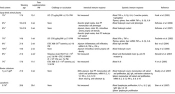
The effects of SDP on local intestinal immune responses have been widely studied in pigs (Table 5). Results are concordant and reveal that SDP prevents the infiltration of gut-associated lymphoid tissue by immune cells, including macrophages and lymphocytes (Jiang et al., Reference Jiang, Chang, Stoll, Fan, Arthington, Weaver, Campbell and Burrin2000; Nofrarias et al., Reference Nofrarias, Manzanilla, Pujols, Gibert, Majo, Segales and Gasa2006 and Reference Nofrarias, Manzanilla, Pujols, Gibert, Majo, Segales and Gasa2007). This decreased infiltration is likely to be a reflection of a lower antigenic challenge (Nofrarias et al., Reference Nofrarias, Manzanilla, Pujols, Gibert, Majo, Segales and Gasa2007). Additionally, the jejunal expression of the pro-inflammatory cytokines IL-8 and TNF-α was decreased in 6% SDP-fed piglets 15 days after a challenge with ETEC K88, irrespective of their status for K88 receptor (Bosi et al., Reference Bosi, Casini, Finamore, Cremokolini, Merialdi, Trevisi, Nobili and Mengheri2004). This decreased inflammatory response was associated with a lower histopathological score in SDP-fed piglets, thus suggesting that SDP would be efficient in helping piglets fight against infections.
Immune parameters measured in in vivo experiments where pigs are dietary supplied with animal by-products

CFU = colony forming unit; PW = post-weaning; d = day; TNF-α = tumour necrosis factor-α; IL = interleukin; wk = week; PP = Peyer’s patches; LPS = lipopolysaccharide; PP = Peyer’s patches; IEL = intraepithelial lymphocytes; LPS = lipopolysaccharide; IFN-γ = interferon-γ; Ig = immunoglobulin; ETEC = EnteroToxinogenic Escherichia coli; MLN = mesenteric lymph nodes.
aAppetein (SDPP), APC-Europe, Barcelona, Spain; bMP722, Merrick’s, Union Center, WI, origin unknown; cAPC-Europe, Barcelona, Spain; dPorcine plasma protein concentrate from unknown origin, one group restricted to control group feed intake in this study; eporcine SDP produced from non-immunised and immunised (Porcilis coli, Intervet International BV, Boxmeer, The Netherlands) pigs; fUnknown origin; gBy daily gavage, spray-dried bovine colostrum (CER, Marloie, Belgium); hInstitute of Feed Science (Zhejiang University, Hangzhou, China); iSupplemented diet from d-3 post-weaning.
Concerning systemic immune responses, a 6% or 10% supplementation of the diet with porcine plasma did not modulate the increase in blood white blood cell count that normally occurs during the 2 weeks following weaning (Jiang et al., Reference Jiang, Chang, Stoll, Fan, Arthington, Weaver, Campbell and Burrin2000; Nofrarias et al., Reference Nofrarias, Manzanilla, Pujols, Gibert, Majo, Segales and Gasa2006 and Reference Nofrarias, Manzanilla, Pujols, Gibert, Majo, Segales and Gasa2007). SDP did not influence serum IFN-γ or TNF-α under basal conditions (Touchette et al., Reference Touchette, Carroll, Allee, Matteri, Dyer, Beausang and Zannelli2002), but when stimulated with an intraperitoneal LPS injection, SDP-fed piglets showed exacerbated increases in serum levels of IFN-γ and TNF-α associated with severe intestinal damage, suggesting that SDP-fed piglets would be more susceptible to certain immunological challenges (Touchette et al., Reference Touchette, Carroll, Allee, Matteri, Dyer, Beausang and Zannelli2002). Similarly, in piglets challenged intravenously with LPS, the increase in serum IL-6 and IL-1β was exacerbated in SDP-fed piglets, but there was no evidence of an effect on mRNA cytokine levels in the liver, thymus or spleen (Frank et al., Reference Frank, Carroll, Allee and Zannelli2003). Conversely, SDP supplementation led to a decrease in C-reactive protein concentration but not in haptoglobin (Frank et al., Reference Frank, Carroll, Allee and Zannelli2003). In those models, the extremely high dose of LPS (75–150 μg/kg of BW) as well as the route of administration (intraperitoneally or intravenously) preclude from concluding on the plasma effects on inflammatory responses when piglets are exposed to pathogens via the enteral route.
The growth-promoting properties of SDP that are usually reported (Jiang et al., Reference Jiang, Chang, Stoll, Fan, Arthington, Weaver, Campbell and Burrin2000; Bosi et al., Reference Bosi, Casini, Finamore, Cremokolini, Merialdi, Trevisi, Nobili and Mengheri2004; Nofrarias et al., Reference Nofrarias, Manzanilla, Pujols, Gibert, Majo, Segales and Gasa2006 and Reference Nofrarias, Manzanilla, Pujols, Gibert, Majo, Segales and Gasa2007; Niewold et al., Reference Niewold, van Dijk, Geenen, Roodink, Margry and van der Meulen2007) are more commonly observed with SDP from porcine than bovine origin (Hansen et al., Reference Hansen, Nelssen, Goodband and Weeden1993; van Dijk et al., Reference van Dijk, Everts, Nabuurs, Margry and Beynen2001). Moreover, their anti-microbial/immunomodulatory properties would be more likely to be beneficial when piglet growth may be compromised, such as in conventional on-farm nursery setting as compared to ‘cleaner’ off-site nursery (Coffey and Cromwell, 1995), or in pigs weaned early v. a late weaning (Torrallardona et al., Reference Torrallardona, Conde, Esteve-Garcia and Brufau2002). This could be related to the health-promoting properties of SDP observed in challenged piglets. Indeed, in piglets challenged via the oral route with an ETEC strain, inclusion of porcine SDP at an 8% level in diet can reduce post-weaning diarrhoea and increase growth of piglets that are positive for F4 receptor (Niewold et al., Reference Niewold, van Dijk, Geenen, Roodink, Margry and van der Meulen2007). This effect was enhanced in piglets fed a diet containing an SDP obtained from pigs previously immunised with a vaccine against neonatal E. coli, where a concomitant decreased ETEC excretion was observed. These results confirm that protection conferred by SDP would be mainly due to specific antibodies, but also highlight the implication of less-specific components. Indeed, the decreased intestinal inflammatory response observed in SDP-fed pigs challenged with ETEC K88 may be explained by a lower colonisation or binding of E. coli to intestinal receptors as assessed by their lower serum IgA titre and histopathological score (Bosi et al., Reference Bosi, Casini, Finamore, Cremokolini, Merialdi, Trevisi, Nobili and Mengheri2004). For instance, the glycan moieties of glycoproteins would compete with natural intestinal receptors, thus reducing bacterial adhesion to the gut (Nollet et al., Reference Nollet, Deprez, Van Driessche and Muylle1999). Indeed, the protective effect of SDP may also be non-specific as the use of a plasma powder depleted of antibodies directed towards adhesion factors of the challenge strain of E. coli (O141:K85ab expressing the F18ac fimbriae) was efficient in protecting piglets against oedema disease (Nollet et al., Reference Nollet, Deprez, Van Driessche and Muylle1999). In piglets orally challenged with different strains of pathogenic E. coli (van Dijk et al., Reference van Dijk, Enthoven, Van den Hoven, Van Laarhoven, Niewold, Nabuurs and Beynen2002; Bosi et al., Reference Bosi, Casini, Finamore, Cremokolini, Merialdi, Trevisi, Nobili and Mengheri2004; Yi et al., Reference Yi, Carroll, Allee, Gaines, Kendall, Usry, Toride and Izuru2005; Torrallardona et al., Reference Torrallardona, Conde, Badiola and Polo2007), SDP generally prevents growth retardation and clinical signs, a phenomenon inconstantly associated with decreased circulation or excretion of pathogenic strains. This supports the theory of a direct competition of SDP with intestinal receptors for pathogenic E. coli, more than a direct anti-microbial effect (Bosi et al., Reference Bosi, Casini, Finamore, Cremokolini, Merialdi, Trevisi, Nobili and Mengheri2004). Both theories concerning mechanisms of action of SDP are likely, and may act synergistically to decrease disease susceptibility of piglets. Glycoproteins, but probably other components, may prevent attachment of pathogens to intestine in a non-specific way, which could still be reduced by specific antibodies linking to pathogens. The elimination of undesired pathogens from the gastro-intestinal tract would be facilitated.
Other animal by-products
Bovine colostrum has a critical role in postnatal health as an immune booster. Apart from the passive transfer of antibody, it provides growth- and anti-microbial factors (IGF, transforming growth factor, epidermal growth factor, lactoferrin, lysozyme, etc.), which will be beneficial to the neonate (Pakkanen and Aalto, Reference Pakkanen and Aalto1997). In piglets, bovine colostrum has been shown to enhance mucosa restoration by stimulating migration of epithelial cells along the crypt–villous axis in the intestine, and by decreasing apical cell apoptosis (Huguet et al., Reference Huguet, Le Normand, Fauquant, Kaeffer and Le Huerou-Luron2007). Recent studies would suggest that bovine colostrum has immunomodulatory properties that are directly related to a specific region of the porcine gut-associated lymphoid tissue. Depending upon the region studied, bovine colostrum could lead to Th1 (IL-2, IFN-γ and IL-12) or Th2 (IL-4 and IL-10) cytokine profiles (Boudry et al., Reference Boudry, Buldgen, Portetelle, Collard, Thewis and Dehoux2007). In ileal PP, the increase in Th2 mRNA expression (IL-4 and IL-10) was associated with an impaired Th1 profile (IFN-γ), suggesting that bovine colostrum supplementation may cause a more marked Th2 immune response (Boudry et al., Reference Boudry, Buldgen, Portetelle, Collard, Thewis and Dehoux2007). In contrast, a Th1 profile was more pronounced in the jejunum. This bipolarity activities associated with bovine colostrum would be of importance in the context of exposure to a wide range of antigens (Boudry et al., Reference Boudry, Buldgen, Portetelle, Collard, Thewis and Dehoux2007). Bovine colostrum significantly decreased the total number of mononuclear cells in ileal PP, but their proliferative responses were increased (Boudry et al., Reference Boudry, Buldgen, Portetelle, Collard, Thewis and Dehoux2007). Phenotyping of the ileal PP cells showed a very significant dose-related decrease in CD21+ cell count, concomitant with an increase in the CD3+ cell population (involving mainly the CD4+ subpopulation) 21 days post-weaning. Perhaps not surprisingly, the piglets mounted a strong local antibody response to the bovine colostral Igs by 21 days post-weaning (Boudry et al., Reference Boudry, Buldgen, Portetelle, Collard, Thewis and Dehoux2007). The activities associated with fed bovine colostrum were mainly associated with the intestinal tract, and systemic immune responses were not altered. To our knowledge, its health benefits that could result from its orchestration of immune responses have not yet been studied and could be the subject of further investigations.
Lactoferrin is a member of the transferrin family of iron-binding glycoproteins and is ubiquitous in animal secretions (colostrum, milk, tears). It displays a wide range of physiological functions including protection against microbial infections, regulation of non-specific immune response and modulation of the inflammatory response (see Levay and Viljoen (Reference Levay and Viljoen1995) for review). Both specific and innate systemic immune responses seem dependent on lactoferrin. In pigs, it has been shown that a 15-day lactoferrin treatment increased the concentration of IL-2 in blood as well as the C4 component of complement, at levels much higher than those reached by positive controls receiving AB (Shan et al., Reference Shan, Wang, Liu and Xu2007). Conversely, lactoferrin did not seem to have any impact on IL-1α or C3 blood concentrations. Lactoferrin enhanced the proliferation of peripheral blood and spleen lymphocytes after stimulation with phytohemagglutinin and concanavalin A (for spleen lymphocytes only). Serum concentrations of IgG, IgA and IgM were also increased in lactoferrin-supplemented piglets. To our knowledge, its impact on local intestinal immunity as well as its potential to enhance specific immune responses has not yet been investigated in pigs. The use of lactoferrin is promising, as it seems efficient in preventing diarrhoea in piglets (Shan et al., Reference Shan, Wang, Liu and Xu2007; Wang et al., Reference Wang, Shan, Xu, Feng and Wang2007).
Concluding remarks
Literature dealing with natural alternatives to in-feed AB and their impact on pig immunity is still scarce. In our opinion, two main reasons are responsible for this lack of knowledge. First, the total ban of in-feed AB is recent and published works on that topic may arise in the following years. Secondly, in-feed alternatives that can act directly on immunity, and not through the control of the microflora, have only recently gained interest. Indeed, the main mechanism proposed for the action of AB as growth-enhancers was only linked to their ability to control flora. It was suggested that AB improve the efficiency of animal growth via the inhibition of intestinal microflora, leading to a reduction in the maintenance costs of the gastro-intestinal system including immune responses (Gaskins et al., Reference Gaskins, Collier and Anderson2002). However, new data are emerging and suggest that AB may directly interact with host cells (mainly inflammatory ones), and decrease their production and excretion of catabolic mediators (Niewold, Reference Niewold2007). Niewold (Reference Niewold2007) argues that the changes in microflora would be more likely the consequence of the altered condition of the intestinal wall and that this may explain the highly reproducible effects of AB, as opposed to the inconstant results obtained with alternatives aiming at controlling the microflora. However, data obtained with different classes of alternative substances able to directly interact with natural defences of the host are not so conclusive.
As highlighted in this review, substances whose immunomodulatory properties often issue from in vitro experiments are not so potent when tested in vivo as feed additives. The heterogeneity of experiments aiming at studying the effects of feed additives on pig immunity can partly explain the discrepancies on their efficacy: variable composition of additives, different time for supplementation, diversity of experimental designs or measured parameters, etc. Indeed, from the feed processing to the intestinal absorption, the different compounds will be submitted to a myriad of events that can reduce their activity before they reach the cells of the gut-associated immune system (feed storage, interactions with other nutrients, digestive processes, absorption kinetics, etc.). Moreover, the level of the feed additive in the final diet often remains unknown, and in some cases, the composition of the additive itself is not revealed. This is particularly true for yeast extracts that are mainly commercial products, but also for plant extracts whose active principle(s) content is highly dependent on environmental growth conditions, harvesting time and state of maturity or storage management (Wenk, Reference Wenk2003). Independent of these basic information that should be mentioned in every scientific article, some experimental designs do not seem to be fully adapted to the study of the immunomodulatory potential of in-feed additives. Indeed, this review highlights that effects of in-feed supplements on systemic immunity are quite well documented, whereas local intestinal immune responses have only received little attention. It can be understood from both a practical and an ethical point of view as the study of systemic immunity via the blood does not require the slaughter of animals, and offers the possibility of kinetic studies. However, mucosal responses can occur independently of systemic immunity (Cunningham-Rundles and Lin, Reference Cunningham-Rundles and Lin1998; Hannant, Reference Hannant2002). Thus, the study of systemic immune responses may not reflect immune functions and dysfunctions occurring in the gut.
In spite of all these experimental design considerations, in the context of testing immunomodulatory compounds, one main problem is to define what ‘desired’ immune functions should be targeted. If there is evidence that immunosuppressive effects are expected to suppress potentially damaging reactions (chronic inflammation for instance), promotion of the active immunity is conversely required for development and education of the immune system for both short-term and long-term health (Kelly and Coutts, Reference Kelly and Coutts2000). This is particularly true for mucosal surfaces where a homeostatic balance has to be reached to both tolerate antigens from commensal bacteria or feed, and eliminate invading pathogens (Sansonetti, Reference Sansonetti2004). The definition of a ‘desired’ immune function is thus highly complex, and in the context of animal productions, it could be defined as the one that offers both the best growth and the best health status to animals. This requires, however, that piglets are not kept under very clean conditions but are exposed to immune/infectious challenges to assess the impact of in-feed immunomodulators simultaneously on immunity and on performances and health.
In conclusion, the effects of in-feed additives on the development of immune responses in gut-associated lymphoid tissues should be more largely documented in the future, as their effects are mainly expected in this compartment. Concomitantly, pharmacokinetic studies are necessary to know the fate of these compounds in the organism, in order to precise their site of action and biological effects. Finally, relationships between immunomodulations induced by in-feed supplements and health status of animals have to be clearly stated, as the final aim of using such compounds in animal nutrition is to promote piglet performance and health.
Acknowledgments
The authors thank the European Union for its financial support for research in gut health and nutrition in piglets (Feed for Pig Health project no. FP6-FOOD-1-506144). ‘The authors are solely responsible for the work described in this article, and their opinions are not necessarily those of the European Union’.