INTRODUCTION
The hallmark motor signs of Huntington's disease (HD), chorea and dystonia, are usually accompanied by perturbations of cognition and affect. The cognitive deficits seen in HD include executive dysfunction (e.g., impaired set-shifting, working memory, and verbal fluency), as well as impairments in psychomotor speed and learning and memory functions that require strategic encoding and effortful retrieval (Lawrence et al., 1996; Bauchoud-Levi et al., 2001; Joel, 2001; Mahant et al., 2003). Like other aspects of the HD phenotype, these cognitive impairments are progressive (Bamford et al., 1995; Bauchoud-Levi et al., 2001; Snowden et al., 2001; Ho et al., 2003).
While severity of motor dysfunction has been the most consistently used marker of disease onset and progression, cognitive decline has been found to occur early in HD (Bamford et al., 1995; Foroud et al., 1995; Hahn-Barma et al., 1998; Witjes-Ane et al., 2003) and to have an independent, negative impact on everyday functioning (Rothlind et al., 1993; Mahant et al., 2003; Nehl & Paulsen, 2004). Cross-sectional studies of cognitive functioning in HD reveal deficits in attention, executive function, memory, and visuospatial skills (Butters et al., 1978; Brandt, 1991; Mohr et al., 1991; Lawrence et al., 1996). More recently, longitudinal studies have confirmed that declines in attention and executive control are prominent, early-appearing features of cognitive impairment in HD and that measures of these functions are sensitive to disease progression (Bauchoud-Levi et al., 2001; Snowden et al., 2001; Ho et al., 2003).
The factors that influence the rate of cognitive decline in HD are relatively unexplored. Although it is well established that the disease is caused by a mutated (expanded) trinucleotide repeat coding for glutamine (CAG)N (Huntington's Disease Collaborative Research Group, 1993), and that the size of mutation (i.e., repeat length) is negatively correlated with age at symptom onset (Stine et al., 1993; Brandt et al., 1996; Brinkman et al., 1997), the effect of repeat length on progression of HD is less clear. While several longitudinal studies of cognitive decline in HD are currently underway, the only studies reported thus far have been of small samples of patients followed for only a few years (Brandt et al., 1996; Snowden et al., 2001; Bauchoud-Levi et al., 2001; Ho et al., 2003). In 1996, we reported that 46 HD patients with longer CAG mutations (i.e., ≥47 trinucleotide repeats) exhibited greater decline in global cognitive capacity (a factor derived from neuropsychological tests) over two years than those with shorter mutations (Brandt et al., 1996). Bauchoud-Levi and associates (2001) studied 22 patients annually for at least two years (i.e., three evaluations), and some for as many as four years (i.e., five evaluations), and found significant declines in attention and executive functioning (i.e., three-digit cancellation task, Trail Making Test Part A, verbal fluency, color naming and word reading from the Stroop Color-Word Interference Test, and visual span), as well as auditory verbal comprehension. Improved performance over time was noted on the Wisconsin Card Sorting Test (WCST), which was attributed to a practice effect. Similarly, Ho and colleagues (2003) found decline on most tests of attention and executive functioning, auditory verbal comprehension, and three-dimensional block construction in their sample of 61 patients studied over a three-year period. They too found improvement in WCST performance.
In the present study, we investigated the sources of variability in cognitive functioning and rate of cognitive decline in 70 early- to mid-stage HD patients studied at baseline and serially over four years. Random effects modeling allowed for random intercepts to reflect the fact that patients entered the study at varying stages of illness and accounted for correlations in the data gathered from the same participants at serial time points.
METHODS
Research Participants
The data analyzed here are derived from patients with HD who are followed in the clinical core longitudinal study of the Baltimore Huntington's Disease Project (BHDP) at the Johns Hopkins University School of Medicine. The parent study has previously been described in detail (Brandt et al., 1996). To date, 157 patients with genetically-confirmed HD have been evaluated at baseline with the following neuropsychological tests: Mini-Mental State Exam (MMSE), Vocabulary and Block Design subtests of the Wechsler Adult Intelligence Scale-Revised (WAIS-R), Brief Test of Attention (BTA), Controlled Oral Word Association Test (COWAT), Developmental Test of Visual-Motor Integration (VMI), Hopkins Verbal Learning Test (HVLT), Stroop Color-Word Test (Stroop), Trail Making Test (TMT), and Wisconsin Card Sorting Test (WCST).a
This study began before version three of the WAIS (WAIS-III) and the revised version of the Hopkins Verbal Learning Test (HVLT-R) were available.
No significant differences in age of onset, age at baseline visit, duration of illness at study entry, CAG repeat length, or years of education completed, were found between the study sample and the remaining 87 patients who had baseline data (see Table 1). Of the 87 patients not included in the current sample, 38 entered the study after the time period during which the current subjects entered (i.e., between 1991 and 1999) and therefore did not have year-4 (i.e., visit 5) data at the time of data analysis. One subject had only baseline and year-4 data but no interim visits and therefore did not meet our inclusion criteria. Three subjects had missed their year-4 visit but had baseline and at least year-5 visit data, and so had not actually been lost to long-term follow-up but could not be included in the current sample. Thirteen of the 87 subjects not included were deceased, 6 were in nursing facilities, 7 had moved to other states, and 2 had withdrawn their research participation. The remaining 17 subjects who entered the study between 1991 and 1999, but who were not included in the current sample, simply did not have year-4 data, sufficient interim data, or data from later visits. The reasons why they stopped returning for follow-up are unknown. Considering those subjects who were residing in care facilities, deceased, or lost to follow-up for unknown reasons, there is the possibility that as many as 36 of 87 participants (not included in the present analyses) may have had a more rapid disease course, which precluded their inclusion in the current study. However, there were no significant baseline differences between the current sample and the other 87 participants.
Means and standard deviations for baseline data on current sample (N = 70) and 87 other participants with baseline data not included in the current study. No significant differences were found between groups on any variable.

Statistical Analyses
Random effects modeling was used to investigate the relationship between patients' cognitive functioning and demographic, neurological, and genetic variables over the four years of study participation. The benefits of using random effects modeling for our analyses were three-fold: (1) it allowed for random intercepts that acknowledge the fact that participants entered the study at varying stages of their illness; (2) correlation across multiple observations of the same individual were addressed in the covariance structure; and, (3) it allowed for missing data, which invariably occurs in longitudinal research protocols involving serial observations.
Covariates, including time (i.e., follow-up year), age, education, gender, CAG repeat length, and QNE score, as well as the interactions between disease duration and age, education, gender, and CAG repeat length, were entered in the analysis of each cognitive (i.e., dependent) variable. Disease duration was calculated by subtracting age at onset from age at each study visit. In other words, disease duration represented duration of illness at each study visit (not just disease duration at study entry). Inclusion of interaction terms allowed for investigation of the slope (i.e., rate of change) of cognitive scores over time associated with each of the covariates. The dependent variables were HVLT total learning (sum of trials 1–3), BTA total score, TMT (time to complete Parts A & B), Stroop (t scores for the word reading, color naming, and color-word trials), WCST efficiency score (sorts/cards), COWAT (raw score), VMI (raw score), and the WAIS-R Vocabulary and Block Design subtest scale scores. Scores for the TMT (Parts A and B), and the WCST efficiency score were nonnormally distributed and were therefore log transformed.
Two general models were used to investigate cognitive functioning over time. Model I included most of the mentioned covariates. However, rather than entering age and total QNE score, which are time-varying covariates, into the model, age at baseline, baseline QNE score, and the interaction between baseline QNE score and disease duration were entered. Therefore, Model I allowed investigation of whether certain patient characteristics at the time of study entry (i.e., baseline visit) were predictive of cognitive functioning over time.
In Model II, the time-varying covariates, age (at each study visit) and total QNE score (at each study visit), were entered along with the other demographic and neurologic covariates previously mentioned. In Model II, interaction terms addressing disease duration and age and disease duration and total QNE score were not necessary since age and total QNE score were time-varying covariates (i.e., were measured at each study visit over the course of study participation). Of note and as previously mentioned, time varying covariates (i.e., age and total QNE score at each study visit) and the interactions between covariates and disease duration allowed for assessment of the relationship between these variables and the rate of change (i.e., slope) in cognitive performance over the course of study participation. Conversely, covariates measured at only one time point (e.g., age at baseline, educational attainment, gender) that were found to be significant predictors of cognitive functioning could be said to be related to poorer (or better, as the case may be) performance, but not to rate of change in performance. Models I and II were also recomputed excluding the nine patients whose age of onset was ≤ 25 years of age, as they represented cases of juvenile onset of HD.
In light of the consistent role of total QNE score in predicting cognitive functioning, post hoc analyses were also undertaken. Factors affecting neurological severity over the course of study participation were analyzed in a model with total QNE (at each visit) as the dependent variable. Additionally, regression models with only time (i.e., follow-up year) and the QNE Eye Movement, Chorea, and Motor Impairment subtest scores (rather than total QNE score) were computed in order to investigate which aspects of neurologic impairment were most predictive of cognitive functioning.
RESULTS
Demographic and basic clinical characteristics of the study sample are shown in Table 1. Random effects model I revealed that time (i.e., follow-up visit) was significantly related to poorer performance on all of the cognitive measures, with the exception of the Stroop color-word trial, WCST efficiency, and WAIS-R Vocabulary scores (Table 2). Although statistically significant, the decline in cognitive functioning was nonetheless modest (see Table 3). Older age at baseline visit was associated with poorer performance on the BTA and WAIS-R Block Design subtest and with longer task completion time on TMT Part A. Higher educational level at study entry was related to better performance on the COWAT and WAIS-R Vocabulary subtest, and the interaction between education and disease duration was related to a less precipitous decline in TMT Part A and VMI test performance (see Figures 1 and 2).
Regression coefficients for Model I (with probability values in parentheses if statistically significant)

Means and standard deviations for cognitive variables at baseline and each follow-up visit


Predicted means for Trail Making Test (TMT) Part A performance by educational level.

Predicted means for the Developmental Test of Visual-Motor Integration (VMI) raw scores by educational level.
Sex was related to performance on the WAIS-R Block Design subtest, with men performing better than women. Interestingly, the interaction between sex and disease duration indicated a less steep decline for women than men. Figure 3 indicates that while female participants had a lower WAIS-R Block Design performance at study entry, they experienced less decline over time and that scores for male participants were equivalent to those of female participants by their fifth study visit (i.e., year 4 of follow-up).

Predicted means for WAIS-R Block Design scaled scores by sex.
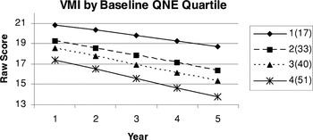
Baseline QNE score, and its interaction with disease duration, was also significantly related to change in a number of the cognitive variables. Not surprisingly, higher baseline QNE score (i.e., greater neurologic dysfunction) was related to poorer performance on the BTA, TMT Part B, and all Stroop trials. Poorer QNE performance at study entry (i.e., baseline QNE × disease duration) resulted in a faster rate of decline over time for TMT Part A and the VMI (see Figures 4 and 5, respectively).

Predicted means for Trail Making Test (TMT) Part A time to completion by baseline Quantified Neurologic Examination (QNE) quartile.

Predicted means for the Developmental Test of Visual-Motor Integration (VMI) raw score by baseline Quantified Neurologic Examination (QNE) quartile.
Given the importance of baseline QNE score in predicting neuropsychological test performance in Model I, the role of total QNE score at each visit (i.e., a time varying covariate) and change in cognition was investigated in a second model. Model II also utilized age at each study visit (rather than solely at baseline, as done in Model I) as another time-dependent covariate. All previously investigated interaction terms were also included. Results of this model are depicted in Table 4.
Regression coefficients for Model II (with probability values in parentheses if statistically significant)

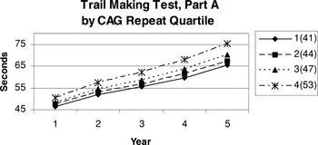
The relationship between total QNE score measured at each study visit was even more strongly related to decline in cognitive functioning than was seen for baseline QNE score or the interaction between baseline QNE score and disease duration. Higher QNE scores (i.e., greater neurologic dysfunction) were related to a more rapid rate of decline for all neuropsychological variables except the WCST efficiency score. Higher educational attainment was related to better performance on the Stroop word and color-word trials and for the COWAT and WAIS-R Vocabulary scores. A more gradual decline over time was seen on TMT Part A and COWAT performances of individuals with higher levels of education at study entry. As seen in Model I, sex was similarly related to WAIS-R Block Design, and women were also noted to exhibit better performance on all Stroop trials across study participation. Finally, the interaction between CAG repeat length and disease duration resulted in a faster rate of decline only for the TMT Part A (see Figure 6).

Predicted means for Trail Making Test (TMT) Part A time to completion by CAG repeat length quartile.
Models I and II were also recomputed excluding the nine subjects with juvenile onset HD (i.e., age of onset prior to age 26; results not shown). In Model I, the most notable changes involved the baseline QNE score becoming a significant predictor of HVLT learning and the interactions between education and disease duration and CAG repeat length and disease duration becoming significant predictors of rate of decline on the WCST and COWAT performance. In Model II, education and total QNE score were no longer significant predictors of WCST efficiency score, but the interactions between education and disease duration and CAG repeat length and disease duration were predictive of rate of change on this measure. Additionally, time (i.e., follow-up year) and educational attainment at study entry became significant predictors of performance on the TMT Part B. Despite these modest changes noted earlier, the most consistent finding of the current study (i.e., the significant value of the total QNE score in predicting cognitive functioning) remained stable when the models were recomputed excluding participants with juvenile onset HD.
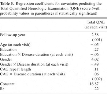
In light of the consistently significant relationship between neurologic functioning and cognitive decline, random effects models were also computed in which only the QNE Eye Movement, Chorea, and Motor Impairment subtest scores (rather than the total QNE score) and time, served as covariates (results not shown). Only the Motor Impairment score was a significant predictor of rate of change in all cognitive variables. The Chorea score was significantly predictive of decline only for the TMT Parts A and B, and the Eye Movement score was not significantly predictive of performance on any of the cognitive variables. Finally, a model was computed to investigate factors associated with rate of change in QNE score. Model III included all of the previous covariates, but the QNE score now served as the dependent variable. As can be seen in Table 5, both time (i.e., follow-up year) and the interaction between CAG repeat length and disease duration were significantly related to rate of decline in QNE score over time, together accounting for 22% of the explained variance.
Regression coefficients for covariates predicting the Total Quantified Neurologic Examination (QNE) score (with probability values in parentheses if statistically significant)

DISCUSSION
Random effects modeling was used to investigate predictors of cognitive functioning and rate of cognitive change in 70 early- to mid-stage patients with HD who were studied prospectively over a four-year period (i.e., five assessments). Performance on all cognitive variables declined significantly over time with the exception of the color-word trial of the Stroop, the WCST, and the WAIS-R Vocabulary subtest. However, the decline in cognitive functioning was modest (see Table 3), consistent with prior longitudinal research (Kirkwood et al., 2001; Snowden et al., 2001). Kirkwood and associates (2001) investigated disease progression in a sample of 1,238 individuals with a minimum 6-year history of HD. Symptom ratings made by caregivers (i.e., spouses or first-degree relatives) indicated that decline in intellectual functioning was first recognized in 18.8% of the sample within 2–5 years of disease onset and in 22.1% of patients during the 6–10 year interval of manifest disease. Worsening of memory was seen within 2–5 years of onset for 17% of individuals with HD and during 6–10 years of disease for 22.5% of the sample. These investigators noted that while subtle changes in cognitive functioning may be identified through careful neuropsychological assessment prior to or soon after patients exhibit sufficient neurological deficits to meet diagnostic criteria for HD, such changes do not become evident to family members until later in disease progression. Snowden and her colleagues (2001) noted only subtle changes in their sample of individuals with HD over one and three years of follow-up.
It is important to consider that the slow progression seen in the current sample may have also been related, in part, to a form of survival bias. Follow-up neuropsychological evaluations would not have been possible in cases where disease had progressed to a level that precluded clinic visits (i.e., nursing home placement and death). In other words, it is possible that the current sample over-represents individuals with more slowly progressive HD. However, there were no differences at baseline between the 70 patients in our study sample and the 87 who did not have sufficient longitudinal data for inclusion.
The lack of significant decline in the WAIS-R Vocabulary score is consistent with the notion that word knowledge is a crystallized cognitive ability, which remains relatively stable in the face of neurological insult. A practice effect related to annual testing may also provide an explanation for the relatively modest decline in some aspects of cognitive functioning. Other investigators have expressed concern that the risk of practice effects inherent in longitudinal research may serve to mask the degree of change in functioning over time. While Snowden and associates (2001) found no change in performance on a modified WCST over a three-year period, other teams of investigators found improvements in WCST performance over a four-year follow-up interval, which they attributed to practice effect (Bauchoud-Levi et al., 2001; Ho et al., 2003). Consistent with these earlier findings, performance on the WCST efficiency score did not significantly decline over time in the current sample. This confluence of findings strongly suggests that use of the WCST in longitudinal research may be of limited utility (Snowden et al., 2001) and that other measures of complex problem-solving, preferably with alternate forms, should be considered.
Interestingly, performance on the Stroop interference trial did not significantly decline over time, while performance on the word reading and color naming subtests did. Decline on the less demanding speed of reading and color naming trials and relatively stable performance on the more challenging color-word interference condition has been noted by other researchers (Snowden et al., 2001) and suggests that a global slowness in processing speed and response execution precludes detection of impaired inhibition of prepotent response tendencies.
Of all covariates included in the random effects regression models other than time (i.e., follow-up year), the total QNE score, both at baseline and at follow-up visits, was most predictive of cognitive functioning for all neuropsychological test variables, again with the exception of the WCST. Among the components of the QNE, only the Motor Impairment score (MIS) was significantly predictive of rate of decline on all cognitive measures. QNE Chorea score was significantly predictive of decline only for time to complete the TMT Parts A and B; and, the Eye Movement score was not significantly associated with any measure of neuropsychological test performance. We have previously found such a relationship between neurological functioning and cognition at our center (Brandt et al., 1984). Snowden et al. (2001) also found that the MIS was the best predictor of change in cognition in their sample of 87 patients studied at one-year follow-up and a smaller sample of 31 participants evaluated two years later. Neurological functioning assessed using the MIS of the QNE may be related to cognition in several ways. First, deficits in purposive motor movements may affect performance on some (e.g., TMT Parts A & B, VMI, Block Design), but certainly not all (e.g., HVLT and BTA), tests of cognitive functioning. Additionally, feedback loops between the prefrontal cortex and basal ganglia are known to affect both cognitive functioning and the coordination of motor movements (Middleton & Strick, 2002), which could also account for the relationship seen between the QNE MIS and all measures of cognitive functioning. While these neural circuits appear to affect involuntary movements (i.e., chorea) and oculomotor control as well, it may be that the effects of disrupted cortico-striatal feedback is more notable for purposive movements and higher-order processing assessed via neuropsychological assessment.
Interestingly, CAG repeat length was not generally associated with cognitive functioning over the course of study participation after adjusting for QNE. This is somewhat in contrast to our findings at two-year follow-up, which revealed a significant relationship between CAG repeat length and a composite score (i.e., factor score) of general cognitive functioning. However, the difference in findings between these studies is not completely surprising given that the current sample size was larger and further along in the progression of their illness, more sophisticated statistical analyses were employed, and the data were not reduced by means of factor analysis. Additionally, participants in the current sample were not grouped by CAG repeat length (i.e., short vs. long CAG repeat length determined by a median split of the sample), as was done in the previous study. In the current study, CAG repeat length was predictive of a more precipitous rate of decline only on a single task of visual scanning, mental tracking, and psychomotor speed (i.e., TMT Part A). The lack of a significant relationship between CAG repeat length and most measures of cognition is not completely unexpected given previous findings. Gomez-Tortosa et al. (1998) studied the characteristics of disease presentation and disease progression in patients with juvenile-, adult-, and late-onset HD and found that CAG repeat length, while strongly associated with akinetic symptoms, was not significantly related to performance on tests of cognitive functioning for any of the patient groups. As previously noted, motor impairment was significantly predictive of cognitive change in Snowden's sample of patients with early- and mid-stage HD, while such a relationship was not seen for CAG repeat length, age, or sex (Snowden et al., 2001). In assessing factors associated with change in total QNE score, we too found that the interaction between CAG repeat length and disease duration was significantly predictive of the motoric functions assessed using the QNE, although not generally predictive of cognition. A recent investigation at our center also revealed that the interaction between CAG repeat length and disease duration was a significant predictor of neurologic dysfunction, adaptive functioning, and cognition assessed globally (i.e., Mini Mental State Examination) in a sample of 413 patients followed annually for up to seven years (Rosenblatt et al., 2006).
Education was variably associated with performance on several neuropsychological measures, including the COWAT, WAIS-R Vocabulary, and the Stroop color and color-word trials. Lower educational attainment at study entry was predictive of a faster rate of decline in performance on the TMT Part A, VMI, and the COWAT. The association between education and word knowledge (many measures of which are used to estimate baseline verbal intellect) is expected, as was the relative stability of performance on the WAIS-R Vocabulary subtest over time in the current sample. Finally, the relationship between older age and poorer performance on tests of speeded visual scanning (i.e., TMT Part A), divided auditory attention (i.e., BTA), complex problem solving (i.e., WCST efficiency score), and three-dimensional block construction (i.e., WAIS-R Block Design) is a pattern often noted in healthy aging and could speculatively be related to the perturbation of dopamine function secondary to basal ganglia degeneration associated with HD, that is, a more malignant form of dopamine depletion than seen in normal aging.
There are several limitations of the current study. While the current sample size is generally larger than that seen in prior longitudinal studies of cognitive change associated with HD, it is nonetheless small given the numerous analyses undertaken. While the relatively small sample size limits statistical power (i.e., increasing the risk of Type II error), the multiple comparisons likely increased the risk of Type I error. Despite these possibilities, the relationship between QNE score and cognition was consistent across all analyses, strongly indicating that this is not a spurious finding. Additionally, modeling of a possible indirect relationship between CAG repeat length and cognition (via QNE score) using structural equation modeling, for example, was precluded due to the relatively small sample size. It is hoped that such analyses will become possible as additional data are collected.
In summary, the degree of neurologic dysfunction, as assessed using the QNE, was most predictive of cognitive decline in early- to mid-stage patients with HD evaluated annually from study entry to 4-year follow-up. The Motor Impairment score, which assesses purposive movement, was the only QNE subscore that was significantly associated with rate of decline on all cognitive variables. Chorea, an early occurring motor dysfunction of HD, was significantly related only to rate of decline on the TMT Parts A and B, which measure speeded visual scanning and sequencing. While not generally related to change in cognition (with the exception of TMT Part A), CAG repeat length was significantly predictive of QNE ratings across study participation. Snowden et al. (2001) proposed that significant changes in simple versus more effortful cognitive tasks may be associated with HD-related dysfunction in the basal ganglia, which are important in mediating automatic motor tasks rather than effortful tasks under conscious control. We suggest that CAG repeat length is more closely linked with initial changes in the basal ganglia that predominate in early- to mid-stage HD than with cortical degeneration seen later in disease progression. Such a relationship would explain the predictive value of CAG repeat length in changes associated with automatic motor response programs (e.g., QNE and Trail Making Test Part A) but not with tasks that require more effortful processing.
ACKNOWLEDGMENTS
Supported by grant P01-NS16375 from the National Institutes of Health to the Johns Hopkins University. The authors wish to thank Ms. Deborah Pollard, Dr. Abhijit Agarwal, and Dr. Christopher Ross. Dr. Brandt is an author of the Hopkins Verbal Learning Test–Revised, one of the neuropsychological tests used in this study. He receives royalties on its sale from the test publisher (Psychological Assessment Resources, Inc.).