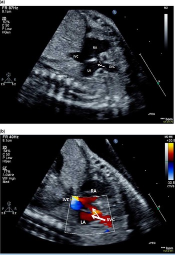
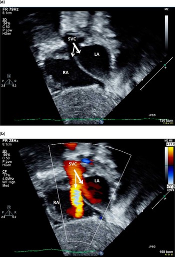
We report the first published pictures of superior-type caval vein defect – sinus venosus atrial septal defect – in a foetus. The foetus was 25 weeks and 6 days old and referred for “echogenic foci” and bilateral pyelectasis in a mother who had an abnormal quadruple screen test – α-fetoprotein, human chorionic gonadotropin, unconjugated oestriol, and inhibin A. The echocardiogram was suspicious for a small patent foramen ovale (1.5 mm). Upon further examination in the bicaval view it was confirmed by two-dimensional echocardiogram (Fig 1a) and colour mapping (Fig 1b) that the superior caval vein overrode the left and right atria. The bicaval view is an important view and can be found near the long-axis aortic arch view in a parasagittal plane of the right chest.Reference Chiappa, Cook, Botta and Silverman 1 Postnatal echocardiogram confirmed the diagnosis (Fig 2a and b) and additionally showed the commonly associated partially anomalous pulmonary venous return of the right-upper and right-middle pulmonary veins.
Bicaval view – inferior and superior caval veins in long-axis view – in a 25-week-old foetus with superior-type caval vein defect. ( a ) Two-dimensional foetal echocardiogram showing the overriding superior caval vein and its connection to the right atrium (thin arrow) and the defect allowing connection to the left atrium (thick arrow). ( b ) On colour flow mapping, foetal blood flow from the superior caval vein is seen draining both into the right atrium (thin arrow) and the left atrium (thick arrow).

Subxiphoid sagittal view in the same patient as 1-day-old neonate. ( a ) Two-dimensional neonatal echocardiogram confirms the superior-type caval vein defect. ( b ) Colour flow mapping demonstrates the bi-atrial drainage of the superior caval vein in the neonate. As the superior caval vein’s blood flow enters both atria, that which goes to the right atrium has a smaller orifice and is more directly aligned with the transducer; this flow is seen aliasing.

Financial Support
This research received no specific grant from any funding agency, commercial or not-for-profit sectors.
Conflicts of Interest
None.