Introduction
The likelihood of second primary tumours in patients with squamous cell carcinoma (SCC) of the head and neck has long been recognised, with the 20-year risk being as high as 36 per cent.Reference Chuang, Scelo, Tonita, Tamaro, Jonasson and Kliewer1 Second primary tumours occur in these patients as a result of field cancerisation of the upper aerodigestive tract,Reference Slaughter, Southwick and Smejkal2, Reference Braakhuis, Leemans and Brakenhoff3 with the lung being the most common site for a synchronous second primary tumour.Reference Chuang, Scelo, Tonita, Tamaro, Jonasson and Kliewer1, Reference Graff, Schipman, Desandes, Mecellem, Toussaint and Cortese4
Patients presenting with head and neck SCC should have their full history taken, and undergo examination including fibre-optic laryngoscopy.Reference Pfister, Ang, Brizel, Burtness, Busse and Caudell5 Patients with oral SCC should also undergo head, neck and chest imaging, usually either neck computed tomography (CT) with contrast and chest CT, or whole body positron emission tomography (PET)-CT, as part of their staging.
Panendoscopy performed under general anaesthesia, consisting of rigid bronchoscopy, oesophagoscopy and direct laryngoscopy, is standard practice at many centres for the assessment of primary tumours and for concurrent screening of second primary tumours of the upper aerodigestive tract.Reference Stoeckli, Zimmermann and Schmid6 With improvements in in-office assessment, including better fibre-optic laryngoscopy and transnasal oesophagoscopy,Reference Postma, Bach, Belafsky and Koufman7 and advancements in multi-modality imaging, in particular thin-slice CT, magnetic resonance imaging and PET-CT, the question remains as to whether panendoscopy improves the detection of second primary tumours.Reference Davidson, Gilbert, Irish, Witterick, Brown and Birt8, Reference Rodriguez-Bruno, Ali and Wang9 The need for panendoscopy in lower risk groups, especially the non-smoking, non-drinking subgroup considered at lowest risk for second primary tumours, is debatable.
Tobacco and alcohol are known to be risk factors for second primary tumour development.Reference Day, Blot, Shore, McLaughlin, Austin and Greenberg10 However, while a recent study observed no second primary tumours in non-smoking, non-drinking patients presenting with oral cavity and oropharyngeal SCCs,Reference Rodriguez-Bruno, Ali and Wang9 other authors have reported that the risk of second primary tumours is similar to that in the general population.Reference Farshadpour, Hordijk, Koole and Slootweg11 Previously published data from our institution revealed that the rate of second primary tumour in non-smoking, non-drinking patients was similar to that in patients who were smokers and/or drinkers, with all second primaries presenting in the oral cavity or oropharynx; no synchronous primaries were noted in the non-smoking, non-drinking group.Reference Koo, Barrowman, McCullough, Iseli and Wiesenfeld12
Given the conflicting evidence, this study aimed to review the incidence and prevalence of second primary tumours, specifically in early stage oral tongue carcinomas. The study compared non-smoking, non-drinking patients to those with known risk factors, in order to evaluate the need for separate panendoscopy in this subgroup.
Materials and methods
This project was approved by the Human Research Ethics Committee of the Office for Research at Melbourne Health (QA2011036). Internal review board ethics approval was obtained.
All patients presenting with T1 or T2 oral tongue SCCs to the Royal Melbourne Hospital, from 1 January 1991 to 31 July 2010, were identified from a prospectively maintained head and neck database (from 1 January 2007) and from medical records (prior to 1 January 2007). Demographic, disease characteristics and survival data were collected from a review of the patient records.
Tumours were classified according to the International Classification of Diseases for oral cavity subsites, and retrospectively updated to reflect the American Joint Committee on Cancer staging system.Reference Greene, Page, Fleming, Fritz, Balch and Haller13 Second primary tumours and recurrences were classified as described by Warren and Gates.Reference Warren and Gates14 The census date was set at 1 July 2012.
Patients were classified as ‘smokers’ if they had a smoking history of greater than one pack year. To be classified as a ‘drinker’, a patient was required to have an average weekly alcohol intake of greater than 20 standard units.
Statistical analysis was performed using the IBM SPSS® Statistics software package, version 19. Chi-square tests were employed for the analysis of disease characteristics and demographics, while Student's t-tests were utilised for age analysis. Statistical significance was set at p < 0.05.
Results
A total of 119 consecutive patients with T1 or T2 oral tongue carcinomas were identified. Seven patients were excluded as a result of incomplete records. Of the 112 remaining patients, only 14 presented with nodal disease (12.5 per cent), while none presented with distant metastases. Mean follow-up duration was 71.7 months (range, 3.6–238.3 months). Thirty-five patients (31.2 per cent) died within the follow-up period.
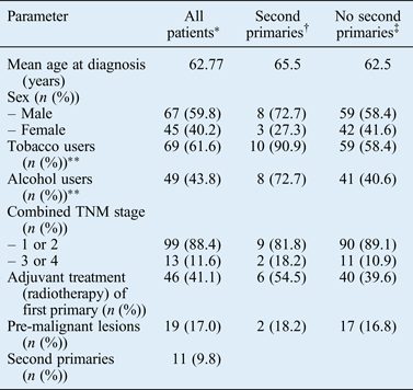
Apart from tobacco and alcohol use, there were no significant demographic differences between the patients with second primary tumours and those without (Table I). In particular, there was no difference in the mean age at presentation between the two groups. While more men than women had second primary tumours, this difference was not statistically significant.
Distribution of second primaries by time and subsite. Each dot represents a new primary, with red dots representing females. Dots left of the axis denote tumours predating care at our institution.

Patient characteristics
*n=112; †n = 11; ‡n = 101. **A significant difference was found between having a second primary and not having one, depending on use of alcohol or tobacco (p < 0.05; chi-square test). TNM = tumour–node–metastasis
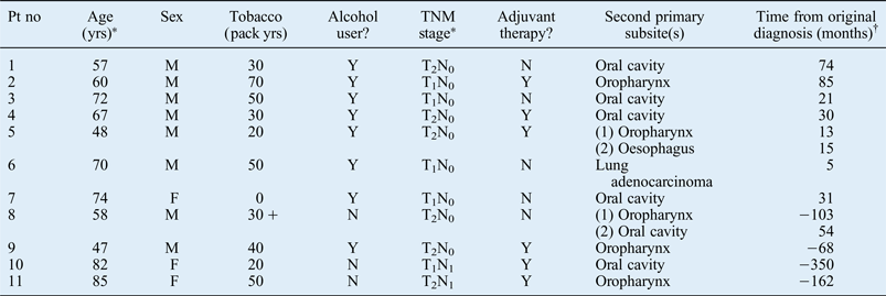
Thirteen second primary tumours were identified in 11 patients (9.7 per cent), with 4 events predating care at our institution. The majority of second primary tumours occurred in either the oral cavity (46.2 per cent) or the oropharynx (38.5 per cent); only two patients (15.4 per cent) presented with tumours in other sites, namely the oesophagus and lung (Figure 1). All patients presenting with a second primary tumour had a history of tobacco or alcohol use, with the only non-smoker (patient 7) consuming four standard drinks per day (Table II).
Patients with second primary tumours
*At first presentation. †Negative numbers denote primaries prior to initial presentation to our institution. Pt no = patient number; yrs = years; TNM = tumour–node–metastasis; M = male; Y = yes; N = no; F = female
Tobacco and alcohol were found to increase the relative risk of second primary development (relative risks of 1.140 and 1.138 respectively), while non-smoking, non-drinking status led to a decreased risk (relative risk of 0.861). Adjuvant treatment with radiotherapy, tumour–node–metastasis stage at initial diagnosis, gender and a history of pre-malignant lesions did not modify the risk of second primary development (Figure 2).
Relative risk of second primary development after an initial T1 or T2 oral tongue primary, categorised by risk factors (*p < 0.05). CI = confidence interval; TNM = tumour–node–metastasis
Discussion
In this cohort of 119 early stage oral tongue SCC patients, no synchronous lesions were detected on panendoscopy. The findings revealed a significantly lower rate of metachronous primaries in the non-smoking, non-drinking group (0 per cent) versus the smokers (14.5 per cent) and alcohol users (16.3 per cent). For patients in this cohort, most second primary tumours occurred in the oral cavity (46.2 per cent) or oropharynx (38.5 per cent).
Examination under anaesthesia, including panendoscopy, aids direct surgical management in selected oral cavity SCC cases; for example, when used to assess for tongue base involvement. The two areas most difficult to assess in-office – the hypopharynx and tongue base – should be checked at the time of primary resection of the oral tongue SCC. However, panendoscopy is associated with a small but real risk of oesophageal perforation,Reference Wennervaldt and Melchiors15 and has disadvantages associated with the expenditure of time and equipment costs. Appropriate patient selection prior to performing a panendoscopy would help with risk and cost minimisation. The findings of the current study suggest that there is no need for separate panendoscopy to be conducted in patients presenting with T1 or T2 oral tongue SCC, and pre-operative assessment with current examination methods including fibre-optic endoscopy and chest CT may be sufficient in such cases.
There is the potential for PET-CT to fulfil this role of second primary tumour detection. This modality allows clinicians to search for second primary tumours and metastases simultaneously over a large field. However, PET-CT does increase patients’ radiation exposure and is not as freely available as panendoscopy.
The current study found that most second primary tumours in early tongue SCC patients occurred in the oral cavity and oropharynx. These regions can be adequately assessed in-office, without formal panendoscopy. Nevertheless, the risks and costs of panendoscopy must be weighed against the potentially devastating consequences of a missed synchronous primary. In addition, panendoscopy performed prior to resection may still have value for the assessment and anatomical appreciation of a known tumour in three dimensions, and for the delineation of margins. The information provided also assists in reconstructive planning. Pre-operative understanding of expected resection margins and the type of reconstruction planned allows patients to give consent in a more informed situation.
• There is a significant risk of second primary malignancies in head and neck squamous cell carcinoma (SCC) patients
• Panendoscopy is commonly used to assess primary tumours and screen for second primary tumours
• In this study, tobacco and alcohol increased the relative risk of second primary tumour development in patients with T1 or T2 oral tongue SCC
• Most second primary tumours were in the oral cavity and oropharynx; no second primary tumours were found in non-smoking, non-drinking patients
• Panendoscopy may have less value in non-smoking, non-drinking patients with early stage oral tongue SCC
• Panendoscopy remains a useful assessment tool in smokers and heavy drinkers
The findings of the current study indicate a lower rate of second primary tumour in patients who do not smoke or consume alcohol, which is consistent with current evidence.Reference Chuang, Scelo, Tonita, Tamaro, Jonasson and Kliewer1 Aetiopathogenesis of oral SCC in the non-smoking, non-drinking group is currently poorly understood. Field cancerisation from an as yet undefined carcinogen may play a role in these patients. Therefore, despite a statistically significant lower rate of second primary tumours in non-smoking, non-drinking patients, information on other potential carcinogens or population-based data sets that support our data would be helpful.
In summary, we have identified that non-smoking, non-drinking patients with early stage tumours of the oral tongue have a lower risk of second primary tumours, and panendoscopy may have a lower utility in this particular subgroup. For patients with oral tongue SCCs, second primary tumours most commonly occur in the oral cavity and oropharynx; these regions can be adequately assessed in-office, without formal panendoscopy. Selective panendoscopy may provide information about the extent of mandibular and tongue base tumours, and aid surgical and reconstructive decision-making.
Acknowledgements
Funding was provided by the Price Family Foundation (the study sponsors had no involvement in the study design or production of the manuscript). The authors would like to acknowledge the assistance of Craig Love for his assistance with data acquisition.