Introduction
Delusional thoughts are common among patients with Alzheimer's disease (AD; Jeste et al., Reference Jeste, Wragg, Salmon, Harris and Thal1992; Paulsen et al., Reference Paulsen2000). Although delusion is regarded as a psychotic symptom of AD patients (Cummings et al., Reference Cummings, Mega, Gray, Rosenberg-Thompson, Carusi and Gornbein1994), the merit of antipsychotic agents remain controversial (Kales et al., Reference Kales2007; Ballard et al., Reference Ballard2009). Meanwhile, studies investigating the specific brain dysfunctions underlying the delusions of AD patients have shown varying findings; many studies have suggested important roles for the right hemisphere of the brain (Staff et al., Reference Staff, Shanks, Macintosh, Pestell, Gemmell and Venneri1999; Reference Staff, Venneri, Gemmell, Shanks, Pestell and Murray2000; Mega et al., Reference Mega, Lee, Dinov, Mishkin, Toga and Cummings2000; Fukuhara et al., Reference Fukuhara2001; Lopez et al., Reference Lopez, Smith, Becker, Meltzer and DeKosky2001; Sultzer et al., Reference Sultzer2003; Nakano et al., Reference Nakano, Yamashita, Matsuda, Kodama and Yamada2006; Matsuoka et al., Reference Matsuoka2010), the temporal lobe (Starkstein et al., Reference Starkstein1994; Hirono et al., Reference Hirono1998; Lopez et al., Reference Lopez, Smith, Becker, Meltzer and DeKosky2001; Sultzer et al., Reference Sultzer2003; Nakano et al., Reference Nakano, Yamashita, Matsuda, Kodama and Yamada2006), or the frontal lobe (Mega et al., Reference Mega, Lee, Dinov, Mishkin, Toga and Cummings2000; Staff et al., Reference Staff, Venneri, Gemmell, Shanks, Pestell and Murray2000; Lopez et al., Reference Lopez, Smith, Becker, Meltzer and DeKosky2001; Sultzer et al., Reference Sultzer2003; Bruen et al., Reference Bruen, McGeown, Shanks and Venneri2008). The reproducibility of previous studies may have been limited by the heterogeneity of delusional phenomena, because uniform account of delusion may conceal the phenomenological and neurobiological heterogeneity (Shanks and Venneri, Reference Shanks and Venneri2004). Moreover, the naive use of the term “delusion” may potentially mislead the understanding of these phenomena, since this term has historically been used for describing the pathological ideations of schizophrenic patients that exhibit clinical pictures that are essentially different from those of AD patients (Burns et al., Reference Burns, Jacoby and Levy1990; Jeste and Finkel, Reference Jeste and Finkel2000).
A promising key to solving this seemingly exhaustive issue may be available within the content of the delusional thoughts. Despite the vast array of delusional episodes, clinical observations have suggested that these episodes demonstrate typical patterns of themes; “People are stealing things,” “One's spouse is unfaithful,” “One's house is not one's house,” etc. (Monteiro et al., Reference Monteiro, Boksay, Auer, Torossian, Ferris and Reisberg2001). These empirical findings may suggest particular neurological bases underlying specific patterns of delusional ideas.
The aim of this study was to clarify the potentially discrete subtypes that have been included in the delusions of AD. We hypothesized that categorization of the content of delusional ideas would correspond to category-specific sets of neural substrates. To further clarify this hypothesis, we enrolled consecutive first-visit outpatients and administered the Frequency-Weighted Behavioral Pathology in Alzheimer's Disease Rating scale (BEHAVE-AD-FW; Monteiro et al., Reference Monteiro, Boksay, Auer, Torossian, Ferris and Reisberg2001) to assess delusional ideas and applied the Statistical Parametric Mapping (SPM) method to analyze Single Photon Emission Computed Tomography (SPECT) images.
Methods
Patients
The study was performed at the Tajiri Clinic, an integrated institute for stroke and dementia, which is situated in Osaki, Miyagi Prefecture, in northern Japan. All patients suspected of having dementia underwent routine medical checkups, including head magnetic resonance imaging (MRI), head SPECT (see below), electrocardiogram, chest X ray, and laboratory urine and blood tests to allow differentiation of the diagnoses. The Mini-Mental State Examination (MMSE; Folstein et al., Reference Folstein, Folstein and McHugh1975), the Cognitive Abilities Screening Instrument (Teng et al., Reference Teng1994), and the standardized Japanese version of the BEHAVE-AD-FW (Asada et al., Reference Asada, Homma, Kimura and Uno1999) were also routinely administered.
Consecutive first-visit outpatients who were seen at the Tajiri Clinic from February 2006 to July 2010 were enrolled. The inclusion criteria were: (1) probable AD according to the National Institute of Neurological and Communicative Disorders and Stroke-Alzheimer's Disease and Related Disorders Association (McKhann et al., Reference McKhann, Drachman, Folstein, Katzman, Price and Stadlan1984); (2) free of any psychotropic medication during the six weeks before the research assessment and SPECT acquisition; and (3) the ability to follow the patient for three months or more, to confirm the inclusion and/or exclusion criteria. The exclusion criteria were (1) past medical histories of stroke, depression or schizophrenia, and other psychiatric diseases; (2) lacunar infarctions, as shown by MRI; (3) presence of early signs of Lewy body disease, rapid eye movement sleep behavior disorder or restless legs syndrome; and (4) MMSE scores lower than 6 at the first visit evaluation.
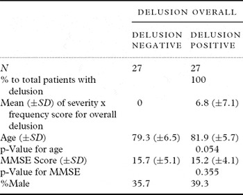
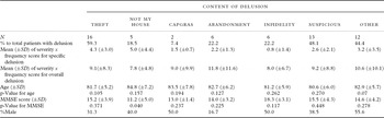
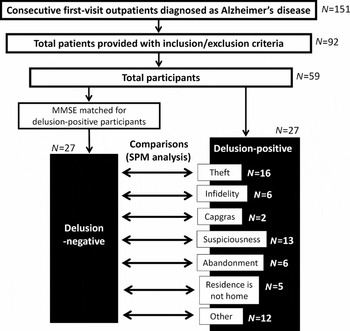
During the study period, 396 patients consulted the clinic for their first visit. Of these patients, 151 were diagnosed as having AD, 69 as AD with cerebrovascular diseases, 42 as vascular dementia, and 21 as dementia with Lewy bodies. Of the 151 AD patients, 92 cases fulfilled the inclusion criteria and did not fit the exclusion criteria. Of the 92 cases, 59 participated in the study. Of the 59 participants, 27 were positive for one or more categories of delusion. For the delusion-negative control subjects, MMSE scores were semirandomly matched with the delusion-positive subjects. Figure 1 and Table 1 show the patient selection procedure and demographics of the participants, respectively. Table 2 shows the demographics of the patients according to the content of delusion. Age, gender, and MMSE scores were not significantly different between those with and without delusion.
Demographic data of the subjects with and without delusion

Note: p-Values for t-test comparisons with the delusion-negative group are shown.
Demographic data for the subjects with specific content of delusion

Note: p-Values for t-test comparisons with the delusion-negative group are shown.
Patient selection procedure and number of patients for each step of the process are shown.

Informed consent was obtained from the patients and their family prior to enrolment to the study. The study was approved by the Ethical Committees of the Osaki-Tajiri SKIP Center and Tohoku University Graduate School of Medicine.
Delusion analyses
In assessing delusion, BEHAVE-AD-FW collects information from informants, i.e. family members, regarding the presence, severity, and frequency of seven categories that constitute most typical delusional episodes found in AD patients: delusion of theft, “one's house is not one's house” delusion (“not home” delusion), Capgras syndrome, delusion of abandonment, delusion of infidelity, suspiciousness/paranoia, and delusions other than those described above. These elements include assessment over the previous two-week interval. This time frame is sufficiently long to give an accurate view of the symptoms and is sufficiently brief for informants to be able to accurately report on the events (Monteiro et al., Reference Monteiro, Boksay, Auer, Torossian, Ferris and Reisberg2001).
Each group of any given category of delusion consisted of patients who were positive finding for the specific delusion category in question. With regard to the procedure for analyzing the functional brain images of a given specific type of delusion (e.g. delusion of theft), there is a problem when considering whether or not the cases with coexisting delusion(s) other than that of interest should be excluded. For the purpose of analyzing “pure” cases with “delusion of theft,” excluding the cases with both delusions of theft and any other type of delusion would cause a bias originating from the assessment method, because the reproducibility of the judgment about “delusion” could be unstable, whereas it may be easy to agree on what the “delusion of theft” is. In particular, if the proportion of cases with multiple types of delusion is large, relative to the overall cases with delusion of theft, the bias becomes even more critical. Therefore, we did not exclude cases with co-existing delusions.
Spearman's correlation coefficients were calculated between significantly correlating subscores of specific categories of delusion (p < 0.01).
Image acquisition
The SPECT investigation was using technetium-99m-labeled ethyl cysteinate dimer (99mTc-ECD). A SPECT scanner (Millennium MG, GE Yokogawa Medical Systems, Tokyo, Japan) with a 2-head rotating gamma camera with low-energy high-resolution collimator and in-plane resolution of 10 mm full width at half maximum was used for all SPECT studies. For each camera, projection data were obtained in a 128 × 128 format for 24 angles at 50 s per angle. Image reconstruction was performed by filtered back projection with a Butterworth filter, and attenuation correction was made numerically by assuming the object shape to be an ellipse for each slice and the attenuation coefficient as uniform. A Shepp and Logan Hanning filter was used for image reconstruction at 0.7 cycles/cm, with attenuation correction using Chang's method. The SPECT scan was performed 15 minutes after intravenous injection of 740 MBq of ethyl cysteinate dimer. The SPECT acquisition protocol comprised 36 projections at 25 seconds per projection with 360 degrees rotation of the camera. No quantitative analysis was performed for the determination of cerebral blood flow (CBF).
Image analysis
Image analysis was performed using SPM 5 software (Wellcome Department of Cognitive Neurology, Institute of Neurology, University of London, UK) incorporated into MATLAB 7.1 (The MathWorks, Natick, MA, USA). All of the SPECT images were spatially normalized to the template image according to the space described in the atlas of Talairach and Tournoux (Reference Talairach and Tournoux1988) using the default options. Normalized images were represented on a 79 × 95 × 69 matrix with 2 × 2 × 2 mm voxel size. Spatially normalized images were then smoothed by convolution using an isotopic Gaussian kernel with a 10-mm full width at half maximum to increase the signal-to-noise ratio.
For the analysis of “overall delusion,” the SPECT images of the patients with at least one type of delusion were compared with those without any type of delusion using the two-sample t-test. The statistical threshold was set at p < 0.05, and was corrected for family-wise error. The cluster size was greater than 50 voxels. If no significant area was detected, the images were reanalyzed at the thresholds of 0.0002, 0.0005, or 0.001, which is uncorrected. We consider that these thresholds are acceptable and sufficiently safe according to previous studies using SPECT and SPM (Imon et al., Reference Imon, Matsuda, Ogawa, Kogure and Sunohara1999; Staff et al., Reference Staff, Venneri, Gemmell, Shanks, Pestell and Murray2000; Fukuhara et al., Reference Fukuhara2001).
The SPECT images of each group of the patients with specific content of delusion were then compared with those without any type of delusion using the two-sample t-test. The statistical thresholds were applied in the same manner as the analysis of the overall delusion as detailed above. There were six comparisons made for specific types of delusion, however, the p-values were not corrected for these multiple comparisons, because members of the groups with a specific type of delusion overlapped with each other and could not be regarded as statistically independent. Although the threshold of an uncorrected p-value of 0.001 may be rather weak in making conclusive interpretations, we consider that these thresholds are acceptable and may successfully show the heterogeneity of delusion.
Results
Analysis of SPECT images
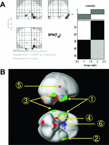
Figure 2(a) indicates the areas of significantly decreased CBF in the overall delusion-positive cases, compared with the delusion-negative cases; the bilateral temporal lobes with right-side dominancy, right inferior temporal gyrus, and right amygdala (p < 0.0002, uncorrected).
(A) Comparison between overall cases with and without delusion. Delusion-positive cases showed lower RI uptake (p < 0.0002, uncorrected) at the bilateral temporal lobes with right-side dominancy, at the right inferior temporal gyrus and the right amygdala. (B) Rendered image illustrating the regions of the brain that correlated with specific types of delusional ideas. Green = delusion of theft; Red = delusion of abandonment; Blue = “Residence is not Home.” 1 = right temporal pole; 2 = left temporal pole; 3 = right inferior temporal gyrus; 4 = right parahippocampal cortex; 5 = right posterior insula; 6 = right amygdala.

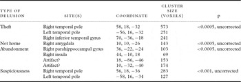
CBF for different regions of the brain correlated with specific categories of delusional ideas; decreased CBF in the bilateral temporal poles (TPs) with right-side dominancy and in the right inferior temporal gyrus correlated with delusion of theft, decreased CBF in the bilateral TPs with right-side dominancy correlated with suspiciousness/paranoia, decreased CBF in the right parahippocampal gyrus and right posterior insula correlated with delusion of abandonment, and decreased CBF in the right amygdala correlated with the delusion of “not home.” No area was detected for the delusions of infidelity or Capgras Syndrome (Table 3, Figure 2(b)).
Results of the SPM analysis (i.e. types of delusion and corresponding brain regions, coordinates, cluster sizes, and p-values) are shown

Correlations between specific delusional thoughts
The Spearman's correlation coefficient between the scores of delusion of theft and abandonment was 0.364 (p < 0.01). Correlations between the scores for other categories of delusion did not reach significance.
Discussion
Limitations of the present study
Despite the limitations below, we consider that our study has persuasively elucidated the heterogeneity of the delusion of AD patients. First, the cross-sectional design of the present study limits the interpretations regarding the mechanisms underlying the delusions. Second, none of the differences were significant using family-wise corrected p-values, which may be a consequence of the small number of patients studied. Given the weakness of the significance level of our image analysis, any conclusions of our study must be considered as tentative. Third, our analysis was based on each specific type of the content of delusions, regardless of other potentially confounding factors, e.g. agitation, apathy, wandering behaviors, etc. However, if any of these factors correlate with any of the specific types of delusional ideas, adjusting for these factors may introduce another bias. Therefore, we consider that our procedure is acceptable. Fourth, our results cannot exclude the effect of the general severity of delusion, since the severity of overall delusion was not controlled among the groups with specific content. With regard to this point, none of the groups with specific content showed significantly different mean values for the total severity x frequency score of delusion. Therefore, the effect of the general severity of delusion may be regarded as minimal in our analysis. Related to this problem, however, our study did not evaluate the effect of the severity of specific types of delusion, which is a weakness that may have originated from the small number of subjects. Fifth, our SPECT analysis may be relatively weak in statistical power because of the poor linearity of ECD and the lack of quantitative evaluation of CBF.
Merit of the BEHAVE-AD-FW
Systematic surveying and the phenomenological approach are the most remarkable features of this scale, which likely prevent missing important events that would otherwise be overlooked.
As an example of other procedures, the Neuropsychiatric Inventory (NPI; Cummings et al., Reference Cummings, Mega, Gray, Rosenberg-Thompson, Carusi and Gornbein1994) determines the presence of “delusion” by explaining to the informants the definition of delusion, and then specifies the frequency, severity, and content. Using this procedure, specific facts demonstrated by the patients can be lost. Furthermore, both informants and interviewers need not only to understand precisely what the term “delusion” stands for, but they must also interpret each event correctly when applying the term to it. These difficulties can be critical when investigating the potential heterogeneity of delusional episodes.
Heterogeneity of delusion
The present study highlighted the heterogeneity of delusion, by clarifying the regions of the brain that correspond to particular types of phenomena according to the classification system of the BEHAVE-AD-FW. Despite the potential heterogeneity of delusion, the right-side dominancy of these findings may support the hypothetical importance of the right hemisphere in delusion formation as suggested by previous studies (Staff et al., Reference Staff, Shanks, Macintosh, Pestell, Gemmell and Venneri1999; Reference Staff, Venneri, Gemmell, Shanks, Pestell and Murray2000; Mega et al., Reference Mega, Lee, Dinov, Mishkin, Toga and Cummings2000; Fukuhara et al., Reference Fukuhara2001; Lopez et al., Reference Lopez, Smith, Becker, Meltzer and DeKosky2001; Sultzer et al., Reference Sultzer2003; Nakano et al., Reference Nakano, Yamashita, Matsuda, Kodama and Yamada2006; Bruen et al., Reference Bruen, McGeown, Shanks and Venneri2008; Matsuoka et al., Reference Matsuoka2010).
For the purpose of clarifying the heterogeneity of delusions of AD, Nomura et al. (Reference Nomura2012) used NPI and conducted a factor analysis for the content of delusion and reported brain dysfunctions correlating to each of the factors. In this previous study, the reproducibility of the data regarding the content of delusion may be weak because of limitations originating from the NPI assessment procedure as noted above. Moreover, factor analysis for the psychological data may conceal neural correlates if any additional mechanisms exist (e.g. social or psychological mechanisms) that could link different content of delusional thoughts. The procedure used in our study may avoid these limitations.
Frontal lobe, memory deficit, and delusion formation
The frontal lobe was not shown to be a significant region in any of our image analyses, which is partly inconsistent with previous findings (Mega et al., Reference Mega, Lee, Dinov, Mishkin, Toga and Cummings2000; Staff et al., Reference Staff, Venneri, Gemmell, Shanks, Pestell and Murray2000; Lopez et al., Reference Lopez, Smith, Becker, Meltzer and DeKosky2001; Sultzer et al., Reference Sultzer2003; Bruen et al., Reference Bruen, McGeown, Shanks and Venneri2008). For example, Sultzer et al. (Reference Sultzer2003) reported that right frontal and temporal hypometabolism correlated with delusion, and argued that dysfunction of critical regions of the frontal lobe may be associated with delusions as a consequence of loss of the frontal systems that monitor reality, link new experiences with memories of the past, or prevent consolidation of inaccurate beliefs. This is a persuasive account, because failure in revising faulty ideas despite evidence to the contrary is, by definition, a key feature of delusion. However, the same failure may also be accounted for by memory deficit alone, because the difficulty in updating memory can impede correcting faulty ideas and cause the thoughts to lose touch with reality. Our results may challenge the primary significance of the frontal lobe, as suggested by these previous studies.
TP, inferior temporal gyrus, and delusion
Our study suggested that the dysfunction of the TPs could be shared by the two categories of delusional thoughts: delusion of theft and suspiciousness/paranoia. The involvement of the temporal lobe is a finding that is partly consistent with several previous studies (Starkstein et al., Reference Starkstein1994; Sultzer et al., Reference Sultzer2003; Nakano et al., Reference Nakano, Yamashita, Matsuda, Kodama and Yamada2006). While any interpretations on causality must be tentative in our study, we infer that there may be a common dysfunction that is shared by delusions of theft and suspiciousness. The common features of these particular delusional thoughts may include a distorted cognition of human relationships. Our findings may support this interpretation, since TP dysfunction may cause a disrupted socioemotional cognition that is similar to the theory of mind (Mychack et al., Reference Mychack, Kramer, Boone and Miller2001; Thompson et al., Reference Thompson, Patterson and Hodges2003; Olson et al., Reference Olson, Plotzker and Ezzyat2007).
Plausible interpretations regarding the involvement of the inferior temporal gyrus in delusion of theft may include poor insight for cognitive impairment (Salmon et al., Reference Salmon2006) and impaired retrieval of autobiographical memory (Addis et al., Reference Addis, McIntosh, Moscovitch, Crawley and McAndrews2004). Clinical observations may support this idea, since patients with the delusion of theft lose their possessions, accuse the people in their surrounding of having stolen these belongings, and are usually unaware that they themselves lost their own items.
Right parahippocampal gyrus, posterior insula, and delusion of abandonment
SPM analysis of this type of delusion resulted in a somewhat noisy finding. While some of the clusters may be artifacts, we infer that those indicated at the right parahippocampal gyrus and the posterior insula should be considered.
The parahippocampal gyrus, which is referred to as a critical site for episodic memory processing (Gabrieli et al., Reference Gabrieli, Brewer, Desmond and Glover1997; Yoo et al., Reference Yoo2012), has also been shown to be involved in emotional processes, especially with respect to negative or unpleasant ones (Wei et al, Reference Wei, Qiu, Du and Luo2011; Aldhafeeri et al., Reference Aldhafeeri, Mackenzie, Kay, Alghamdi and Sluming2012). With regard to delusion, some previous studies have implicated the medial temporal lobe in the persecutory delusion of AD (Ismail et al., Reference Ismail, Nguyen, Fischer, Schweizer, Mulsant and Mamo2011).
A functional imaging study reported the involvement of the posterior insula in both physical pain as well as “emotional pain,” namely, the subjective experience of being intensely rejected by people who are important or beloved (Kross et al., Reference Kross, Berman, Mischel, Smith and Wager2011). Our finding, showing the involvement of the posterior insula, may be consistent with the previous study, since the idea of being abandoned may be translated into that of being rejected.
We infer that involvement of the parahippocampal gyrus and posterior insula suggests that the complaint of “They abandon me” may be a manifestation of an emotional distress. This may be a distress that is similar to what we experience when we are rejected by people we perceive as being important or beloved.
Amygdala and “Residence is not home”
Some AD patients may say “I want to go home,” even though they are already in their own house. Our data suggested that dysfunction of the right amygdala may be associated with this symptom.
The amygdala is a brain region that is speculated to be important for emotional processing, especially that of fear and anxiety (Kalin et al., Reference Kalin, Shelton and Davidson2004; LeDoux, Reference LeDoux2007; Etkin et al., Reference Etkin, Prater, Schatzberg, Menon and Greicius2009). Based upon our data and these previous findings, the “not my home” delusion may be associated with fear or anxiety. In further support of this hypothesis, the score of the subscale “G. Anxiety and Phobia” section of BEHAVE-AD-FW correlated positively with the “not home” delusion (Spearman's correlation coefficient = 0.27, p = 0.044). The “home” mentioned here may not refer literally to their home, but some place where they “feel” at home.
Delusions of theft and abandonment
Our data showed a correlation between the delusions of theft and abandonment. To our knowledge, no previous study has evaluated the correlation between these two elements. While this correlation may partly be consistent with the idea of “persecutory delusion,” as proposed in some previous studies (Hwang et al., Reference Hwang, Tsai, Yang, Liu and Lirng1999; Cook et al., Reference Cook2003), we cannot clearly determine here whether this correlation occurs universally, depending on cultural background, or simply by chance. However, this finding may suggest that it is the presence of certain psychological or social processes, rather than neural dysfunction, that links these phenomena, since the findings of the SPECT images suggested that the relevant brain regions may be discrete between these two elements. One plausible interpretation may be an attempt for the “counterattack” against the surrounding people whom the patients accuse of rejecting them. Further psychological or psychosocial investigations may help elucidate whether there is indeed any mechanism underlying this correlation.
Conclusion
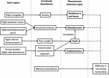
A schema demonstrating interpretations as to the content of delusional ideas and corresponding brain regions is shown in Figure 3. The findings of the present study offer a perspective on the potentially discrete categories of pathological thoughts that have previously been lumped together as “delusions.” Content-based categorization may be a simple and promising method for clarifying the heterogeneity that may critically limit the reproducibility of neurobiological investigations and clinical interventions on delusions in patients with AD.
Schema demonstrating the hypothetical process of delusional ideations and underlying dysfunctions of the regions of the brain. No relevant brain region was detected for delusion of infidelity or Capgras syndrome.

Conflict of interest
None.
Description of authors' roles
Masahiro Nakatsuka formulated the research questions, designed the study, analyzed the data, and wrote the paper. Masahiro Nakatsuka, Kenichi Meguro, Kei Nakamura, Kyoko Akanuma, and Satoshi Yamaguchi were responsible for BEHAVE-AD-FW data collection. SPECT data collection was done by Hiroshi Tsuboi. Kenich Meguro supervised the study design, data analysis, and writing the paper.