Introduction
The emerging technology of endoscopic surgery allows otolaryngologists to perform a variety of surgical procedures in the nose and sinuses. Endoscopic sinus surgery has been used to treat chronic sinusitis. Endoscopic performance of middle meatal antrostomy or inferior meatal nasoantral window has recently been reported.Reference Kamel1–Reference Sato and Nakashima4 Endoscopic sinus surgery has been widely performed to treat not only chronic sinusitis with polyps but also nasal and paranasal neoplasms. However, it is difficult to manipulate anterior wall lesions of the maxillary sinus using conventional surgical instruments.
This paper presents a method for endoscopic sinus surgery for anterior wall lesions of the maxillary sinus, using a 135° reflective CO2 laser.
Surgical procedure
The endoscopic sinus surgery is performed under local anaesthesia. The nasal mucosa is shrunk and anaesthetised using gauze strips impregnated with 10 per cent cocaine. After ethmoidotomy performed as primary endoscopic sinus surgery, the maxillary ostium is widened using cutting and backward-cutting forceps. In this procedure, the following technique is used to remove the antral lesion, especially its base, located at the anterior wall of the maxillary sinus. Healthy sinus mucosa is left intact.
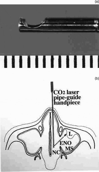
A CO2 laser (Nippon Infrared Industries, Tokyo, Japan) with the 135° reflective tip on the pipe-guide handpiece and a CO2 laser angulated to the same degree are used (Figure 1a). The pipe-guide handpiece with reflective tip is inserted into the nasal cavity, and the base of the maxillary sinus anterior wall lesion is vaporised and removed via an enlarged natural ostium (Figure 1b). The maxillary antrum is viewed during the procedure using a 70° endoscope.
(a) Tip of the CO2 laser pipe-guide handpiece with 135° reflective tip, shown beside 1-mm graduated rule. (b) Schematic representation of the CO2 laser surgical procedure, using the pipe-guide handpiece with 135° reflective tip, and another CO2 laser angulated to the same degree. L = antral lesion located at the anterior wall of the maxillary sinus (MS); ENO = enlarged natural ostium; NC = nasal cavity

Results
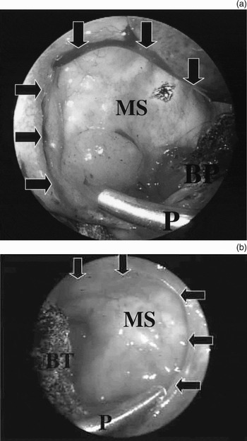
Nine adults and one child, ranging in age from 13 to 76 years, underwent the above surgical procedure as primary or secondary endoscopic sinus surgery for treatment of anterior wall lesions of the maxillary sinus. Four cases of antral polyp were treated with primary endoscopic sinus surgery, and two cases of antral polyp were treated with secondary endoscopic sinus surgery. One antrochoanal polyp was treated with primary endoscopic sinus surgery. One case of antrochoanal polyp (Figure 2a), one of cylindrical cell papilloma (Figure 2b) and one of inverted papilloma were treated with secondary endoscopic sinus surgery. In patients requiring secondary endoscopic sinus surgery, the base of the maxillary sinus anterior wall lesion was not completely removed in the initial operation. Over a 12–18 month course of follow-up endoscopy, no patient required re-operation for antral portion recurrence, and there were no operative complications.
Seventy-degree endoscopic views, via an enlarged left (a) or right (b) natural ostium (indicated by arrows). In both cases, the base of the lesion, located at the anterior wall of the maxillary antrum, is vaporised and removed by CO2 laser via an enlarged natural ostium. P = pipe with 135° reflective tip; MS = maxillary sinus; BP = vaporised base of antrochoanal polyp; BT = vaporised base of tumour

Discussion
Conventional surgical procedure for endoscopic removal of antral lesions, visualisation via endoscope and instrumentation via forceps, laser and microresector were performed through an enlarged natural ostium (via a middle meatal antrostomy) or an inferior meatal nasoantral window.Reference Kamel1–Reference Sato and Nakashima4 Another approach involves insertion of a trocar into the maxillary sinus through the canine fossa.Reference Hong, Min, Kim and Byun5
Recurrence of antrochoanal polyps or neoplasms is relatively frequent following limited surgical procedures, such as nasal polypectomy or tumour resection with the base of the lesion left intact. In the present series, in cases requiring secondary endoscopic sinus surgery, the base of the antral portion of the lesion was not completely removed. The same author has also reported a method for endoscopic sinus surgery using a CO2 laser and/or microresector for paediatric and adult chronic sinusitis with antrochoanal polyps.Reference Sato and Nakashima3, Reference Sato and Nakashima4 In this previous series, the bases of the antrochoanal polyps were located at the medial or lateral posterior wall. The greater portion of the antral part of the antrochoanal polyp could be removed, leaving healthy antral mucosa intact; consequently, the base of the polyp was removed completely and polyp recurrence avoided.Reference Sato and Nakashima3, Reference Sato and Nakashima4
It is essential to completely remove the antral portion of the lesion in order to avoid recurrence. Even though such new instruments as the CO2 laser and microresector now allow otolaryngologists to perform a variety of surgical procedures in the nose and sinuses, the question of how best to surgically manipulate the maxillary sinus anterior wall lesion is still controversial.
The Caldwell–Luc operation via a canine fossa approach ensures complete removal of the maxillary sinus anterior wall lesion, as well as of sinus mucosa. However, this surgical approach is difficult in children. Furthermore, functional preservation and maintenance of maxillary antrum capacity after surgery are difficult.
In the above surgical procedure, the maxillary sinus anterior wall lesion is removed using a 135° reflective CO2 laser; consequently, the base of the lesion is removed completely and recurrence avoided. The present case series suggests that this method is a reliable technique for endoscopic sinus surgery of maxillary antrum anterior wall lesions. The principle of surgery is that the antral portion of the lesion, and especially its base, is completely removed, with healthy antral mucosa left intact, while at the same time keeping surgical invasion to a minimum.