Ventricular fibrillation is a cause of sudden death in patients with Wolff-Parkinson-White syndrome, and has been linked to rapid antegrade accessory pathway conduction to the ventricle during atrial fibrillation.Reference Klein, Bashore, Sellers, Pritchett, Smith and Gallagher1
Chaotic atrial tachycardia is a rare tachyarrhythmia in infants and children, characterised by atrial rates greater than 100 beats per minute, more than three different P-wave morphologies with a discrete isoelectric baseline, and variable PP, RR, and PR intervals. Recordings suggestive of atrial fibrillation in the context of the chaotic atrial tachycardia have been reported.Reference Bradley, Fischbach, Law, Serwer and Dick2 High atrial rates or atrial fibrillation with concurrent chaotic atrial tachycardia may contribute to the ventricular fibrillation associated with Wolff-Parkinson-White syndrome.
We report the occurrence of ventricular fibrillation in a 2-month-old baby with Wolff-Parkinson-White syndrome possibly associated with concurrent chaotic atrial tachycardia.
Case report
The patient, a boy, was born with a birth weight of 3.2 kilograms and was healthy before this event. Since the age of 58 days, his activity and feeding decreased and 3 days later he exhibited signs of sudden onset cyanosis in his face and developed respiratory difficulty. He was brought to a general hospital near his home where ventricular fibrillation was detected and terminated by electrical cardioversion. Subsequently, narrow QRS tachycardia developed, and before being transferred to Asan Medical Center the patient’s supraventricular tachycardia was barely controlled by adenosine and propranolol. On arrival at the centre, his body weight was 5.4 kilograms and his respiration and heart rate were 36 per minute and 230 beats per minute, respectively. His blood pressure was 80/50 millimetres of mercury.
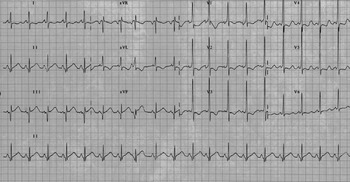
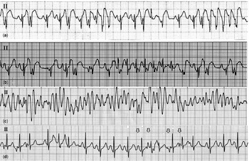
Repeated administration of adenosine resulted in transient termination of the tachycardia. Continuous infusion of procainamide at 60 micrograms per kilogram per minute terminated the tachycardia. During sinus rhythm, echocardiography disclosed a structurally and functionally normal heart and electrocardiogram revealed the delta waves, which suggested that the accessory pathway was located at the left posterior free wall (Fig 1). After discontinuing procainamide, oral flecainide was initiated and a week later sotalol was added; however, both combined drugs failed to provide sustained control of tachycardia and medications were switched to amiodarone. After an intravenous loading dose of amiodarone at 5 milligrams per kilogram, a maintenance dose of amiodarone at 5 milligrams per kilogram per day was given orally. After the administration of amiodarone, electrocardiogram revealed an intermittent irregular rhythm and short runs of wide QRS tachycardia (Fig 2a). Intermittent premature atrial contractions inducing short runs of narrow QRS tachycardia were also observed (Fig 2b). It was suggested that the frequent premature atrial beats and short runs of a concurrent atrial tachycardia conducted to the ventricles with aberrancy or exaggerated pre-excitation and occasionally induced supraventricular tachycardia in this baby. After two days of the amiodarone administration, the patient had a sudden onset of ventricular fibrillation (Fig 2c). We believed that rapid antegrade accessory pathway conduction to the ventricles during the atrial tachycardia resulted in the ventricular fibrillation, and we thus decided to perform radiofrequency catheter ablation.
Surface electrocardiogram showing ventricular pre-excitation. Electrocardiogram during sinus rhythm showed delta waves suggestive of left posterior-free wall accessory pathway.

Electrocardiogram rhythm strips (a) intermittent irregular rhythm and short run of tachycardia with wide QRS morphology were seen. (b) Frequent atrial premature contractions with varying features of P wave were seen and short run of narrow QRS tachycardia was induced by a premature atrial contraction. (c) Recorded polymorphic ventricular tachycardia in the baby. (d) Varying morphologies of P waves (↓) with irregular PP and PR intervals were seen. Some of the early QRS complexes were conducted with aberrancy.

Radiofrequency application to the posterior mitral annulus successfully eliminated the delta wave. The ablation catheter was advanced into the left atrium through a patent foramen ovale. The antegrade effective refratory period of the accessory pathway could not be determined due to the sustained tachycardia.
The next day the patient’s electrocardiogram exhibited an irregular rhythm with intermittent pauses. We confirmed this irregular rhythm as being chaotic atrial tachycardia after detailed review of the electrocardiogram (Fig 2d). After 7 days, the patient was brought home without any medications. Chaotic atrial tachycardia was not recorded on Holter recording after 2 months. The patient’s growth and development remained normal throughout the 4-year follow-up period and his last electrocardiogram showed normal sinus rhythm without delta waves.
Discussion
This case shows that concurrent chaotic atrial tachycardia associated with Wolff-Parkinson-White syndrome may result in life-threatening ventricular fibrillation in young infants.
The occurrence of ventricular fibrillation associated with Wolff-Parkinson-White syndrome has long been recognised; however, it is infrequently observed in infants and children.Reference Vignati, Balla, Mauri, Lunati and Figini3
The majority of children with chaotic atrial tachycardia are under 1 year of age without any underlying illness, and the clinical course in children has been variable, ranging from a benign and transient arrhythmia to sudden death. Similar to the more common forms of supraventricular tachycardia seen in childhood, chaotic atrial tachycardia is rapid, with atrial rates increasing to 400 beats per minute, but unlike most supraventricular tachycardias it is markedly irregular with pauses following blocked premature atrial impulses. At high atrial rates, aberrant ventricular conduction might resemble non-sustained ventricular tachycardia.Reference Bradley, Fischbach, Law, Serwer and Dick2, Reference Dodo, Gow, Hamilton and Freedom4, Reference Fish, Mehta and Johns5 Before amiodarone administration, tachycardia was so incessant in this patient that the chaotic atrial tachycardia could not be recognised. After the recurrence of tachycardia decreased due to amiodarone effects, electrocardiogram suggested chaotic atrial tachycardia and retrospectively, chaotic atrial tachycardia was thought to be the reason for the incessant supraventricular tachycardia in this patient.
Rapid ventricular conduction of chaotic atrial rhythms through the accessory pathway might have resulted in ventricular fibrillation in this case. During short runs of wide QRS tachycardia, the shortest RR interval was 160 milliseconds. However, it was also suggested that although we could not record the onset of the ventricular fibrillation, atrial fibrillation associated with chaotic atrial tachycardia might have contributed to the ventricular fibrillation in this patient. Recordings suggestive of atrial fibrillation in the context of chaotic atrial tachycardia have been reported.Reference Bradley, Fischbach, Law, Serwer and Dick2 Bevilacqua et alReference Bevilacqua, Rhee, Epstein and Triedman6 reported successful focal ablation of chaotic atrial tachycardia in an infant, and suggested that the resemblance of the chaotic atrial rhythm electrocardiogram to that of atrial fibrillation in some cases might be attributed to the fibrillatory conduction from a unifocal ectopic mechanism in a manner similar to that of the focal atrial fibrillation described in adults by Haïssaguerre et al.Reference Haïssaguerre, Jaïs and Shah7
In conclusion, chaotic atrial tachycardia might be attributed to life-threatening ventricular fibrillation in infants with Wolff-Parkinson-White syndrome. Ventricular fibrillation results from rapid antegrade accessory pathway conduction of high atrial rates or atrial fibrillation in the setting of chaotic atrial tachycardia.