Introduction
The tundra or root vole, Microtus oeconomus (Pallas), is widespread in the Holarctic region (Shenbrot & Krasnov, Reference Shenbrot and Krasnov2005; Tinnin et al., Reference Tinnin, Ganzorig and Gardner2011). According to Tinnin et al. (Reference Tinnin, Ganzorig and Gardner2011), ‘In the Old World, it (distribution) ranges from Scandinavia south to the Baltic and east across Siberia, south into China (…). In North America they range into north central Canada.’ In the Magadan region in the north-east of Russia, these animals inhabit open, moist biotopes such as floodplain meadows and bogs. They are numerous in the forbs–sedge–graminoid maritime meadows and the sedge–graminoid plant associations in river valleys along the coast of the Sea of Okhotsk (Kostenko, Reference Kostenko2000). According to Chernyavskii (Reference Chernyavskii1984) and Kostenko (Reference Kostenko2000), the local population of the tundra vole is represented by M. oeconomus koreni Allen.
The fauna of internal parasites of several subspecies of M. oeconomus has been studied in northern and eastern Europe, Alaska, Western Siberia, China and Mongolia, and representatives of several nematode orders have been found.
So far, only three species from the order Spirurida were found associated with a tundra vole: Mastophorus muris (Gmelin, 1790) of Spirocercidae Chitwood & Wehr, 1932; Rictularia microti (McPherson & Tiner, 1952); and Pterygodermatites kolimensis (Gubanov & Fedorov, 1967) of Rictulariidae Hall, 1913. Surveys carried out in Yakutia, the Far East and the Far North-East of Russia reported the presence of M. muris and P. kolimensis in a tundra vole (Gubanov & Fedorov, Reference Gubanov and Fedorov1965, Reference Gubanov and Fedorov1967; Nadtochi, Reference Nadtochi1966; Yudin et al., Reference Yudin, Krivosheev and Belyaev1976; Domnich, Reference Domnich1984, Reference Domnich1985) (taxonomy is consistent with Anderson et al., Reference Anderson, Chabaud and Wilmott2009).
A heavy nematode infection was found in tundra voles collected in the Magadan region, in the vicinity of the city of Magadan. The nematodes inhabited the anterior portion of the small intestine of rodents (duodenum). Certain morphological traits of these medium-sized nematodes indicated that they may belong to the family Acuariidae (Spirurida) (Anderson et al., Reference Anderson, Chabaud and Wilmott2009). The family Acuariidae generally comprises bird parasites. However, four genera (Stammerinema Osche, 1955, Tikusnema Hasegawa, Shiraishi & Rochman, 1992, Chandleronema Little & Ali, 1980 and Antechiniella Quentin & Beveridge, 1986) and some species of Skrjabinoclava Sobolev, 1943 and Paracuaria Rao, 1951 as well as Synhimantus australiensis Johnston & Mawson, 1952 are known to parasitize mammals (Bain et al., Reference Bain, Mutafchiev, Junker and Schmidt-Rhaesa2014). Stammerinema contains four species parasitic in Holarctic shrews (Mutafchiev et al., Reference Mutafchiev, Mariaux and Georgiev2015); three species of Tikusnema are parasitic in murid rodents with Indomalayan and Australasian distribution (Hasegawa et al., Reference Hasegawa, Shiraishi and Rochman1992; Smales, Reference Smales1995, Reference Smales2006; Bain et al., Reference Bain, Mutafchiev, Junker and Schmidt-Rhaesa2014); the monotypic Chandleronema was described based on material from the common raccoon in Mexico and Florida (Little & Ali, Reference Little and Ali1980); two species of Skrjabinoclava were described from the crab-eating raccoon and the rice rat in the New World (Teixeira de Freitas, Reference Teixeira de Freitas, Dayal and Singh1953; Yamaguti, Reference Yamaguti1961; Anderson & Wong, Reference Anderson and Wong1996); and two species of Paracuaria were described from European shrews (Jančev, Reference Jančev1972) and the Pyrenean desman (Alvarez et al., Reference Alvarez1994).
Further morphological study of the nematodes from a tundra vole, along with phylogenetic analysis based on three DNA loci showed that they represented yet another species of Antechiniella Quentin & Beveridge, 1986, which is described below.
Materials and methods
Parasitological procedures
Forty specimens of Microtus oeconomus (Muridae Illiger, 1811) (ten underyearlings and 30 overwintered individuals) were collected using snap traps at the coast and islands of Tauyskaya Bay in the northern part of the Sea of Okhotsk during June–August 2015 and June–August 2016. Dissections were made shortly after collection of the hosts. The nematodes were difficult to recover because of their attachment to host tissues by the mid-body parts. As a result, the material examined comprised anterior and posterior body fragments of nematodes. The worms were preserved in 4% formaldehyde solution for the morphological study and in 70% ethanol for the molecular study. Formalin-preserved material was processed to anhydrous glycerol according to Seinhorst (Reference Seinhorst1959) and mounted on slides. Light microscopy studies were carried out and drawings were prepared using an Eclipse microscope (Nikon, Tokyo, Japan) equipped with a camera lucida. Measurements are presented as mean values followed by the range in parentheses. Illustrations were finalized using an Intuos A4 USB drawing tablet (Wacom, Kazo, Japan) and Adobe Illustrator CS5 according to Coleman (Reference Coleman2003). Several nematode fragments were used for the scanning electron microscopy (SEM) study; these were rehydrated after formaldehyde solution, dehydrated in a graded ethanol series, critical-point dried using a HCP-2 HITACHI dryer, mounted on aluminium stubs and coated with gold in a BIO-RAD SC502 sputter coater. Specimens were studied under a JCM-6380 LA SEM.
Molecular characterization and DNA analysis
DNA was extracted according to Holterman et al. (Reference Holterman2006) in the worm-lysis solution containing 950 μl of a mixture of 2 ml of 1M NaCl, 2 ml of 1M Tris-HCl (pH 8) and 5.5 ml of deionized water plus 10 μl of mercaptoethanol and 40 μl of proteinase K (20 mg/ml). Fragments of the reproductive system of males and females were excised and digested in a solution containing 25 μl sterile water and 25 μl worm-lysis solution at 65°C (90 minutes). After deactivation of proteinase K (99°C, 5 minutes), 1.2 μl of the digest was used as a polymerase chain reaction (PCR) template.
The pair of nematode-specific primers, Nem18SF (5′-CGC GAA TRG CTC ATT ACA ACA GC-3′) and Nem18SR (5′-GGG CGG TAT CTG ATC GCC-3′), was used to amplify the 5′ portion of the 18S rRNA gene (Floyd et al., Reference Floyd2005). PCR cycling parameters included an initial denaturation at 95°C for 5 minutes, followed by 35 cycles of 95°C for 45 s, 51°C for 60 s and 72°C for 60 s, followed by a post-amplification extension step at 72°C for 5 minutes.
The pair of primers LSU391 (5′-AGC GGA GGA AAA GAA ACT AA-3′) and LSU501 (5′-TCG GAA GGA ACC AGC TAC TA-3′) were used to obtain a partial sequence of the 28S rRNA gene fragment (Nadler et al., Reference Nadler2006). PCR cycling parameters included an initial denaturation at 94°C for 4 minutes, followed by 35 cycles of 94°C for 30 s, 56°C for 30 s and 72°C for 60 s, followed by a post-amplification extension step at 72°C for 5 minutes.
The primer pair COI-F Acu2 (5′-TTT CCT CGT GTT AAT GCT TT-3′) and COI-R Acu3 (5′-CAA ACA AAC GCT CCT TAT CAG A-3′) was used to amplify a partial fragment of the mitochondrial gene cox1 (Mutafchiev et al., Reference Mutafchiev, Mariaux and Georgiev2015). PCR cycling parameters were 94°C for 120 s, followed by five cycles of 94°C for 50 s, 47°C for 50 s and 72°C for 60 s, followed by 30 cycles of 94°C for 50 s, 50°C for 50 s, 72°C for 60 s, and a post-amplification extension step at 72°C for 5 minutes.
PCR products were sequenced directly with Genotech Ltd, Moscow. The newly generated sequences for A. septentrionalis n. sp. were deposited in the GenBank database under accession numbers KY984296 (18S rRNA gene), KY984295 (28S rRNA gene) and MF125274 (coxI mtDNA gene).
The sequences of phylogenetically related nematodes were detected in GenBank using BLAST (Altschul et al., Reference Altschul1990), retrieved and used for a comparative analysis. In most cases, all of the spirurid nematodes with similar nucleotide sequences identified by BLAST were included in the phylogenetic analyses. When several sequences for the same species were available for the locus under study, a few (1–4) showing distinctive intraspecific variability were selected and used for analysis. The sequences of related groups of parasitic nematodes (members of the Ascaridoidea Baird, 1853, Cosmocercoidea Travassos, 1925, Panagrolaimoidea Thorne, 1937) were used as the outgroups. The sequences were aligned using Clustal X with default values for gap opening and gap extension penalties. Different methods of phylogenetic analysis, i.e. maximum parsimony, neighbour-joining and maximum likelihood, were performed with MEGA7.0.14 (Kumar et al., Reference Kumar, Stecher and Tamura2016). Analyses of 18S and 28S rRNA were performed using the CIPRES Science Gateway V. 3.3 (Miller et al., Reference Miller, Pfeiffer and Schwartz2010). Prior to analyses, the models of nucleotide substitution were selected using JModelTest2 (Darriba et al., Reference Darriba2012). The following settings were applied for Bayesian analysis on CIPRES platform: number of generations = 10,000,000; number of runs = 4; number of chains = 4; ‘burnin’ = 0.25.
Results
Antechiniella septentrionalis n. sp.
Type host. Microtus oeconomus koreni Allen (Rodentia, Muridae).
Type locality. Maritime marsh, Tauyskaya Bay (59°34.117′N, 151°19.107′E).
Other localities. Maritime marshes, Tauyskaya Bay (59°34.100′N, 151°20.068′E; 59°34.786′N, 151°20.913′E); Sikulun isle (59°33.596′N, 151°20.204′E) by Ola village; Vdovushka isle (59°29.751′N, 150°55.116′E), Veselaya Bay.
Type habitat. Duodenum.
Prevalence. All underyearlings were free of infection, whereas 13 out of 30 overwintered voles (43.3%) were infected by A. septentrionalis n. sp.
Intensity. 1–60 specimens (mean 24 specimens).
Type material. The holotype, the anterior fragment of a female (catalogue number 1284), a male paratype, a tail and a head end (on the same slide, catalogue number 1285), and a female paratype, tail (catalogue number 1286), are deposited in the Museum of the Helminthological Collections of the Centre of Parasitology at the Severtsov Institute of Ecology and Evolution, Moscow. The rest of material is in the collection of the first author and is available on request.
Etymology. The species name refers to the northern position of the type locality.
Description
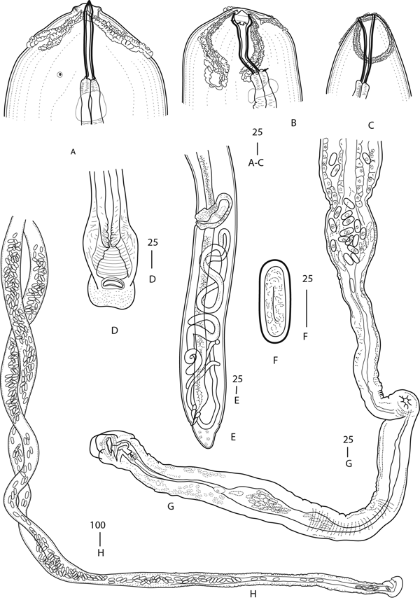
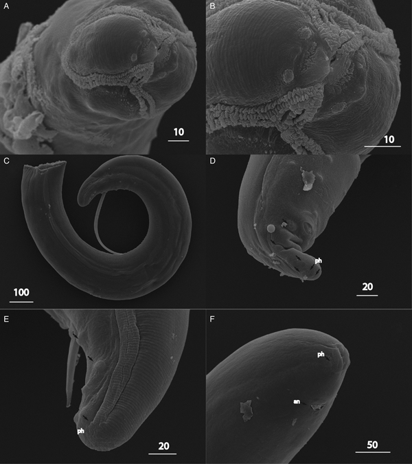
Adults. Cuticle thick, annulated. Body with maximum diameter in anterior part and narrowest in its middle. Head not inflated in relation to dilated anterior body portion, bluntly rounded, with two pseudolabia with apical protuberance; each pseudolabium bearing a pair of papillae and an amphid located between papillae, all situated at the same level close to mouth aperture (fig. 3A, B). Cordons arise dorsally and ventrally between pseudolabia, not extending beyond base of buccal cavity, curved in lateral direction, anastomosing in pairs or not; each cordon consists of two rows of irregularly shaped cuticular plates divided by a narrow groove (fig. 1A–C and 3B). Deirids plain, inconspicuous, situated at base of buccal cavity or just anterior to nerve ring. Lateral fields poorly expressed, seen in posterior body part as relatively thin cuticular fold with central incision (fig. 3E). Spines absent on cuticle. Oral opening small, flattened laterally. Buccal cavity long, strongly sclerotized, anterior part expanded in dorso-ventral direction (fig. 1A–C). Muscular part of oesophagus 1.5 times longer than buccal cavity, cylindroid, slightly tapered anteriorly (fig. 2A). Glandular part of oesophagus massive, greatly exceeding muscular oesophagus in length and width. Nerve ring surrounding anterior part of muscular oesophagus. Excretory pore situated at two body diameters from anterior extremity or opposite anterior to glandular oesophagus. Tail in both sexes short, rounded.
Female. Holotype: length of fragment = 6100 μm; maximum diameter = 620 μm; buccal cavity length = 152 μm; muscular oesophagus length = 280 μm and width = 44 μm; glandular oesophagus length = 2140 μm and width = 270 μm; nerve ring = 250 μm; distance from apex to posterior of cordons = 84 μm.
Paratypes (n = 12): length of fragments = 221–8270 μm (n = 9); maximum diameter = 284 (103–620) μm (n = 12); buccal cavity length = 148 (135–160) μm and width = 11 (10–12) μm (n = 5); muscular oesophagus length = 240 (220–294) μm and width = 46 (42–49) μm (n = 5); glandular oesophagus length = 1130–2600 μm and width = 180–270 μm (n = 2); nerve ring = 208 (171–250) μm (n = 5); distance from apex to posterior of cordons = 116 (84–167) μm (n = 5); vulva, distance from posterior body end = 2765 (2350–3520) μm (n = 4); anal diameter = 182 (56–380) μm (n = 4); tail length = 286 (166–520) μm (n = 4); egg length = 61 (61–62) μm and width = 24 (23–25) μm (n = 9).
Ratio: length of muscular oesophagus/length of glandular oesophagus 0.143 (0.131–0.195) μm (n = 5). Excretory pore observed in a single specimen situated at 870 μm from anterior extremity. Vulva a transverse slit with flat lips, posterior, situated at level of last quarter of body. Vagina vera short, with thick muscular walls (fig. 1D). Vagina uterina thick-walled, 908 (230–1500) μm long, 75 (59–93) μm wide (n = 4), leading to two uteri directed anteriad, filled with embryonated eggs and grain-shaped objects slightly smaller than eggs (fig. 1G, H). Eggs in ovejector and distal parts of uteri with juveniles. Egg shells oblong with rounded ends and with thick, translucent walls (fig. 1F). Tail widely conical, rounded (fig. 3F).
Juvenile III–IV stage of female. Length of fragments 3620 (1202–5930) μm (n = 5); maximum width 75 (47–102) μm (n = 4); width at anus 42 (25–56) μm (n = 3). Buccal cavity 157‒184 μm long, 9‒12 μm wide (n = 2). Muscular oesophagus 189‒230 μm long, 42‒45 μm wide; glandular oesophagus 840‒1500 μm long, 180‒230 μm wide (n = 2). Nerve ring at 191‒207 μm from apex (n = 2). Genital tube developed in posterior region between vulval primordium and anus (fig. 1E).
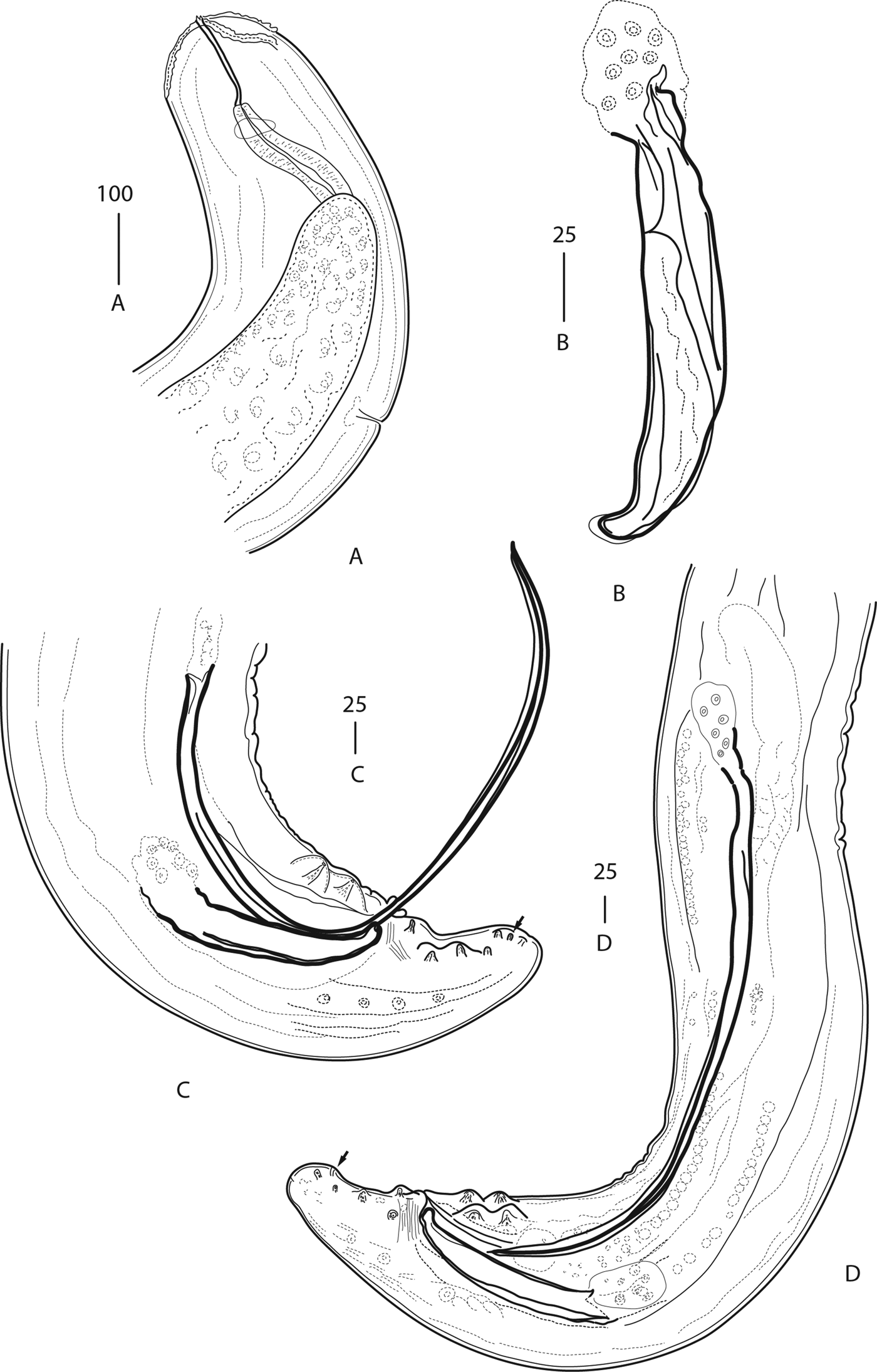
Male. Paratypes (n = 7): length of fragments = 637–9410 μm; maximum diameter = 222 (150–321) μm; anal diameter = 122 (94–160) μm; tail length = 156 (124–198) μm (n = 7); left spicule length = 679 (590–747) μm and width = 11 (8–17) μm; right spicule length = 216 (164–262) μm and width = 28 (10–37) μm.
Fig. 1.Antechiniella septentrionalis n. sp. Female. (A) Anterior extremity, ventral view; (B, C) anterior extremity, lateral view; (D) vulva and vagina vera, ventral view; (E) posterior extremity of young immature female; (F) egg; (G, H) complex vagina. Deirid indicated by an arrow. All scale bars are in μm.
 Fig. 2.
Antechiniella septentrionalis n. sp. Male. (A) Anterior extremity; (B) right spicule; (C, D) tail, lateral view. Arrow indicates phasmid. All scale bars are in μm.
Fig. 3.Antechiniella septentrionalis n. sp. SEM images. (A, B) Anterior extremity of a female; (C, E) male tail, lateral view; (D) male tail, subventral view; (F) female tail. Abbreviations: an, anus; ph, phasmid. Arrows indicate precloacal (C, D) and postcloacal (D, E) papillae. All scale bars are in μm.


Smaller in size than female. Ratio length of muscular oesophagus/length of glandular oesophagus 0.109 (n = 1) (fig. 2A). Excretory pore observed in a single specimen, situated at 610 μm from anterior extremity. Caudal alae poorly developed, incorporating two pairs of sublateral precloacal papillae and two pairs of sublateral postcloacal papillae. Ventral median papilla in front of anus, and four pairs of large precloacal papillae present: two pairs precloacal incorporated into alae and two pairs subventral (fig. 2C). Postcloacal papillae 5–6 pairs: a pair situated immediately posterior to anus in ventral position; two pairs further posteriad incorporated into alae and 2–3 pairs of smaller papillae situated close to tail tip (figs 2C, D and 3C–E). Phasmids subterminal, located between the two posteriormost pairs of papillae. Anus surrounded by pericloacal disk (fig. 3C–E). Area rugosa absent. Spicules dissimilar; right spicule short (fig. 2B), robust, with rounded, slightly separated distal tip and not distinctly expressed handle 40 (17–49) μm long and 46 (48–51) μm wide. Left spicule approximately 3× longer (ratio left spicule length/right spicule length 3.14 (2.62–3.4) and c. 2× thinner than right, with acute distal tip and slightly expanded handle 26 (17–32) μm long and 29 (25–34) μm wide (fig. 2C–D); narrow ala seen in longer spicule. Tail tapering, slightly curved ventrally, bluntly rounded.
Juvenile IV stage of male. Developed spicules and papillae present in the advanced IV stage juvenile. Left spicule 495–545 μm long, right spicule 150–187 μm long; body width at anus 75–87 μm (n = 2).
Diagnosis and relationships
The genus Antechiniella Quentin & Beveridge, 1986 currently includes two species, A. suffodiax (Beveridge & Barker, 1975) and A. sertatum Smales, 1991 from the Australian marsupial Antechinus stuartii Macleay, 1841 and native Australian rodents Rattus fulcipes (Waterhouse, 1839) and R. lutreolus (J.E. Gray, 1841), and an Australian water rat, Hydromys chrysogaster Geoffroy, 1804, respectively (Smales, Reference Smales1991). The former species was originally described as Stammerinema suffodiax by Beveridge and Barker (Reference Beveridge and Barker1975) but later, following the examination of head structures of three species of Stammerinema, the genus Antechiniella was erected to accommodate the species (Quentin & Beveridge, Reference Quentin and Beveridge1986).
The present species was placed in Antechiniella based on similar body proportions, the structure of the anterior extremity and tail in males, the lack of spines posterior to cordons, small/indistinct deirids and the posterior position of the vulva, as well as the similar site of infection in mammals and the host range.
Antechiniella septentrionalis n. sp. is characterized by didelphic females with a long, complex vagina directed anteriad, oblong eggs, and a short, rounded tail, and males possessing 5–6 pairs of postcloacal papillae and a left spicule 590–747 μm long with acute distal tip. The new species is morphologically similar to A. suffodiax in the body proportions, the shape of the buccal cavity, the length of cordons and the absence of distinct deirids. It differs from the latter in having a non-coiled (vs coiled) tail in the male, a somewhat more anterior nerve ring position, more numerous postcloacal papillae (5–6 vs 4 pairs), a differently shaped left spicule with a prominent acute distal tip without alae (vs with alae), less expressed caudal alae, and in the disposition of both ovaries anterior to vulva (vs disposition of ovaries in different directions) as well as a shorter tail in females. Females of both species have a complex vagina (a vagina and an ovejector as described in the original description by Beveridge & Barker (Reference Beveridge and Barker1975)); however, in A. suffodiax it leads posteriad (vs anteriad in the new species). Eggs of A. septentrionalis n. sp. are oblong (62 μm × 24 μm) vs oval (39 μm × 27 μm) in A. suffodiax.
From A. sertatum, to which the new species can be compared in having a similarly sized right spicule and anterior direction of vagina, it differs in the presence of plain (vs bicuspid) deirids, shorter cordons (average 114 μm vs 320 μm), which are not anastomosing/imperfectly anastomosing (vs anastomosing), longer buccal cavity (average 154 μm vs 92 μm, females), shorter left spicule (average 679 μm vs 927 μm) and its acute (vs flared) distal tip and five to six (vs four) pairs of postcloacal papillae. The females of the new species differ additionally from A. sertatum in having a much shorter (average 241 μm vs 886 μm) anterior muscular oesophagus, the smaller distance between vulva and anus (average 2 mm vs 9 mm), a shorter tail (average 204 μm vs 328 μm) and elongated (vs rounded) eggs (62 μm × 24 μm vs 43 μm × 32 μm). No ovejector (vagina uterina) had been described for A. sertatum; ovejector is remarkably long in A. septentrionalis n. sp.
Males of A. septentrionalis n. sp. are similar to other species of the genus in having the same number of precloacal papillae (four pairs). However, in the present species precloacal papillae are situated in two rows: two pairs subventral and two pairs lateral incorporated into the caudal alae. The position of precloacal papillae in A. suffodiax and A. sertatum was not specified by authors (Beveridge & Barker, Reference Beveridge and Barker1975; Smales, Reference Smales1991) but appeared to be lateral on illustrations given in the original descriptions.
By the shape of plates constituting cordons, A. septentrionalis n. sp. is also comparable with the species of Stammerinema as seen from Fig. 7.222 B in Bain et al. (Reference Bain, Mutafchiev, Junker and Schmidt-Rhaesa2014). Although scanning microscopic images for the cordon plates are unavailable for either of the known Antechiniella species, the shape in the drawings of both species is similar to that of Stammerinema. However, the cordons of the new species are shorter and less curved, which is more characteristic for Antechiniella. Also, the new species lacks body spines, a diagnostic trait of Stammerinema.
Molecular characterization and phylogeny
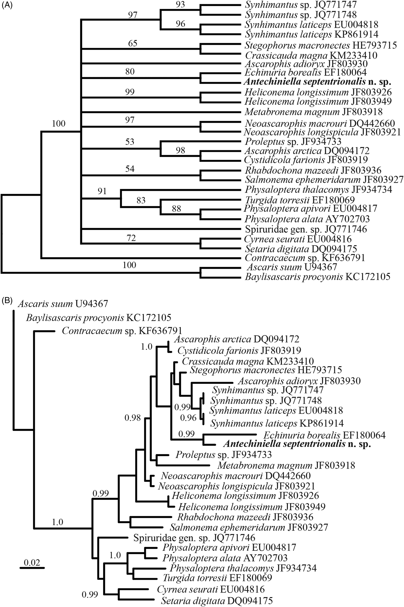
The analysis of partial 18S sequences (835 bp long alignment) demonstrated the affinity of the nematodes described herein to the nematodes of the family Acuariidae and other spirurid nematodes. All the methods of analysis (maximum parsimony, neighbour joining and maximum likelihood) of 18S rRNA in MEGA7 have revealed similar topologies and close relationships of A. septentrionalis n. sp. with Echinuria borealis Mawson, 1956. The phylogenetic relationships of A. septentrionalis n. sp. with other spirurid genera remained unresolved, although the monophyletic status of spirurids involved in the analysis was strongly supported (fig. 4A). The Bayesian analysis of 18S rRNA using CIPRES Science Gateway V. 3.3 has facilitated the resolution of more nodes of the phylogenetic tree leading to A. septentrionalis n. sp. (fig. 4B). In this analysis, A. septentrionalis n. sp. once again was a sister taxon to E. borealis. The monophyly of spirurids was also strongly supported. The high level of posterior probability (>0.95) was characteristic for some inner spirurid clades, e.g. that including A. septentrionalis n. sp. and representatives of several acuariid genera (Synhimantus Railliet, Henry & Sisoff, 1912, Echinuria Soloviev, 1912, Stegophorus Wehr, 1934) as well as the certain spirurid genera: Ascarophis van Beneden, 1871, Cystidicola Fischer, 1798, Metabronema Yorke & Maplestone, 1926 (Cystidocolidae), Crassicauda Leiper & Atkinson, 1914 (Tetrameridae) and Proleptus Dujardin, 1845 (Physalopteridae).
Phylogenetic relationships of Antechiniella septentrionalis n. sp. inferred from analysis of partial 18S rRNA sequences. (A) Maximum likelihood analysis, Kimura 2-parameter model, with gamma distribution and invariant sites (K2 + G + I), bootstrap support indicated near nodes (500 pseudoreplicates); (B) Bayesian inference analysis, transversion model with gamma distribution and invariant sites (TVM + I + G); posterior probability values indicated near nodes.

The analysis of partial 28S rRNA (544 bp long alignment) using CIPRES Science Gateway V. 3.3 demonstrated similar phylogenetic relationships, taking into account that only a single 28S rRNA sequence of acuariid nematodes (that for Stegophorus macronectes (Johnston & Mawson, 1942)) is available in GenBank (fig. 5A). The clade including A. septentrionalis n. sp., S. macronectes, Ascarophis arctica Poljansky, 1952 and Proleptus obtusus Dujardin, 1845 was strongly supported.
There are several partial coxI mtDNA sequences for Acuariidae available in GenBank. The phylogenetic analysis of this quite limited dataset (403 bp long alignment) has revealed strong support for the close relationships of A. septentrionalis n. sp. with Stammerinema hyalinum (von Linstow, 1890) and moderate support for a clade consisting of these two latter species together with three other acuariid species: Acuaria europaea Mutafchiev, Mariaux & Georgiev, 2017, Synhimantus laticeps (Rudolphi, 1819) and Proyseria petterae Mutafchiev, Mariaux & Georgiev, 2014 (fig. 5B).
Phylogenetic relationships of Antechiniella septentrionalis n. sp. inferred from an analysis of partial 28S rRNA and mitochondrial cox1 gene sequences. (A) analysis of partial 28S rRNA; (B) analysis of partial cox1 mtDNA. For both loci: Bayesian inference analysis, general time reversible model with gamma distribution (GTR + G), posterior probability values indicated near nodes.

Discussion
Antechiniella septentrionalis n. sp. is the first member of the genus found outside the Australian continent, in the host with the widest distribution but very different environment. One to three specimens of unidentified nematodes were found in the small intestines of 14–30% (2015 and 2016, respectively) of M. oeconomus infected by A. septentrionalis n. sp. No cestode infestation was found. Antechiniella septentrionalis n. sp. was found only in the overwintered tundra voles inhabiting maritime marshes and shore slopes of the small islands. These habitats, and the whole coast of the Sea of Ochotsk, abound with supralittoral amphipods of the family Talitridae. Earlier, Dokuchaev & Atrashkevich (Reference Dokuchaev and Atrashkevich2015) speculated that in the life cycle of A. septentrionalis n. sp., amphipods Traskorchestia sp. could likely serve as intermediate hosts for the nematode. It is known that acuariid nematodes always complete their development using arthropods as the intermediate hosts (Anderson et al., Reference Anderson, Chabaud and Wilmott2009; Bain et al., Reference Bain, Mutafchiev, Junker and Schmidt-Rhaesa2014). A tundra vole is considered to be strictly vegetarian, maintaining underground winter stores of rhizomes, tubers and bulbs (Yudin et al., Reference Yudin, Krivosheev and Belyaev1976), or seeds and rhizomes (Batzli & Lesieutre, Reference Batzli and Lesieutre1991). The content of such stores demonstrates that tundra voles do not willingly take any but vegetarian food. Hypothetically, amphipods can serve as a proper intermediate host, considering the high density of amphipods in the habitats studied and the possibility of occasionally being swallowed. Whether amphipods serve as intermediate hosts for A. septentrionalis n. sp. is in need of further research.
The formation of fibrous nodules in the stomach wall was reported for both known species of Antechiniella (Beveridge & Barker, Reference Beveridge and Barker1975; Smales, Reference Smales1991). Similar nodules were found in the intestine walls of the infected tundra voles. Similarly, a portion of nematodes had overgrowths at mid-body where they were in contact with the intestine walls.
Smales (Reference Smales1991) discussed the food preferences of the Antechiniella hosts (the marsupial and the rodents) and found them to be similar in consuming large quantities of a variety of arthropods. Such feeding behaviour is expected to ensure that parasites will get to their definitive hosts, although the types of arthropods involved in the transmission of nematodes remains unclear.
The molecular analysis of three DNA loci of A. septentrionalis n. sp. provided some nucleotide information for further phylogenetic and faunistic studies on spirurids. Several sequences of the nematodes of the subfamily Acuariinae Railliet, Henry & Sisoff, 1912 were found to be the most similar to the three sequences of A. septentrionalis n. sp. obtained. Bayesian analysis of the partial 18S rRNA has revealed strong support for the clade composed of the representatives of spirurid superfamilies (Acuarioidea, Habronematoidea, Physalopteroidea), although not all the inner nodes of this clade demonstrated sufficient posterior probabilities (>0.95). Strong or moderate support was only characteristic for subclades including the spirurids belonging to the same family, such as Ascarophis arctica + Cystidicola farionis (Cystidicolidae, Habronematoidea), and two acuariid (Acuariidae, Acuarioidea) subclades: Echinuria borealis + A. septentrionalis n. sp. as well as the subclade including four Synhimanthus sequences. In an earlier analysis of 18S rRNA, Černotiková et al. (Reference Černotíková, Horák and Moravec2011) demonstrated significant support for the clades composed of taxonomically and ecologically distant spirurids: Cystidicolidae and Physalopteroidea of fishes and Acuariidae of birds. The revision of the spirurid taxonomy on the basis of phylogenetic analyses of the nuclear and mitochondrial sequences demands wider representation of different taxonomical and ecological groups of these nematodes. The basal position of fish parasites in one of the spirurid clades inferred from the 18S rRNA analysis was evident (fig. 4B). Yet another clade represented on the tree consisted of forms parasitic mainly in birds and reptiles. The allocation of spirurids parasitic in birds and mammals in different clades speaks in favour of the multiple acquisition of parasitism in higher vertebrates within the order Spirurida.
All three genetically related species (i.e. A. septentrionalis n. sp., Echinuria borealis in the analysis of the partial 18S rRNA and Stammerinema hyalinum in the partial coxI mtDNA) are classified into the subfamily Acuariinae, implying a degree of similarity. As the most similar features, the following ones should be noted: the structure of cordons composed of two rows of plates, small deirids, the presence of weakly developed caudal alae and the absence of area rugosa in males. Whether more molecular data on acuariid nematodes showing the genetic distances is available, in-depth comparative phylogenetic analysis may show a wider set of synapomorphies of these nematodes.
Acknowledgements
We are greatly indebted to Dr Yasen Mutafchiev, who kindly provided the literature on acuariids and gave guidance on the subject of our study. We would also like to acknowledge the referees' contributions.
Financial support
This study was funded by grants from the Russian Fund for Basic Research Nos. 15-04-02668 and 17-04-00095 to NED and SES.
Conflict of interest
None.