Introduction
The global loss of biodiversity is an actual and tragic scenario and has been influenced by a myriad of factors, such as climate change, agricultural land use, deforestation, and infectious diseases (Scheele et al. Reference Scheele, Pasmans and Skerratt2019; Habibullah et al. Reference Habibullah, Din, Tan and Zahid2021; Jaureguiberry et al. Reference Jaureguiberry, Titeux, Wiemers, Bowler, Coscieme, Golden, Guerra, Jacob, Takahashi, Settele, Díaz, Molnár and Purvis2022; Pfenning-Butterworth et al. Reference Pfenning-Butterworth, Buckley, Drake, Farner, Farrell, Gehman, Mordecai, Stephens, Gittleman and Davies2024), with direct negative impacts on ecosystem functioning (Isbell et al. Reference Isbell, Balvanera and Mori2023). Among the various groups of vertebrates, amphibians are considered the most sensitive to anthropogenic environmental changes, facing currently a growing rate of declining populations and a decline in diversity worldwide (Alton & Franklin Reference Alton and Franklin2017; Carrasco et al. Reference Carrasco, Souza and Souza Santos2021; Luedtke et al. Reference Luedtke, Chanson and Neam2023). More importantly, amphibians serve as hosts to thousands of helminth species worldwide (Carlson et al. Reference Carlson, Hopkins, Bell, Doña, Godfrey, Kwak, Lafferty, Moir, Speer, Strona and Torchin2020a), which can also be considered threatened species when considering the process of co-extinction (Carlson et al. Reference Carlson, Dallas, Alexander, Phelan and Phillips2020b). In this scenario, such impact can be more evident in trematodes, which have complex life cycles and are considered biodiversity indicators in ecosystems (Esch et al. Reference Esch, Barger and Fellis2002; Hudson et al. Reference Hudson, Dobson and Lafferty2006; Morley Reference Morley2012; Pinto & Melo Reference Pinto and Melo2013; Sures et al. Reference Sures, Nachev, Schwelm, Grabner and Selbach2023).
Brazil has the major richness of anuran species in the world, with more than 1,100 described species (Segalla et al. Reference Segalla, Berneck, Canedo, Caramaschi, Cruz, Garcia, Haddad, Lourenço, Mângia, Mott, Nascimento, Toledo, Werneck and Langone2021; Boeger et al., Reference Boeger, Valim and Zaher2024). Regarding the trematodes found in these vertebrates in the country, helminthological studies carried out over more than a century revealed about 60 species of trematodes infecting more than 30 amphibian species (Campião et al. Reference Campião, Morais, Dias, Aguiar, Toledo, Tavares and Silva2014, Reference Campião, Silva, Dalazen, Paiva and Tavares2016a,Reference Campião, Ribas, Silva, Dalazen and Tavaresb; Fernandes & Kohn Reference Fernandes and Kohn2014; Graça et al. Reference Graça, Oda, Lima, Guerra, Gambale and Takemoto2017; Lins et al. Reference Lins, Aguiar, Morais, DA Silva, Ávila and Silva2017; Toledo et al. Reference Toledo, Schwartz, Nomura, Aguiar, Velota, Silva and Anjos2017; Queiroz et al. Reference Queiroz, López-Hernández, Locke, Pinto and Anjos2019, Reference Queiroz, Pontes, Neto, Campião and Anjos2020, Reference Queiroz, Alves, López-Hernández, Anjos and Pinto2021; Aguiar et al. Reference Aguiar, Morais, Silva, Anjos, Foster and Silva2021; Alcantara et al. Reference Alcantara, Úngaria, Müllerb, Ferreira-Silva, Emmerich, O’Dwyer and Silva2022, Reference Alcantara, Ebert, Ferreira-Silva, Forti, Morais, Pérez-Ponce de Léon and Silva2024; Benício et al. Reference Benício, Santos, Freire, Ávila, Silva and Fonseca2022; Soares et al. Reference Soares, Passos, Anjos and Wachlevski2022; Sampaio et al. Reference Sampaio, Teixeira, Nascimento, Ribeiro, Almeida and Brito2022; Martins et al. Reference Martins, Almeida-Santos, Ávila, Siqueira and Rocha2024). However, the diversity of anuran trematodes can be underestimated, and even considering the described species, several of them are known only from the original description. Moreover, studies related to molecular phylogeny and life cycle of anuran trematodes in Brazil are scarce and recent, with only four species evaluated so far (Queiroz et al. Reference Queiroz, López-Hernández, Locke, Pinto and Anjos2019, Reference Queiroz, Alves, López-Hernández, Anjos and Pinto2021; Alcantara et al. Reference Alcantara, Úngaria, Müllerb, Ferreira-Silva, Emmerich, O’Dwyer and Silva2022, Reference Alcantara, Ebert, Ferreira-Silva, Forti, Morais, Pérez-Ponce de Léon and Silva2024).
Among the poorly studied anuran trematodes are species of the genus Rauschiella Babero, 1951. This digenean was first described from worms found in the intestine of an unidentified green frog species in Mexico (Babero, Reference Babero1951). Posteriorly, studies throughout time considered Rauschiella as a valid genus (Yamaguti Reference Yamaguti1971; Sullivan Reference Sullivan1977), but Brooks (Reference Brooks1977) and Brooks & McLennan (Reference Brooks and McLennan1993) discorded with this and considered Rauschiella as a synonym of Glypthelmins Stafford, 1905. However, more recently, Razo-Mendivil et al. (Reference Razo-Mendivil, León-Règagnon and Pérez-Ponce de León2006) conducted morphological and molecular analyses of several Glypthelmins species, and the data obtained resulted in the reestablishment of the validity of Rauschiella, in which 18 species were recognised within it, as can be found in the online database WORMS (2025). The taxonomic complexity related to this genus has been verified at the family level of the classification system. Initially, this genus was included in the Plagiorchiinae Pratt, 1902, within Plagiorchiidae Lühe, 1901, but Front & Lotz (Reference Front, Lotz, Bray, Gibson and Jones2008) provisionally transferred it to Macroderoididae McMullen, 1937. The most recent phylogenetic analysis of Macroderoididae by Truong et al. (Reference Truong, Curran, Reyda, Horton and Bullard2023) considered the previous sequences generated for Rauschiella tineri Babero, 1951, and Rauschiella poncedeleoni Razo-Mendivil & León-Règagnon, 2001, and made comments on some morphological features shared with plagiorchids. Based on the analysis, Rauschiella was excluded from Macroderoididae and considered as a genus incertae sedis in the superfamily Plagiorchioidea. Although phylogenetic analysis suggests the need to establish a new family to accommodate species of Rauschiella (Razo-Mendivil et al. Reference Razo-Mendivil, León-Règagnon and Pérez-Ponce de León2006; Pérez-Ponce de Léon & Hernández-Mena Reference Pérez-Ponce de León and Hernández-Mena2019; Truong et al. Reference Truong, Curran, Reyda, Horton and Bullard2023), the number of sequenced species is considered low and restricted to two Mexican species. In fact, seven species of Rauschiella were described in reptiles and anurans from South America (Fernandes & Kohn Reference Fernandes and Kohn2014), but surprisingly, no molecular data have yet been generated for any of them.
In this study, morphological analysis and the first molecular data were generated for Rauschiella proxima (Freitas, 1941), a species known only from its formal description of the species in frogs from Uruguay several decades ago. Besides registering new hosts and locality, we present a detailed morphological characterisation by optical and electronic microscopies. Moreover, the phylogenetic position of this species was evaluated for the first time.
Material and methods
Study area and host sampling
Anurans were collected in a temporary marsh pond located in a riparian forest fragment of Véstia’s stream (20°23′43.4″S 51°23′39.8″W), in the municipality of Selvíria, state of Mato Grosso do Sul, within the Cerrado biome of Brazil. The pond contains water for about ten months per year, with a maximum depth of 1.6 m, perimeter of 314 m, and a surface area of 5000 m2. The hosts were collected using the scan searching method (Halliday Reference Halliday and Sutherland2006), with 1 hour of capture effort during 23 samplings between December 2016 and February 2019. The animals were placed individually in plastic bags and transported to the laboratory, where they were measured (snout-vent length, SVL) and euthanised [sodium thiopental solution (Thiopentax®, Itapira, SP, Brazil), via i.p.]. The animals were subjected to macroscopic inspection, and after this process, the viscera were removed, transferred to Petri dishes containing physiological solution (0.9% NaCl), and examined under a stereomicroscope for detecting the presence of parasites. The taxonomic identification of anurans was based on morphological criteria, according to Provete et al. (Reference Provete, Garey, Silva and Rossa-feres2011). Voucher specimens of the anurans analysed were deposited at the Amphibian Collection of the Universidade Federal do Mato Grosso do Sul (ZUFMS-AMP)
Parasite fixation and morphological analysis
The trematodes collected from anuran hosts were subjected to two different methodologies. Some of the worms were slightly compressed between glass slides and coverslips, in order to compare them with the original description. Other specimens were killed in hot water (70°C) as currently recommended in studies involving trematodes. The flukes were subsequently fixed in a 2.5% glutaraldehyde solution diluted in a buffer with a pH of 7.4, stained with haematoxylin, serially dehydrated in ethanol solutions, cleared in methyl salicylate, and mounted on permanent slides with Canada balsam (adapted from Amato et al. Reference Amato, Boeger and Amato1991). For morphological analysis, the parasites were studied under a Leica DM 2500 optical microscope equipped with a differential phase contrast system and photographed with a camera coupled to the microscope. Measurements were taken using the Leica software. For scanning electron microscopy (SEM), some heat-killed (70°C) and glutaraldehyde-fixed trematodes were processed through increasingly concentrated acetone, which was subsequently removed during critical point drying in a Leica Microsystems model CPD300. The samples were then coated with metal using a Quorum model Q150TE and photographed with a Zeiss microscope model EVO-LS15 using SmartSEM. The taxonomic identification of trematodes was based on morphological traits according to different authors (Travassos et al. Reference Travassos, Freitas and Kohn1969; Fernandes & Kohn Reference Fernandes and Kohn2014). The quantitative descriptors of parasitism used in this study (prevalence and mean intensity of infection) were calculated according to Bush et al. (Reference Bush, Lafferty, Lotz and Shostak1997). Samples of the trematodes studied were deposited at the Collection of Trematodes of the Universidade Federal de Minas Gerais (UFMG-TRE).
Molecular analysis
Specimens of trematodes recovered from naturally infected anurans were fixed in 96% ethanol for molecular analysis. DNA was extracted using the QIAamp DNA micro kit (Qiagen Ltd., Crawley, UK) and quantified with a microvolume spectrophotometer (NanoDrop Lite; Thermo Fisher Scientific). A partial region of the gene 28S rDNA (~1200 bp) was amplified using the primers (DigL2 and 1500R) and PCR conditions described by Tkach et al. (Reference Tkach, Littlewood, Olson, Kinsella and Swiderski2003). PCR reactions were conducted in a final volume of 25 μl, which included 12.5 μl Platinum Hot Start PCR Master Mix, 1.25 pmol of each primer, and approximately 50 ng of template DNA. Amplification products were visualised using 1% agarose gel electrophoresis stained with UniSafe Dye 20.000x (Uniscience) and purified with polyethylene glycol 20% (PEG 8000, Promega, USA). Sequencing was performed in two directions by electrophoresis using the BigDye Terminator Cycle Sequencing Ready Reaction Kit (Applied Biosystems, Inc., Foster City, CA), with the same primers used in PCR.
Contiguous sequences were assembled in ChromasPro (Technelysium Pty Ltd., Australia) and aligned with species of the superfamily Plagiorchoidea available in GenBank. Uncorrected p-distances were calculated using MEGA X. Phylogenetic reconstructions were conducted with the Bayesian inference (BI) in MrBayes ver. 3.2 (Ronquist et al. Reference Ronquist, Teslenko, van der Mark, Ayres, Darling, Höhna, Larget, Liu, Suchard and Huelsenbeck2012). The model GTR+G+I was used according to Abadi et al. (Reference Abadi, Azouri, Pupko and Mayrose2019). The analysis was based on running two independent Monte Carlo simulation runs of four chains, for 1 million generations, sampling tree topologies every 500 generations, with the first 25% of samples discarded as burn-in. The analysis was run in the CIPRES Science Gateway V 3.3. We use FigTree ver. 1.4.4 (Rambaut Reference Rambaut2018) for tree visualisation. A species of Allocreadiidae, Pseudoparacreptotrema macroacetabulatum Pérez Ponce de León et al., 2016, was used as an outgroup based on previously published phylogenetic analyses (Pérez-Ponce de Léon & Hernández-Mena Reference Pérez-Ponce de León and Hernández-Mena2019) Moreover, Adobe® Photoshop® (Adobe Inc., USA) was used for tree phylogenetic tree editing. The sequences generated in the study were deposited in GenBank.
Results
Anuran survey and morphological study
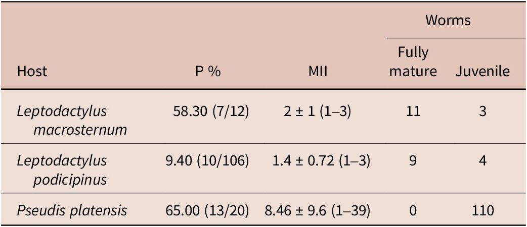
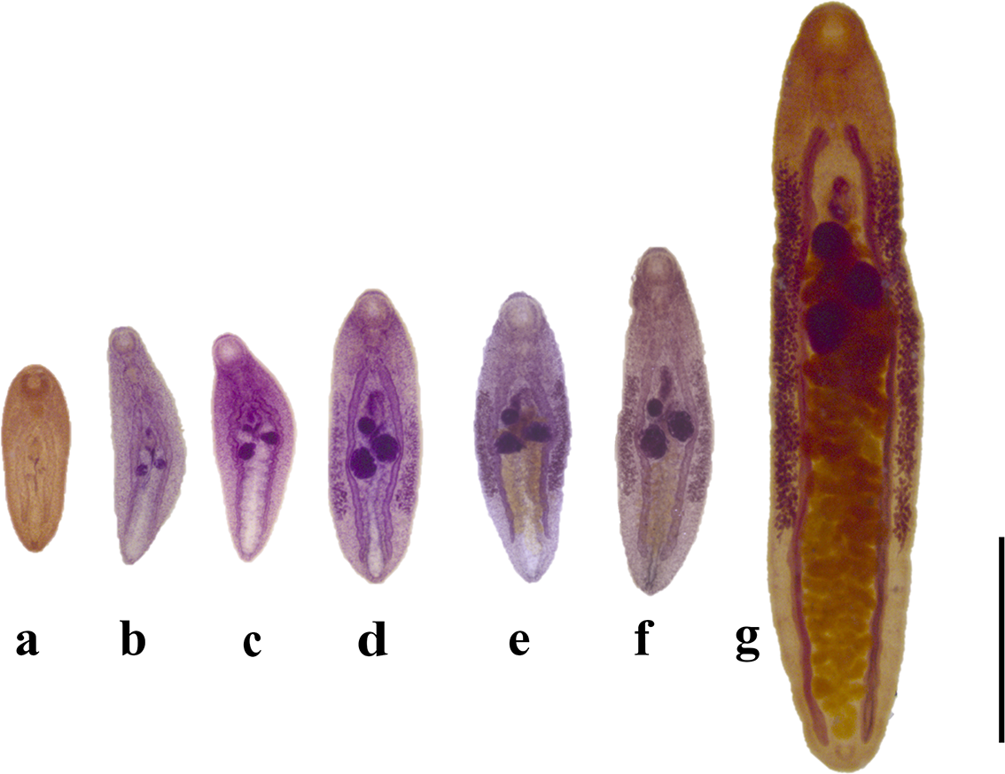
Among the 17 species of adult anurans analysed for infection with trematodes (n = 297), three species, Leptodactylus macrosternum Miranda-Ribeiro, 1926, Leptodactylus podicipinus (Cope, 1862), and Pseudis platensis Gallardo, 1961 were found infected by the species of trematode considered for this study. In total, 30 of these anurans were found to be infected with trematodes morphologically identified as Rauschiella proxima (Freitas, 1941). The ecological data obtained are presented in Table 1. A total of 137 flukes were recovered, of which only 20 found in L. macrosternum and L. podicipinus were classified as fully mature, which were larger and presented the uterus replete with eggs (Figs. 1 and 2g) and were only recovered in large hosts. Worms at this stage were used for morphological description and molecular analysis. Moreover, juveniles and non-fully developed specimens (44 with eggs and 73 without eggs) (Fig. 2a–f) were found in all three anuran specimens, indicating a recent infection. A significant morphological variability was found among these juvenile stages, since a stage with rudimentary reproductive organs and probably very young specimens (Fig. 2a), passing by specimens with developed reproductive organs but without eggs (Fig. 2b–d), and small ovigerous specimens (Fig. 2e, f). In Pseudis platensis, only juvenile specimens were recovered, which were morphologically indistinguishable from those found in the other two anuran species and were therefore also considered as the same species. Additionally, no morphological differences were observed among specimens from different hosts, supporting the taxonomic identification of R. proxima in the three anuran species in the locality.
Ecological data related to the infection of 30 among three anuran species found infected with Rauschiella proxima in a temporary marsh pond from Brazil. The number of worms fully mature or juveniles is also given to each anuran species.

Abbreviations: P%: prevalence in percentage, MII: mean intensity of infection, with standard deviation and range between parentheses.
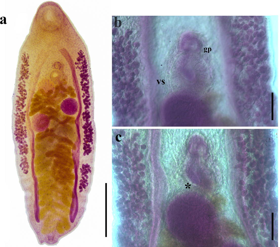
Rauschiella proxima found in Leptodactylus macrosternum from Brazil. (a) Whole view of a haematoxylin-stained specimen. (b) Details of the genital pore (gp) at the upper margin of the ventral sucker (vs); (c) detail of the sacculate seminal vesicle (asterisk). Scale bars: 1 mm (a); 100 μm (b, c).

Sequence of different maturation stages of juvenile specimens of Rauschiella proxima found in anurans from Brazil. (a–d) juveniles without eggs; (e, f) juvenile with eggs; (g) fully mature. Scale bar: 1 mm.

Taxonomic Summary
Superfamily Plagiorchoidea Lühe, 1901
Genus Rauschiella Babero, 1951
Rauschiella proxima (Freitas, 1941) (Figs. 1–3, Table 2)
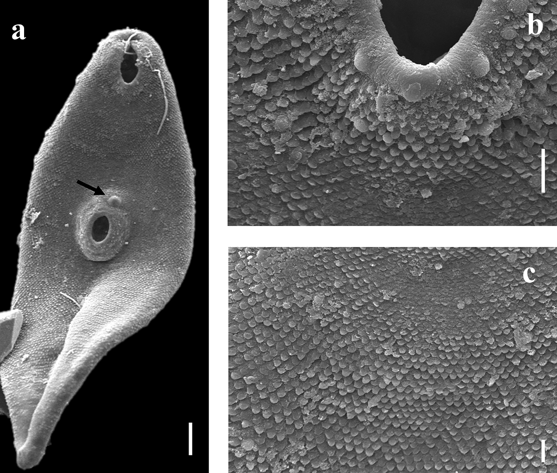
SEM images of Rauschiella proxima found in Leptodactylus macrosternum from Brazil. (a) Whole view, with details of the genital pore (arrow). (b) Details of scale-like spines surrounding the oral sucker. (c) Details of spines in ventral regions of body. Scale bars: 100 μm (a); 10 μm (b, c).

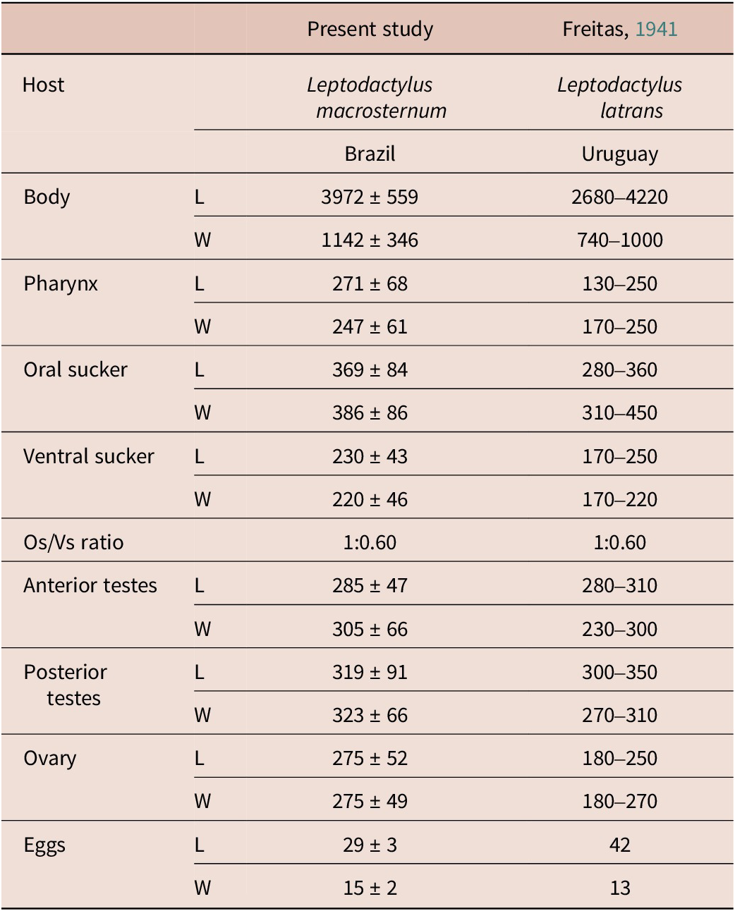
Morphometric data (in micrometres) of fully mature specimens of Rauschiella proxima found in Leptodactylus macrosternum in Brazil and data available in the original description of the species.

Abbreviations: L: length; W: width.
New definitive hosts: Leptodactylus macrosternum, Leptodactylus podicipinus (Anura: Leptodactylidae), Pseudis platensis (Anura: Hylidae).
New Locality: Selvíria, State of Mato Grosso do Sul, Brazil.
Morphological characterisation (based on 12 fully mature specimens from L. macrosternum and L. podicipinus; measures from 6 specimens from L. macrosternum in Table 2): Body elongated, spatuliform, with scale-like spines extending across entire body surface. Oral sucker subterminal, muscular, rounded, larger than ventral sucker; oral sucker/ventral sucker ratio 1:0.6. Pharynx muscular, cylindrical to elongated; oesophagus indistinct leading to caecal bifurcation; intestinal caeca running laterally too posterior body end; all internal organs intercaecal. Ventral sucker muscular, medial, in the anterior thirds of the body. Ovary round, dextral, in the anterior third of the body, posterior or sometimes overlapping to ventral sucker. Two testes, oval, oblique, equatorial, posterior to the ovary and ventral sucker, sometimes overlapping the internal margin of caeca. Uterus sinuous, intracaecal, sometimes surpassing the internal margin of caeca; uterine coils running from the posterior region of the body to the genital pore. Cirrus sac lateral or overlapping the ventral sucker, with cirrus, prostate, and sacular seminal vesicle. Genital pore round, next to the upper margin of the ventral sucker, without papillae. Vitellaria extracecal, bilateral, with clusters of follicules, extending from the level of anterior margin of ventral sucker to posterior third of body. Eggs small, elongated. Excretory vesicle Y-shaped.
Remarks: The diagnostic characteristic of the species of the genus Rauschiella is a Y-shaped excretory vesicle, uterine coils not extending beyond the posterior margins of the caeca, dextral ovary, and a tegument covered by spine-like scales. This last trait, evidenced in the analysis by SEM (Fig. 3), is a diagnostic characteristic that allows us to identify the genus, as this shape of spine is a diagnostic characteristic of species of Rauschiella (Razo-Mendivil et al. Reference Razo-Mendivil, León-Règagnon and Pérez-Ponce de León2006). Rauschiella proxima differs from its other congeners by the presence of a sacculate seminal vesicle, vitelline follicles extending from the anterior margin of acetabular region and ending in the posterior third of the body (Travassos et al. Reference Travassos, Freitas and Kohn1969; Sullivan, Reference Sullivan1977). In South America, another six species of Rauschiella were recorded parasitising anurans: R. chaquensis (Mañé-Garzón & Holcman-Spector 1967), R. lenti (Freitas, 1941), R. linguatula (Rudolphi, 1819), R. palmipedis (Sullivan, 1977), R. repandum (Babero, 1951), and R. robusta (Brooks, 1976) (Fernandes & Kohn Reference Fernandes and Kohn2014). All these species share with R. proxima the Y-shaped excretory vesicle, intercaecal uterus, dextral ovary, and extracecal vitellaria. However, they differ in the extension of the vitellaria in the lateral of body, including the line with beginning and ending of it, and the presence of a coiled seminal vesicle (Sullivan Reference Sullivan1977; Razo-Mendivil et al. Reference Razo-Mendivil, León-Règagnon and Pérez-Ponce de León2006). According to Freitas (Reference Freitas1941), the species more closely related to R. proxima is R. palmipedis, which can be differentiated by the shape of the seminal vesicle, which in the last species is strongly coiled. The morphological analysis of this structure in our adult specimens unequivocally demonstrates the presence of a sacculate seminal vesicle (Fig. 1c), enabling us to make the specific identification. Moreover, the measures obtained in this study from specimens fully mature recovered from L. macrosternum (Table 2) match well with data from the original species description.
Molecular analysis
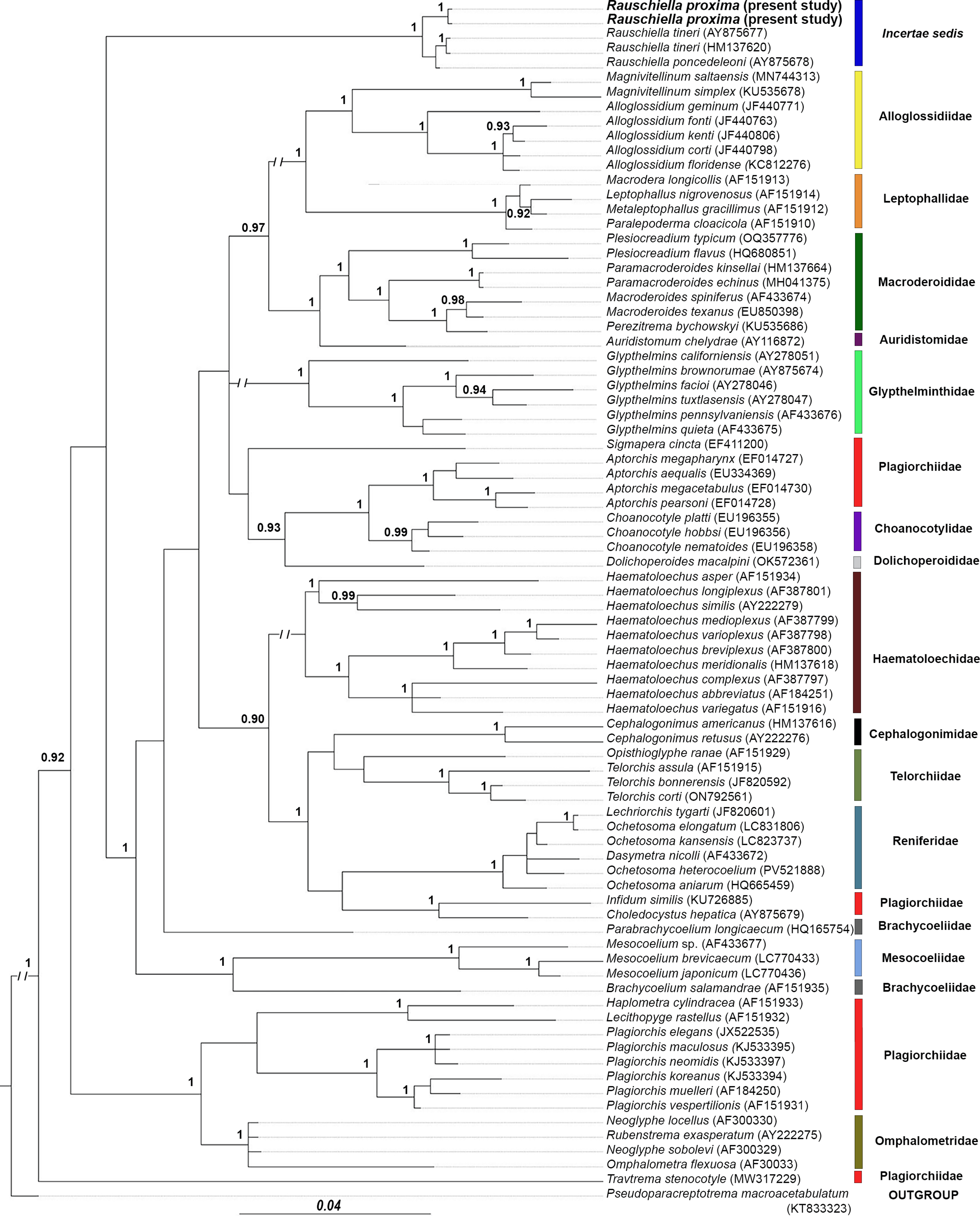
Fragments of 1148 bp of the gene 28S were obtained for two paragenophores (fully mature specimens) of R. proxima from L. podicipinus and L. macrosternum. These sequences were used for the construction of an alignment that presented 78 taxa from 15 families of Plagiorchioidea. After being trimmed to the shortest sequences, the final dataset used in the phylogenetic analysis consisted of 1137 bp. The phylogenetic tree obtained (Fig. 4) shows that our isolates of R. proxima from Brazil grouped in a well-supported clade with R. poncedeleoni and R. tineri, both from Mexico. The genetic divergences among R. proxima and these two Mexican species were low (0.45% and 0.54%, respectively). Taken together, the tree topology and genetic divergences support the congeneric status among these three species of Rauschiella. The clade composed of species of this genus diverges early in the base of the tree, together with most of the plagiorchoids included in the analysis, being separated from a clade composed of species of Plagiorchiidae (Plagiorchis spp. + Haplometra cylindracea + Lecithophyge rastellus) + Omphalotrematidae. Rauschiella spp. emerged as a distinct clade, but the nodal support for this clade to be apart from all other species included in the analysis was very low (posterior probability: 0.76). Overall, the topology obtained was similar to the previous one that showed Rauschiella out of the family Plagiorchiidae or Macroderoididae and in an incertae sedis status in Plagiorchioidea (Pérez-Ponce de León & Hernández-Mena Reference Pérez-Ponce de León and Hernández-Mena2019; Truong et al. Reference Truong, Curran, Reyda, Horton and Bullard2023).
Phylogenetic relationship among Rauschiella proxima (in bold) found in anurans from Brazil and species of the superfamily Plagiorchoidea. The tree was based on 28S rDNA sequences from 80 species belonging to 15 families and was inferred by the Bayesian Inference method (evolutive model GTR+G+I). Posterior probability values >90 were indicated as nodal support. The bar indicates the number of substitutions by site.

Discussion
In this study, we use the integrative taxonomy approach to characterise R. proxima, a parasite of Neotropical anurans. This trematode species was described more than 80 years ago. Since then, the only available record of the species was based on a survey of helminth parasites in anurans from the South region of Brazil (Portela et al. Reference Portela, Santos and Anjos2020). Moreover, for the first time, a South American representative of the genus Rauschiella had its phylogenetic position assessed and data obtained confirmed the distribution of this genus across the American continent. From a nomenclatural perspective, Rauschiella proxima was described by Freitas (Reference Freitas1941) in the intestine of Leptodactylus latrans collected in Uruguay. At that time, the species was included in the genus Glypthelmins, as G. proximus Freitas, 1941. Sometime later, Mañé-Garzón & Holcman-Spector (Reference Mañé-Garzon and Holcman-Spector1974) transferred the species to the genus Margeana Cort, 1919, which is currently a synonym of Glypthelmins, with G. proximus reestablished by some authors (Yamaguti Reference Yamaguti1971). However, the species was initially transferred to the genus Rauschiella in the revision by Sullivan (Reference Sullivan1977), and this status has been followed by more recent authors (Razo-Mendivil & Pérez-Ponce de Léon Reference Razo-Mendivil and Pérez-Ponce de León2008; Fernandes & Kohn Reference Fernandes and Kohn2014).
The familial level of classification of species of Rauschiella has also changed over time. In the revision of the genus by Sullivan (Reference Sullivan1977), the species was considered a member of the family Plagiorchiidae. The more recent revision by Font & Lotz (2008) included the genus in the family Macroderoididae. On the other hand, previous phylogenetic analysis based on 28S rDNA did not recover the two previously sequenced species within a phylogenetically supported family (Pérez-Ponce de León & Hernández-Mena Reference Pérez-Ponce de León and Hernández-Mena2019; Truong et al. Reference Truong, Curran, Reyda, Horton and Bullard2023). In this study, the inclusion of a South American representative and a more robust phylogenetic analysis did not enable the determination of its inclusion in any of the 15 plagiorchoid families included in the analysis. Moreover, the relatively low support for the clade containing the species of Rauschiella and the use of a single genetic region also preclude us from proposing a new family for this genus. Thus, in this scenario, we maintain the incertae sedis status proposed by Pérez-Ponce de León & Hernández-Mena (2019) and Truong et al. (Reference Truong, Curran, Reyda, Horton and Bullard2023) until new sequences and a more elucidative phylogenetic tree become available. A certain level of complexity was also observed in relation to other plagiorchioids included in the phylogenetic analysis. In fact, species of the genera Sigmapera, Aptorchis, Travtrema, Infidum, and Choledocystus (currently included in Plagiorchiidae) also do not group in the clade containing Plagiorchis, the type species of the family. Moreover, we also note the paraphyly in Brachycoelidae, given that Brachycoelium salamandrae (Frölich, 1789) and Parabrachycoelium longicaecum Pérez-Ponce de León et al., 2011 were grouped in distinct clades, indicating the need for new studies to propose a natural classification for this group of trematodes.
In this study, three new host records and one new geographical distribution are presented for R. proxima. The occurrence in Brazil of a fluke described in Uruguay was an expected result, as the anuran trematodes are known to show high plasticity in host occurrence and geographical distribution (Campião et al. Reference Campião, Morais, Dias, Aguiar, Toledo, Tavares and Silva2014, Reference Campião, Dias, Silva, Ferreira and Tavares2016c; Fernandes & Kohn Reference Fernandes and Kohn2014; Graça et al. Reference Graça, Oda, Lima, Guerra, Gambale and Takemoto2017; Hamann et al. Reference Hamann, González and Fernández2020; Queiroz et al. Reference Queiroz, Pontes, Neto, Campião and Anjos2020). Throughout South America, another six species of Rauschiella are reported in 22 anuran species belonging to six families (Bufonidae, Ceratophryidae, Hylidae, Leptodactylidae, Odontophrynidae, and Ranidae) (Campião et al. Reference Campião, Morais, Dias, Aguiar, Toledo, Tavares and Silva2014; Fernandes & Kohn Reference Fernandes and Kohn2014; Draghi et al. Reference Queiroz, Pontes, Neto, Campião and Anjos2020; Hamann et al. Reference Hamann, González and Fernández2020; Queiroz et al. Reference Queiroz, Pontes, Neto, Campião and Anjos2020; Aguiar et al. Reference Aguiar, Morais, Silva, Anjos, Foster and Silva2021; Benício et al. Reference Benício, Santos, Freire, Ávila, Silva and Fonseca2022). Other non-anuran hosts include one reptile species, the snake Palusophis bifossatus Raddi 1820, and the caecilian Chthonerpeton indistinctum Reinhardt & Lütken, 1862, both reported infected with Rauschiella linguatula (Rudolphi, 1819) (Fernandes & Kohn Reference Fernandes and Kohn2014). These findings confirm the nonspecificity of species of this genus of intestinal fluke, which can be found in a broad spectrum of cold-blooded hosts. Molecular studies involving these other species of Rauschiella found in South America are still necessary.
Although no statistical analysis was performed related to host size and parasite developmental level of maturation, we found remarkable results in this regard. Interestingly, juvenile specimens were also found in the original description of the species by Freitas (Reference Freitas1941). The fact that large and full gravid flukes were only recovered in large hosts can be explained by the host body size that provides a big house (intestine of an adult anuran) and major nutrient apport to the fluke by the host diet, a relation mentioned in other studies (Hamann Reference Hamann2006; Bolek & Janovy Jr Reference Bolek and Janovy2008; Campião et al. Reference Campião, Dias, Silva, Ferreira and Tavares2016c). The effect of seasonality in the finding of young and immature flukes without eggs is probably, considering that in the rainy period of the year, there is an increase in the mollusk host population and consequently the peak of cercarial emergence and infection in this temporary pond, as reported for another anuran trematode by Hamann (Reference Hamann2006). Further studies related to the ecology of the parasitism related to R. proxima and its host are necessary.
The life cycle of Rauschiella spp. remains unknown. Although long-term malacological surveys have been carried out in the same study area (Queiroz et al. Reference Queiroz, Alves, López-Hernández, Anjos and Pinto2021), larval stages that could be linked with R. proxima were not encountered. The possibility of other patterns in the life cycle of Rauschiella cannot be ruled out. The only available information on larval stages for these parasites is limited to the life cycle of Rauschiella repanda (Rudolphi 1819), briefly described in Venezuela by Lutz (Reference Lutz and Lutz1928) almost a century ago. A xiphidiocercaria found in the planorbids, Drepanotrema spp., developed as metacercaria in aquatic coleopterans (Lutz Reference Lutz and Lutz1928). This result indicates the possibility of insects acting as a second intermediate host of species of Rauschiella, which needs to be explored in more detail. This shows our lack of knowledge about the biology of anuran trematodes.
In this study, we report new hosts and locality for a South American anuran trematode. The morphological analysis includes, for the first time, SEM images for R. proxima, and the first molecular sequences were generated for the species. Moreover, we report on the different levels of parasite maturation in the species. We believe that the data generated here can support future studies that will contribute to the advancement of the taxonomy and biology of Neotropical anuran trematodes. More studies in this field are necessary to reduce the gap in the knowledge of trematodes existing in anuran biodiversity hotspots, which need to be studied before both hosts and parasites can be lost.
Acknowledgements
The author is grateful to all the participants of the manuscript and the National Council for Scientific and Technological Development (CNPq).
Author contributions
MSQ: Formal analysis, data curation, writing, revision. DLH: Molecular analysis. HAP: Molecular analysis, writing, revision. LAA: Formal analysis, data curation, writing, revision.
Financial support
We thank the National Council for Scientific and Technological Development (CNPq) (research scholarship to L.A.A. and H.A.P.).
Competing interests
The authors declare no competing interests.
Ethical standard
All activities were authorised by the Brazilian Institute of the Environment and Renewable Natural Resources (IBAMA) (research permit SISBIO-58746-4). The experimental procedures were carried out in this study following the recommendation of the local committee of ethics in animal experimentation (FEIS/UNESP – protocol CEUA/FEIS – 12/2018).