Introduction
The Pampas region, located in central Argentina, encompasses the most productive land in the country and is characterised by agricultural activity and extensive livestock production (Cieza et al. Reference Cieza, Mendicino, Larrañaga and Seibane2025). This predominantly flat landscape has historically been shaped by repeated marine transgressions and regressions (Woodward et al. Reference Woodward, Perkins and Brown2010). In addition, temperature fluctuations during glacial and interglacial periods, together with faunal exchanges from both Brazilian and Patagonian regions, have driven shifts in ecotones and transitions between biogeographic zones (Cardoso et al. Reference Cardoso, Rosso, Mabragaña, González Castro, Delpiani, Avigliano, Bogan, Covain, Schenone and Diaz de Astarloa2018, Reference Cardoso, Almirón, Casciotta, Aichino and Říčan2021).
Seasonal killifishes of the family Rivulidae inhabit ephemeral ponds that typically dry out completely for several weeks or months each year. These species persist through the dry season as eggs buried in the soil and hatching shortly after ponds refill during the rainy period. The eggs are highly resistant to desiccation and exhibit multiple developmental arrests (diapauses). Following hatching, these fish display rapid growth and early sexual maturation, enabling them to successfully exploit highly unstable and temporary habitats (Berois et al. Reference Berois, Garcia and De Sá2015; Costa Reference Costa1998; Furness Reference Furness2016; Podrabsky and Hand Reference Podrabsky and Hand2015).
Currently, six species of Rivulidae inhabit temporary ponds across the Pampas of Argentina. Until recently, these species were considered to belong to the same genus; however, Alonso et al. (Reference Alonso, Terán, Serra Alanís, Calviño, Montes, García and Casciotta2023) elevated several subgenera and described new genera. As a result, the Pampean rivulid fauna currently includes Matilebias alexandri (Castello and López), Argolebias nigripinnis (Regan), Austrolebias bellottii (Steindachner), Cypholebias robustus (Günther), Amatolebias patriciae (Huber), and Titanolebias elongatus (Steindachner) – the largest seasonal killifish species in the world, reaching over 22 cm in total length and displaying ichthyophagous habits (Alonso et al. Reference Alonso, Calviño, Serra and García2020). Detailed information on the distribution of these species is provided by Alonso et al. (Reference Alonso, Calviño, Serra and García2020, Reference Alonso, Terán, Serra Alanís, Calviño, Montes, García and Casciotta2023).
Surveys of parasites infecting rivulid fishes have revealed a diverse assemblage of parasites and pathogens, including nematodes (Contracaecum sp., Hedruris iheringi Pereira & Vaz, 1933, Gnathostoma lamothei Bertoni-Ruiz, García-Prieto, Osorio-Sarabia & León-Règagnon, 2005, Serpinema trispinosum (Leidy, 1852), and Eustrongylides sp.), acanthocephalans (Polymorphus sp. and Octospiniferoides chandleri Bullock, 1957), copepods (Caligidae), monogeneans (Leredactylus rivuli Schelkle, Paladini, Shinn, Johnson, van Oosterhout, Mohammed & Cable, Reference Schelkle, Paladini, Shinn, King, Johnson, van Oosterhout, Mohammed and Cable2011), digeneans (Catadiscus uruguayensis Freitas & Lent, 1939, Dendrorchis pampae Monteiro, Wendt & Zebral, Reference Monteiro, Wendt and Zebral2019, and Ascocotyle sp.), cestodes (Spirometra decipiens (Diesing, 1850), Valipora minuta (Coil, 1950), and Valipora campylancristrota (Wedl, 1855) Baer & Bona, 1960), microsporidians (Glugea anomala (Moniez, 1887) Gurley, 1893), ciliates (Trichodina sp. and Ichthyophthirius multifiliis Fouquet, 1876), euglenozoans (Ichthyobodo necator (Henneguy, 1883) Pinto, 1928), oomycetes (Saprolegnia sp.), and apicomplexans (Glosatella sp.) (Pereira and Vaz 1933; Bunkley-Williams and Williams Reference Bunkley-Williams and Williams2002; Bertoni-Ruiz et al. 2005; Lom and Dyková Reference Lom and Dykova2005; Delgado and García Reference Delgado and García2015; Monteiro et al. Reference Monteiro, Wendt and Zebral2019; Vettorazzi et al. Reference Vettorazzi, Norbis and Martorelli2020, Reference Vettorazzi, Norbis, Martorelli, García and Ríos2023). In Argentina, however, parasite records from rivulid fishes are limited to digeneans (Clinostomum sp., members of Telorchiidae, and Posthodiplostomum metacercariae), cestodes (Proteocephalidae), nematomorph larvae, acanthocephalans (Neoechinorhynchus villoldoi Vizcaíno, 1992), and ciliates (Trichodina bellottii Marcotegui, Montes, Barneche, Ferrari & Martorelli, Reference Marcotegui, Montes, Barneche, Ferrari and Martorelli2018) (Montes et al. Reference Montes, Barneche, Garcia, Preisz and Martorelli2017; Marcotegui et al. Reference Marcotegui, Montes, Barneche, Ferrari and Martorelli2018; Di Cesare et al. Reference Di Cesare, Montes, Vargas, Barbeito and Plaul2024; Montes et al. Reference Montes, Alonso, Terán, Serra Alanís, Waldbillig, Cavallo, Balcazar and Reig Cardarella2024, Reference Montes, Barneche, Alonso, Serra, Moncada, Theiller and Cardarella2026).
The genus Posthodiplostomum consists of small digenean trematodes with a complex life cycle: involving aquatic snails as the first intermediate host, freshwater fish as the second intermediate host, and piscivorous birds as definitive hosts (Ferguson 1937). According to WoRMS (2026), the genus currently includes 41 valid species, eight of which have been recorded in South America (Achatz et al. Reference Achatz, Chermak, Martens, Pulis, Fecchio, Bell, Greiman, Cromwell, Brant, Kent and Tkach2021; Fernandes et al. Reference Fernandes, Justo, Cárdenas and Cohen2015). Records of Posthodiplostomum metacercariae in South America remain scarce and are restricted to a few studies reporting infections in fishes belonging to the families Anablepidae, Auchenipteridae, Cichlidae, Galaxiidae, Poeciliidae, and recently Rivulidae (Azevedo et al. Reference Azevedo, Abdallah and Luque2006, Reference Azevedo, Abdallah and Luque2011; Mesquita et al. Reference Mesquita, Azevedo, Abdallah and Luque2011; Montes et al. Reference Montes, Barneche, Alonso, Serra, Moncada, Theiller and Cardarella2026; Neves et al. Reference Neves, Pereira, Tavares-Dias and Luque2013; Ostrowski de Núñez Reference de Núñez M1973; Pinto and Melo Reference Pinto and Melo2012; Rauque et al. Reference Rauque, Viozzi, Flores, Vega, Waicheim and Salgado-Maldonado2018; Ritossa et al. Reference Ritossa, Flores and Viozzi2013; Santos et al. Reference Santos, Oliveira, Neves and Tavares-Dias2018; Tavares-Dias et al. Reference Tavares-Dias, Alves Goncalves, Brito Oliveira and Rigor Neves2017). Although molecular data are available for several species of Posthodiplostomum, the number of sequenced metacercariae is still low (Choudhary et al. Reference Choudhary, Tripathi, Pandey and Agrawal2017), and some available sequences have been shown to be misidentified (Blasco-Costa and Locke Reference Blasco-Costa, Poulin and Presswell2017). Furthermore, Pérez-Ponce de León et al. (Reference de León G, Sereno-Uribe, Pinacho-Pinacho and García-Varela2022) noted a strong geographical bias in sequence availability, with comparatively few records from the Neotropical – a region that nevertheless hosts a high diversity of freshwater fish and bird species (Balian et al. Reference Balian, Segers, Lévêque and Martens2008). Consequently, the diversity of Posthodiplostomum is likely underestimated (Achatz et al. Reference Achatz, Chermak, Martens, Pulis, Fecchio, Bell, Greiman, Cromwell, Brant, Kent and Tkach2021).
Against this background, the objective of the present study was to determine the taxonomic identity and phylogenetic relationships of a diplostomid metacercarial morphotype, herein designated as MetNea1, recovered from the seasonal rivulid fishes Ar. nigripinnis, Au. bellottii, Am. patriciae, and T. elongatus inhabiting temporary ponds of the Pampas region. Using molecular data, we assessed its phylogenetic position within Posthodiplostomum and examined whether it represents a previously recognised species or an undescribed evolutionary lineage.
Materials and methods
Collection of samples and morphological study
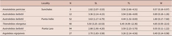
Specimens of Au. bellottii and T. elongatus were collected using hand nets from ponds located in Punta Indio (35°16′30″ S, 57°13′52″ W), whereas Ar. nigripinnis and Au. bellottii were collected in the Punta Lara Nature Reserve (34°52′12″ S, 57°57′32″ W); both sites are situated in Buenos Aires Province, Argentina. Amatolebias patriciae was collected from temporary ponds near Sunchales, Santa Fe Province (30°52′23″ S, 61°34′02″ W; Figs. 1 and 2). Parasite samples were collected between 2016 and 2025, mostly during the winter season, which corresponds to the rainy period when ponds are filled. Fish were transported alive to the field laboratory in sealed plastic bags containing water from the sampling site and supplemented with oxygen. Upon arrival, fish were euthanized by cervical transection. Total length, standard length, and body weight were recorded (Table 1), and the specimens were immediately necropsied (Protocol No. 016.05.2024, approved by the Institutional Committee for the Care and Use of Study Animals [CICUAE], Faculty of Natural Sciences and Museum, National University of La Plata).
Sampling sites of temporary ponds inhabited by seasonal rivulid killifishes. 1 = Punta Indio, Buenos Aires Province (Austrolebias bellottii, Titanolebias elongatus); 2 = Punta Lara Nature Reserve, Buenos Aires Province (A. bellottii, Argolebias nigripinnis); 3 = Sunchales, Santa Fe Province (A. bellottii, Amatolebias patriciae).

Hosts infected with Diplostomidae metacercariae. (A) Titanolebias elongatus from Punta Indio (site 1); (B) Austrolebias bellottii from Punta Indio, Punta Lara Nature Reserve, and Sunchales (site 1–3); (C) Argolebias nigripinnis from Punta Lara Nature Reserve (site 2); (D) Amatolebias patriciae from Sunchales (site 3).

Figure 2. Long description
A four-panel photo grid displays four distinct fish species.
* Panel A, top-left: Titanolebias elongatus, a slender, golden-brown fish with rounded fins and visible white parasitic cysts on its body and fins.
* Panel B, top-right: Austrolebias bellottii, a light blue fish with dark vertical banding and numerous small white spots representing metacercariae across its scales and fins.
* Panel C, bottom-left: Argolebias nigripinnis, a dark, almost black fish covered in bright iridescent blue and white spots, with parasites visible as distinct white nodules among the natural coloration.
* Panel D, bottom-right: Amatolebias patriciae, a silvery-grey fish with faint vertical stripes and prominent white parasitic cysts concentrated on its dorsal and anal fins.
Host species and localities examined, including sample size (N), standard length (SL), total length (TL), and body weight (W)

Table 1. Long description
The table consists of six columns: Host Species, Locality, N (sample size), S L (standard length), T L (total length), and W (body weight). Lengths and weights are presented as mean values followed by the range in parentheses.
* Row 1: Amatolebias patriciae from Sunchales. N is 9. S L is 2.82 (2.07–3.53). T L is 3.56 (2.66–4.31). W is 0.57 (0.18–0.97).
* Row 2: Austrolebias bellottii from Sunchales. N is 12. S L is 3.36 (2.14–4.15). T L is 3.94 (2.66–4.69). W is 0.89 (0.18–1.26).
* Row 3: Austrolebias bellottii from Punta Indio. N is 52. S L is 3.63 (1.17–6.79). T L is 4.44 (1.32–8.00). W is 1.80 (0.07–7.60).
* Row 4: Titanolebias elongatus from Punta Indio. N is 40. S L is 5.50 (3.25–10.53). T L is 6.45 (4.09–12.36). W is 4.85 (0.59–22.6).
* Row 5: Austrolebias bellottii from Punta Lara. N is 94. S L is 2.88 (1.90–4.20). T L is 3.59 (0.23–5.70). W is 0.55 (0.11–1.22).
* Row 6: Argolebias nigripinnis from Punta Lara. N is 27. S L is 2.70 (1.83–3.58). T L is 3.26 (2.14–4.56). W is 0.49 (0.14–0.98).
Internal organs and muscle tissues were examined under a stereomicroscope. Parasites were carefully removed from host tissues and placed in Petri dishes containing saline solution. Metacercariae were tentatively identified as members of the family Diplostomidae based on morphological characteristics, including the bipartite body and the presence of a well-developed holdfast organ, following Niewiadomska (Reference Niewiadomska, Gibson, Jones and Bray2002). All recovered specimens belonged to a single morphotype (MetNea1) and were preliminarily assigned to Diplostomidae based on morphology.
Metacercariae were found encysted in muscle tissue; some were excysted using fine needles, then killed by immersion in near-boiling water and fixed in 10% formalin for morphological analyses. Some specimens were preserved in 96% ethanol and stored at −20°C for DNA extraction.
Morphological analysis
Whole-mount metacercariae were processed using standard parasitological techniques and stained with either hydrochloric carmine or aqueous alum carmine (Pritchard and Kruse Reference Pritchard and Kruse1982). Digital images were captured using an Olympus BX51 microscope equipped with an AmScope MU1000 10 MP digital camera. Morphometric measurements were taken using ImageJ software (Schneider et al. Reference Schneider, Rasband and Eliceiri2012), and illustrations were prepared from specimens stained with aqueous alum carmine. Measurements are presented as the mean followed by the range (minimum–maximum) and are given in micrometres (μm). Population parameters (prevalence, mean intensity, and abundance) were calculated according to Bush et al. (Reference Bush, Lafferty, Lutz and Shostak1997).
DNA extraction, amplification, and sequencing
Total genomic DNA was extracted from diplostomid metacercariae using the PURO-Genomic DNA Kit (PB-L Productos Bio-Lógicos® S.A., Argentina), following the manufacturer’s instructions. To assess whether the same metacercarial morphotype occurring in different host species corresponded to a single genetic lineage, DNA was extracted and sequenced from four pooled samples, each comprising five metacercariae recovered from a single host individual of each species, representing one of the four rivulid fish species.
A fragment of the 28S rRNA gene (28S) was amplified using the forward primer LSU-5 (5′-TAG GTC GAC CCG CTG AAY TTA AGC A-3′) and the reverse primer 1500R (5′-GCT ATC CTG AGG GAA ACT TCG-3′), as described by Tkach et al. (Reference Tkach, Littlewood, Olson, Kinsella and Swiderski2003), following their published thermocycling conditions.
The ITS2 rDNA region was amplified using the forward primer 3S (5′-GGT ACC GGT GGA TCA CGT GGC TAG TG-3′; Morgan and Blair Reference Morgan and Blair1995) and the reverse primer ITS2.2 (5′-CCT GGT TAG TTT CTT TTC CTC CGC-3′; Cribb et al. Reference Cribb, Anderson, Adlard and Bray1998). ITS2 PCR conditions were adapted from Muñoz and George-Nascimento (Reference Muñoz and Nascimento2019) and consisted of an initial denaturation at 95°C for 10 min, followed by 10 touchdown cycles (95°C for 15 s, annealing for 30 s with the annealing temperature decreasing from 60°C to 50°C (1°C per cycle), and 72°C for 45 s), followed by 35 additional amplification cycles (95°C for 15 s, 50°C for 30 s, and 72°C for 45 s), and a final extension at 72°C for 10 min.
Reactions were performed in a Trident 960 thermal cycler (Heal Force, Shanghai, China) in a total volume of 50 μl containing 25 μl of PB-L Master Mix (PB-L Productos Bio-Lógicos, Quilmes, Argentina), 0.4 μM of each forward and reverse primer, and 4 μl of template DNA. Amplicons were sequenced in both directions on an ABI 3730XL sequencer (Macrogen Inc., Seoul, South Korea). Sequences were assembled and edited in Geneious v.6.0.5 (http://www.geneious.com) and compared with homologous sequences available in GenBank (Supplementary material S1).
Sequence comparison and phylogenetic analysis
Sequences were aligned using MAFFT v.7 (Katoh and Rozewicki Reference Katoh, Rozewicki and Yamada2019). The best-fit substitution models were selected using the Bayesian Information Criterion (BIC; Schwarz Reference Schwarz1978) as implemented in MEGA X (Kumar et al. Reference Kumar, Stecher, Li, Knyaz and Tamura2018). The optimal nucleotide substitution models were GTR+G+I for the 28S dataset and K2+G+I for the ITS2 dataset.
Phylogenetic analyses were conducted using Bayesian Inference (BI) implemented in MrBayes v.3.2.3 (Ronquist et al. Reference Ronquist, Teslenko, Van der Mark, Ayres, Darling, Höhna, Larget, Liu, Suchard and Huelsenbeck2012). Two independent Metropolis-coupled Markov chain Monte Carlo (MCMC) analyses were run for 20 million generations, sampling every 1,000 generations. Convergence was assessed by monitoring the average standard deviation of split frequencies (<0.01). Clade support was evaluated using posterior probabilities (PP), with values >0.90 considered strong support. A majority-rule consensus tree with branch lengths was obtained after discarding the first 25% of sampled trees as burn-in.
Pairwise genetic distances (uncorrected p-distances; Nei and Kumar Reference Nei and Kumar2000) were calculated to compare divergence among lineages. A p-distance matrix was generated in MEGA X (Kumar et al. Reference Kumar, Stecher, Li, Knyaz and Tamura2018) using 500 bootstrap replicates and considering both transitions and transversions under a uniform substitution rate.
Results
Morphological results
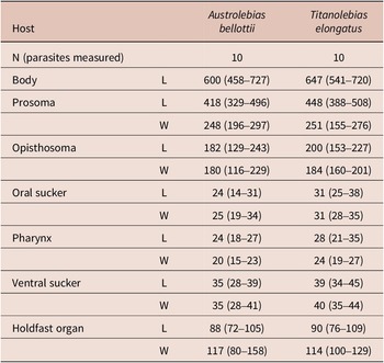
Only the metacercariae from two host species were measured because specimens recovered from the remaining hosts were either fixed in ethanol, failed to excyst, or were partially desiccated, preventing reliable morphometric analysis.
Posthodiplostomum sp.
Morphometric measurements of Posthodiplostomum sp. recorded from Austrolebias bellottii and Titanolebias elongatus

Table 2. Long description
The table compares 10 parasites from each host. Measurements are provided as a mean followed by the range in parentheses.
* Body Length: 600 (458–727) for A. bellottii and 647 (541–720) for T. elongatus.
* Prosoma Length: 418 (329–496) for A. bellottii and 448 (388–508) for T. elongatus.
* Prosoma Width: 248 (196–297) for A. bellottii and 251 (155–276) for T. elongatus.
* Opisthosoma Length: 182 (129–243) for A. bellottii and 200 (153–227) for T. elongatus.
* Opisthosoma Width: 180 (116–229) for A. bellottii and 184 (160–201) for T. elongatus.
* Oral sucker Length: 24 (14–31) for A. bellottii and 31 (25–38) for T. elongatus.
* Oral sucker Width: 25 (19–34) for A. bellottii and 31 (28–35) for T. elongatus.
* Pharynx Length: 24 (18–27) for A. bellottii and 28 (21–35) for T. elongatus.
* Pharynx Width: 20 (15–23) for A. bellottii and 24 (19–27) for T. elongatus.
* Ventral sucker Length: 35 (28–39) for A. bellottii and 39 (34–45) for T. elongatus.
* Ventral sucker Width: 35 (28–41) for A. bellottii and 40 (35–44) for T. elongatus.
* Holdfast organ Length: 88 (72–105) for A. bellottii and 90 (76–109) for T. elongatus.
* Holdfast organ Width: 117 (80–158) for A. bellottii and 114 (100–129) for T. elongatus.
Cysts rounded; metacercariae brownish, opalescent, occupying ~40% of cyst volume. Excysted metacercariae bipartite; prosoma larger than opisthosoma. Calcareous corpuscles distributed throughout body. Forebody leaf-shaped; oral sucker terminal; pharynx elongate-oval; intestinal caeca visible in fresh specimens; ventral sucker larger than oral sucker. Holdfast organ well developed, round to oval, with median slit, located near posterior constriction between prosoma and opisthosoma. Genital primordia located in hindbody; details not observed. Copulatory bursa eversible, terminal to slightly posterodorsal (not drawn).
Taxonomy summary
Hosts: Austrolebias bellottii (Steindachner, 1881), Titanolebias elongatus (Steindachner, 1881), Argolebias nigripinnis (Regan, 1912), Amatolebias patriciae (Huber, 1995)
Site of infection: Muscle tissue
Localities: Punta Indio (35°16′30″ S, 57°13′52″ W, Buenos Aires Province), Punta Lara (34°52′12″ S, 57°57′32″ W, Buenos Aires Province), and Sunchales (30°52′23″ S, 61°34′02″W, Santa Fe Province).
Prevalence, Mean intensity, and Mean abundance: In Sunchales values were 11.11%, 6.00, and 0.66 in Am. patriciae, and 66.67%, 11.13, and 7.42 in Au. bellottii. In Punta Indio values were 28.85%, 154.00, and 44.56 in Au. bellottii, and 60.00%, 28.75, and 17.25 in T. elongatus. In Punta Lara values were 44.68%, 56.54, and 25.27 in Au. Bellottii, and 18.52%, 2.00, and 0.37 in Ar. nigripinnis
Voucher specimens: MLP 8366, MLP 8367, MLP 8368
GenBank accession numbers: 28S: PZ277383 (Au. bellottii) ITS2: PZ282867, PZ282868 (Am. patriciae), PZ282869 (T. elongatus), PZ282870 (Au. bellottii), PZ282871 (Ar. nigripinnis).
Molecular results
A single partial 28S rDNA sequence (1091 bp) was obtained from metacercariae infecting Au. bellottii. Five ITS2 sequences were obtained: two from Am. patriciae (481–482 bp), one from Au. bellottii (395–482 bp), one from Ar. nigripinnis (615 bp), and one from T. elongatus (482 bp). The ITS2 sequences obtained from metacercariae recovered from the four rivulid host species were identical in the overlapping region, supporting their assignment to a single genetic lineage. The 28S dataset comprised 43 taxa (1120 bp), whereas the ITS2 dataset comprised 42 taxa (401 bp).
In the 28S phylogeny (Fig. 3), the newly generated sequence clustered with Posthodiplostomum macrocotyle Dubois, 1937, although with low posterior probability. Both taxa were included within a strongly supported clade (PP = 0.99) that also contained Posthodiplostomum sp. 2, Posthodiplostomum aztlaensis González-García, López-Jiménez, Ortega-Olivares, Sereno-Uribe, Pérez-Ponce de León and García-Varela, 2024, Posthodiplostomum spp. 9, 21, and 22, and Posthodiplostomum microsicya Dubois, 1936. Genetic distance estimates (Table 3; Supplementary material S2) showed low divergence from P. macrocotyle (1.26%), followed by P. aztlaensis and Posthodiplostomum sp. (OR799967) (1.58%), then Posthodiplostomum sp. 9 (1.74%) and sp. 2 (1.90%).
Phylogram inferred by Bayesian inference (20 million generations) based on partial 28S rDNA sequences, showing the relationships of the Posthodiplostomum sp. metacercariae recovered from Austrolebias bellottii (in bold) with congeneric sequences retrieved from GenBank. Branch support values represent posterior probabilities; only values >0.90 are shown.

Figure 3. Long description
A phylogram rooted by the outgroup Orientocreadium pseudobagri P V 9 8 4 3 0 4 at the bottom. The tree branches into two main lineages. The lower lineage includes Posthodiplostomum kinsellae, P. cuticola, and several Posthodiplostomum species numbered 1 and 2. Within this lower clade, a bolded entry Posthodiplostomum sp. P Z 2 7 7 3 8 3 is nested among P. macrocotyle, P. erickgreenei, and P. eurypygae. The upper major lineage contains a dense cluster of species including P. brevicaudatum, P. nanum, P. minimum, and P. centrarchi. At the very top of the tree, a closely related group includes P. scardini, P. ptychocheilus, and P. recurvirostrae. Branch support values, representing posterior probabilities greater than 0.90, are positioned at the nodes, such as 1.00 at the primary divergence point and various values like 0.97 and 0.99 throughout the terminal clusters. A scale bar at the bottom indicates a genetic distance of 0.04.
Genetic divergence (uncorrected p-distances) for the 28S rDNA dataset, among digenean species related to Posthodiplostomum sp. metacercariae recovered from Austrolebias bellottii and included in the phylogenetic analyses. Newly analysed sequence is shown in bold

Table 3. Long description
The table presents uncorrected p-distances for 13 taxa. The rows and columns are indexed 1 to 13 as follows.
1: M Z 7 1 0 9 5 6 Posthodiplostomum erickgreenei.
2: P P 8 6 2 7 8 0 Posthodiplostomum sp. 1.
3: M Z 5 7 0 0 8 3 Posthodiplostomum sp. F C 1.
4: M Z 7 1 0 9 5 8 Posthodiplostomum macrocotyle.
5: M Z 7 1 0 9 6 0 Posthodiplostomum microsicya.
6: M Z 7 1 0 9 9 1 Posthodiplostomum sp. 22.
7: M Z 7 1 0 9 8 9 Posthodiplostomum sp. 21.
8: P P 8 6 2 7 7 6 Posthodiplostomum sp. 2.
9: M K 6 0 4 8 2 3 Posthodiplostomum sp. 9.
10: P P 7 1 8 5 9 7 Posthodiplostomum aztlanensis.
11: O R 7 9 9 9 6 7 Posthodiplostomum sp.
12: Posthodiplostomum sp. from Austrolebias bellottii (bolded in original).
13: Posthodiplostomum sp.
Data points include.
Row 2: 2.21 vs 1.
Row 3: 2.21 vs 1; 0.00 vs 2.
Row 4: 2.37 vs 1; 2.69 vs 2; 2.69 vs 3.
Row 5: 2.37 vs 1; 2.69 vs 2; 2.69 vs 3; 0.63 vs 4.
Row 6: 2.37 vs 1; 2.69 vs 2; 2.69 vs 3; 0.63 vs 4; 0.00 vs 5.
Row 7: 2.21 vs 1; 2.53 vs 2; 2.53 vs 3; 0.79 vs 4; 0.16 vs 5; 0.16 vs 6.
Row 8: 2.37 vs 1; 2.69 vs 2; 2.69 vs 3; 1.26 vs 4; 0.63 vs 5; 0.63 vs 6; 0.47 vs 7.
Row 9: 2.21 vs 1; 2.53 vs 2; 2.53 vs 3; 1.42 vs 4; 0.79 vs 5; 0.79 vs 6; 0.63 vs 7; 0.79 vs 8.
Row 10: 2.05 vs 1; 2.37 vs 2; 2.37 vs 3; 1.26 vs 4; 0.63 vs 5; 0.63 vs 6; 0.47 vs 7; 0.63 vs 8; 0.16 vs 9.
Row 11: 2.05 vs 1; 2.37 vs 2; 2.37 vs 3; 1.26 vs 4; 0.63 vs 5; 0.63 vs 6; 0.47 vs 7; 0.63 vs 8; 0.16 vs 9; 0.00 vs 10.
The ITS2 topology (Fig. 4) grouped all Posthodiplostomum sp. metacercariae from rivulid hosts into a strongly supported clade (PP = 1.00), which was recovered as sister to Posthodiplostomum sp. LVI and P. macrocotyle within a highly supported node (PP = 0.95). Genetic distances among ITS2 sequences of Posthodiplostomum sp. infecting rivulid fishes from the Pampean region were zero, indicating complete sequence identity (Table 4; Supplementary material S3). The closest related taxa were P. macrocotyle (1.42–2.00% divergence) and Posthodiplostomum sp. 9 (1.62–2.24% divergence).
Phylogram inferred by Bayesian inference (20 million generations) based on partial ITS2 rDNA sequences, showing the relationships of the metacercariae Posthodiplostomum sp. found in rivulid fishes from the Pampean region (in bold) with congeneric sequences retrieved from GenBank. Branch support values represent posterior probabilities; only values >0.90 are shown.

Figure 4. Long description
The phylogram branches from a root on the left, anchored by the outgroup Alaria alata M K 1 6 8 6 9 9 at the bottom.
Moving from top to bottom along the right-hand terminal taxa:
* The uppermost clade includes Posthodiplostomum sp. 3 M G 8 5 7 1 0 3, P. cf. minimum M F 1 7 0 9 9 8, and P. centrarchi K X 9 3 1 4 4 1.
* Below this is a cluster containing U G 1, U G 2, and U G 3 (L C 5 1 1 1 8 3 to L C 5 1 1 1 8 5).
* A large central clade follows, containing various species including P. pricei H M 0 6 4 9 5 9 and several numbered species marked with asterisks (sp. 1, 2, 3, 4, 8).
* A distinct lower clade contains the bolded sequences of interest: Posthodiplostomum sp. P Z 2 8 2 8 6 7 through P Z 2 8 2 8 7 1. This bolded group is sister to a clade containing P. macrocotyle P P 7 1 8 6 6 6 and P. nanum M H 3 5 8 3 9 2.
* The bottom-most branches of the Posthodiplostomum genus include P. cuticola M N 0 8 0 2 8 5 and Posthodiplostomum sp. K F 7 3 8 4 4 7.
Branch support values (posterior probabilities) are indicated at major nodes, with values such as 1.00, 0.99, 0.97, 0.95, and 0.94. A scale bar at the bottom represents 0.02 genetic distance.
Line drawings of Posthodiplostomum sp. recovered from Austrolebias bellottii.
Abbreviations: Bc = bursa copulatrix; Ca= caecum; Gp = genital primordium; Hf = holdfast; Hg = holdfast gland; Os = oral sucker; Ph = pharynx; Vs = ventral sucker. Scale bars: A = 50 μm.

Genetic divergence (uncorrected p-distances) for the ITS2 rDNA dataset among digenean species related to Posthodiplostomum sp. metacercariae recovered from killifish in the Pampean region. Newly analysed sequences are shown in bold

Table 4. Long description
The table lists 15 species/sequences on the vertical axis and corresponding numbers 1 through 15 on the horizontal axis.
Row 1: O K 3 1 5 6 9 7 Posthodiplostomum sp. L I I I.
Row 2: O R 7 9 9 9 6 6 Posthodiplostomum sp. has a distance of 2.94 from species 1.
Row 3: O R 7 9 9 9 7 0 Posthodiplostomum sp. has distances of 2.90 from species 1 and 0.00 from species 2.
Row 4: P P 7 1 8 6 3 9 Posthodiplostomum aztlanensis has distances of 7.13 from species 1, and 0.53 from species 2 and 3.
Row 5: O K 3 1 5 7 5 4 Posthodiplostomum sp. L V has distances of 7.31 from species 1, 0.53 from species 2 and 3, and 0.10 from species 4.
Row 6: O K 3 1 5 7 0 4 Posthodiplostomum sp. L V I has distances of 6.61 from species 1, 0.80 from species 2, 0.79 from species 3, 2.01 from species 4, and 2.08 from species 5.
Row 7: O K 3 1 5 7 2 5 Posthodiplostomum sp. L I V has distances of 6.81 from species 1, 0.80 from species 2, 0.79 from species 3, 1.41 from species 4, 1.48 from species 5, and 1.88 from species 6.
Row 8: M H 3 5 8 3 9 2 Posthodiplostomum nanum has distances of 6.91 from species 1, 0.80 from species 2, 0.79 from species 3, 1.51 from species 4, 1.58 from species 5, 1.97 from species 6, and 0.10 from species 7.
Row 9: M K 6 0 4 8 8 1 Posthodiplostomum sp. 9 has distances of 6.83 from species 1, 0.30 from species 2 and 3, 1.37 from species 4, 1.45 from species 5, 1.86 from species 6, 0.83 from species 7, and 0.93 from species 8.
Row 10: P P 7 1 8 6 6 6 Posthodiplostomum macrocotyle has distances of 6.53 from species 1, 0.80 from species 2, 0.79 from species 3, 2.31 from species 4, 2.41 from species 5, 1.51 from species 6, 2.21 from species 7, 2.31 from species 8, and 2.00 from species 9.
Rows 11 through 15 represent newly analyzed sequences (P Z 2 8 2 8 6 7 through P Z 2 8 2 8 7 1). These sequences show 0.00 distance between each other (except for slight variations in row 14) and distances ranging from 1.01 to 4.49 from species 1 through 10.
Discussion
Morphologically similar diplostomid metacercariae (designated herein as MetNea1) were recorded infecting Au. bellottii and T. elongatus. However, morphological measurements were only possible for specimens from these two host species, as material from Ar. nigripinnis and Am. patriciae was unsuitable for morphometric analyses. Based on molecular evidence, all sequenced metacercariae from the four rivulid host species appear to represent a single genetic lineage of Posthodiplostomum. Although this lineage may ultimately correspond to an undescribed species, the limited molecular data and life-cycle information currently available for Posthodiplostomum in Argentina preclude formal taxonomic assessment. Nonetheless, this study contributes to filling important knowledge gaps regarding larval diplostomids in South American seasonal ponds and provides new records of Posthodiplostomum infecting rivulids in Argentina. Recently, Montes et al. (Reference Montes, Barneche, Alonso, Serra, Moncada, Theiller and Cardarella2026) reported a member of this genus parasitizing Argolebias guarani Alonso, Calviño, Terán, Serra, Montes, García, Barneche, Almirón, Ciotek, Giorgis & Casciotta, 2023 from Puerto Iguazú, Misiones. Unfortunately, only COI sequences are currently available for that material, preventing direct molecular comparisons with our samples.
The parasite appears capable of infecting multiple killifish species, suggesting low host specificity at the level of the second intermediate hosts. Digeneans possess indirect life cycles, with cercariae emerging from molluscan first intermediate hosts and actively penetrating the second intermediate hosts. In this context, infection levels in fishes are likely influenced by environmental exposure, including pond surface area (Paull and Johnson Reference Paull and Johnson2018), host density (Johnson et al. Reference Johnson, Stewart Merrill, Dean and Fenton2024), the probability of encounter between free-swimming cercariae and fish hosts (Gopko et al. Reference Gopko, Mironova, Pasternak, Mikheev and Taskinen2020), and the distribution and abundance of definitive hosts (Lafferty Reference Lafferty2008).
Samples from Am. patriciae and Ar. nigripinnis were limited, and metacercariae from these hosts were fixed directly in ethanol without prior excystation, restricting their use to molecular analyses. In contrast, only a limited number of specimens from Au. bellottii and T. elongatus were suitable for detailed morphological examination. Consequently, comparisons involving these hosts should be interpreted with caution, particularly when assessing subtle morphological variation. In addition, the number of specimens available for integrative analyses was further reduced due to deterioration of part of the stored material during long-term preservation, which prevented successful excystation or molecular characterisation of some metacercariae. Furthermore, logistical and funding constraints limited the scope of additional field surveys and molecular analyses.
Although measurements of Posthodiplostomum metacercariae from Au. bellottii and T. elongatus are largely similar, specimens reported from Ar. guarani in Puerto Iguazú were generally larger in most morphometric variables, although substantial overlap in ranges was observed. The most notable differences involved prosoma width and holdfast organ length, traits which tended to be greater in the Iguazú specimens.
Among the species examined, Au. bellottii, at sites where it co-occurred with other rivulid species, consistently exhibited higher prevalence, mean intensity, and abundance values than Am. patriciae and T. elongatus (from Sunchales and Punta Indio, respectively). These patterns suggest that Au. bellottii may be particularly susceptible to infection or may experience higher exposure to cercariae, although additional sampling is required to evaluate these possibilities. In contrast, T. elongatus showed comparatively high prevalence but low mean intensity and abundance. This pattern may be related to its benthic habits (Froese and Pauly Reference Froes and Pauly2021), which likely increase exposure to infective stages, particularly in vegetated or bottom substrates where snails are abundant. However, despite its larger body size, the lower parasite burdens observed in T. elongatus suggest the presence of physiological or immunological mechanisms that may limit parasite establishment and survival. These interpretations should be considered preliminary, and broader sampling is needed to evaluate population parameters across different sites, host sexes, and rivulid species.
Phylogenetic analyses indicate that the closest relatives of the metacercariae reported here include P. macrocotyle, P. microsicya, and several unnamed Posthodiplostomum lineages whose adult stages parasitize herons and other piscivorous birds. This relationship suggests that the life cycle of the Pampean Posthodiplostomum sp. may involve ardeid birds as definitive hosts, although confirmation will require recovery of adult specimens and complete elucidation of the life cycle. Ponds at Punta Lara, although located within a Provincial Reserve, are close to roads and urban areas near La Plata and experience frequent human activity. In contrast, ponds at Punta Indio and Sunchales occur in cattle-dominated pastures, isolated from urban centres and with low human disturbance. These contrasting conditions may influence the abundance and diversity of avian definitive hosts, including herons.
Because these ponds are temporary and dry out during summer, the persistence of the parasite in fish populations may occur through two non-exclusive scenarios. First, infected snails may remain buried in the mud and survive the dry season until the rainy period, when cercariae are released into the water (Chantima et al. Reference Chantima, Suk-ueng, Tananoi and Prasertsin2025). Alternatively, adult parasites inhabiting the intestines of herons may release eggs into the ponds during the rainy season; miracidia then infect snails and reinitiate the life cycle (Kvach et al. Reference Kvach, Jurajda, Bryjová, Trichkova, Ribeiro, Přikrylová and Ondračková2017). The latter scenario highlights the importance of temporary ponds with low levels of human disturbance, which allow birds access to aquatic environments with relatively uncontaminated waters that favour the survival of intermediate hosts and free-living parasite stages (Pietrock and Marcogliese Reference Pietrock and Marcogliese2003; Thieltges et al. Reference Thieltges, Jensen and Poulin2008).
Studies on temporary ponds and their parasite communities are particularly relevant in the context of ongoing climate change in the Pampean region, an area characterized by altered rainfall regimes and increasing climatic variability (Brendel Reference Brendel2024; Müller et al. Reference Müller, Lovino and Sgroi2021). The expansion of agricultural frontiers and increasing livestock pressure may both affect the environmental conditions and alter the transmission dynamics (Monte Reference Monte2017; Schmutzer et al. Reference Schmutzer, Gray, Burton and Miller2008). For example, environmental contamination of ponds may negatively impact host populations or parasite survival, including both intra-host stages and free-living stages (Pietrock and Marcogliese Reference Pietrock and Marcogliese2003; Thieltges et al. Reference Thieltges, Jensen and Poulin2008; Monte Reference Monte2017). Conversely, eutrophication may promote snail population growth, leading to increased parasite intensities and abundances in fish hosts (Johnson et al. Reference Johnson, Chase, Dosch, Hartson, Gross, Larson, Sutherland and Carpenter2007). Elevated parasite loads may compromise fish survival (Guitard et al. Reference Guitard, Chrétien, De Bonville, Roche, Boisclair and Binning2022); however, if infected fishes persist, they may in turn pose increased risks to avian definitive hosts (Dyer et al. Reference Dyer, Williams, Mignucci-Giannoni, Jiménez-Marrero, Bunkley-Williams, Moore and Pence2002), including resident and migratory birds that rely on temporary ponds as feeding or stopover habitats (Martínez-Curci et al. Reference Martínez-Curci, Isacch, Fernández, Bogel, Ruiz and Navedo2025; Skagen et al. Reference Skagen, Granfors and Melcher2008;)
In the present study, MetNea1 metacercariae likely represent a single lineage of Posthodiplostomum infecting four rivulid species across the Pampean region. However, given the limited availability of comparative molecular and life-cycle data for South American representatives of the genus, its specific identity remains unresolved. Additional metacercarial morphotypes were also observed during the surveys, but these were not included in this work because insufficient material was available for detailed morphological and molecular characterisation. Considering that at least 19 rivulid species occur within the Rio de la Plata basin (Alonso et al. Reference Alonso, Calviño, Serra and García2020, Reference Alonso, Terán, Serra Alanís, Waldbillig, Montes, Bugeau, Barneche, Ibañez Shimabukuru Gonzalez, Táboas and Maroli Almeida2025), additional Posthodiplostomum lineages are likely to occur in the region. Further integrative studies incorporating metacercariae, adult stages, and molecular data from both fish and bird hosts will be necessary to better understand the diversity and transmission dynamics of Posthodiplostomum in South America.
Supplementary material
The supplementary material for this article can be found at http://doi.org/10.1017/S0022149X26101862.
Availability of data and material
All the materials are deposited in museums and the sequences are deposited in GenBank.
Code availability
Not applicable.
Acknowledgements
We are grateful to Consejo Nacional de Investigaciones Científicas y Técnicas (CONICET), to CEPAVE for providing facilities and equipment, and to Ministerio de Medio Ambiente de la Provincia de Buenos Aires for providing collection permits (DI-2019-49-GDEBA-DANPOPDS).
Financial support
This work was supported by the Consejo Nacional de Investigaciones Científicas y Técnicas (CCT-CONICET-La Plata; M.M.M., grant number 11220200101713CO) and Fondo para la Investigación Científica y Tecnológica (FONCyT) (M.M.M., grants number PICT-2020-SERIEA-01531).
Competing interests
On behalf of all authors, the corresponding author states that there is no conflict of interest.
Competing interests declaration
Martin M. Montes: Investigation, Writing – Original Draft. Jorge Barneche: Methodology, Formal Analysis. Tomas Acuña González: Methodology. Formal Analysis. Ignacio García: Methodology, Writing – Review and Editing. Felipe Alonso: Writing – Review and Editing. Marina Ibáñez Shibamukuro: Writing – Review and Editing. German Reig Cardarella: Supervision, Visualization.
Ethical standards
The authors assert that all procedures contributing to this work comply with the ethical standards of the relevant national and institutional guides on the care and use of laboratory animals.
Consent to participate
All the authors give their consent to participate in this work.
Consent for publication
All the authors give their consent to the publication of this work.
Use of artificial intelligence
We used AI tools during the preparation of this manuscript exclusively for English language correction. This use has been transparently declared in accordance with the journal’s policy and ethical requirements.