Introduction
Comprising c. 265 species (Kirk et al. Reference Kirk, Cannon, Minter and Stalpers2008), the cosmopolitan genus Rinodina (Ach.) Gray is widely distributed in both Hemispheres from polar to tropical latitudes. Detailed taxonomic treatments are available for saxicolous Rinodina species from Europe (e.g. Mayrhofer & Poelt Reference Mayrhofer and Poelt1979; Mayrhofer Reference Mayrhofer1984a; Giralt Reference Giralt2001), Africa (e.g. Mayrhofer Reference Mayrhofer1984a; Matzer & Mayrhofer Reference Matzer and Mayrhofer1996), Asia (e.g. Mayrhofer Reference Mayrhofer1984a), Australasia (e.g. Mayrhofer Reference Mayrhofer1983, Reference Mayrhofer1984b; Kaschik Reference Kaschik2006), and for epiphytic Rinodina species in Europe (e.g. Ropin & Mayrhofer Reference Ropin and Mayrhofer1993, Reference Ropin and Mayrhofer1995; Giralt & Mayrhofer Reference Giralt and Mayrhofer1994, Reference Giralt and Mayrhofer1995; Giralt & Matzer Reference Giralt and Matzer1994) and North America (e.g. Sheard & Mayrhofer Reference Sheard and Mayrhofer2002). Other treatments focus on a single species or a species group on a worldwide scale (e.g., Mayrhofer et al. Reference Mayrhofer, Scheidegger and Sheard1990; Matzer & Mayrhofer Reference Matzer and Mayrhofer1994; Matzer et al. Reference Matzer, Mayrhofer and Elix1998; Mayrhofer et al. Reference Mayrhofer, Sheard, Grassler and Elix2001). Identification keys to Rinodina species were provided for Great Britain and Ireland (Giavarini et al. Reference Giavarini, James, Purvis, Smith, Aptroot, Coppins, Fletcher, Gilbert, James and Wolseley2009), south-western Germany (Wirth Reference Wirth1995), Scandinavia (Mayrhofer & Moberg Reference Mayrhofer and Moberg2002), the Iberian Peninsula (Giralt Reference Giralt2001), the extended Sonora Desert region (Sheard Reference Sheard, Nash, Ryan, Diederich, Gries and Bungartz2004) and Russia (Kotlov Reference Kotlov and Golubkova2008).
Rinodina species usually have crustose thalli, lecanorine apothecia, 2-celled brown ascospores with inner wall thickenings and Lecanora-type asci. The most important character complexes for determining Rinodina species are those of the proper excipula, the ascospores and the asci. The different phenotypes of these characters were classified as categories or “types” (Zahlbruckner Reference Zahlbruckner and Engler1926;Poelt & Mayrhofer Reference Poelt and Mayrhofer1979; Rambold et al. Reference Rambold, Mayrhofer and Matzer1994). The correlation of ascus types with the other categories agreed with a concept of two main groups in Physciaceae Zahlbr., i.e. the Physcia group (including Rinodina and also the foliose genera Physcia (Schreb.) Michx., Phaeophyscia Moberg, Physconia Poelt among others) and the Buellia group (including Buellia De Not., Diploicia A. Massal., Dirinaria (Tuck.) Clem., Pyxine Fr., Santessonia Hale & Vobis among others). However, there are some interesting exceptions which combine characters of Buellia and Rinodina, for example, Buellia lindingeri Erichsen, Buellia parvula (Mayrhofer & Poelt) Mayrhofer & Scheid. and Rinodina insularis (Arnold) Hafellner.
The current generic concept of Rinodina s. lat. is polymorphic with respect to ascospore pigmentation and internal wall formation (e.g., Hafellner et al. Reference Hafellner, Mayrhofer and Poelt1979; Mayrhofer Reference Mayrhofer1982; Kaschik Reference Kaschik2006). Initial phylogenetic analyses of the Physciaceae suggested that differences in these characters do not reflect larger lineages in Rinodina, but might rather be specific at species level or below (Grube & Arup Reference Grube and Arup2001; Helms et al. Reference Helms, Friedl and Rambold2003; Kaschik Reference Kaschik2006). These studies also raise the hypothesis that Rinodina is a paraphyletic genus, with foliose and fruticose lineages probably originating from various crustose ancestors. All of these investigations, however, relied on ITS data only, which limits the resolution of deeper branches. MtSSU and nrITS rDNA sites were used for studying the natural relationships between the lichen families Physciaceae and Caliciaceae Chevall. (Wedin et al. Reference Wedin, Baloch and Grube2002). These analyses include only three Rinodina species which were close to the foliose genera Physcia and Anaptychia Körb. within the Physcia group. Helms et al. (Reference Helms, Friedl and Rambold2003) included six Rinodina species in their phylogeny of selected Physciaceae and twenty species in their phylogeny of all Physciaceae using ITS data only. Variation among the non-coding ITS regions is useful for delimitations at species level (Myllys et al. Reference Myllys, Tehler and Lohtander2001; Kaschik Reference Kaschik2006), whereas the mtSSU rDNA sequence is a more conserved gene locus and allows the investigation of relationships between genera (Crespo et al. Reference Crespo, Blanco and Hawksworth2001; Wedin et al. Reference Wedin, Baloch and Grube2002). A combined phylogenetic analysis using both nrITS and mtSSU rDNA genes should be valuable in distinguishing genera or species groups. The aim of this study is to test the phylogenetic importance of the main traits which are used for the delimitation of Rinodina species as well as to check the phylogenetic positions of some ‘extraneous’ Rinodina and Buellia species (Buellia lindingeri, Buellia parvula and Rinodina insularis) using nrITS and mtSSU rDNA molecular data.
Materials and Methods
Taxon sampling
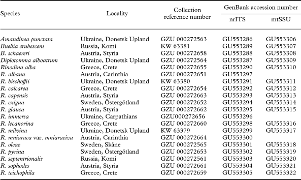
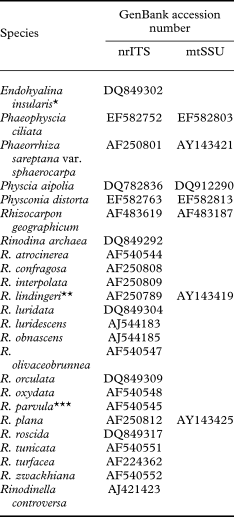
Lichen material used for sequencing is listed in Table 1 and is deposited in GZU or KW. Most samples selected for molecular analyses were collected during 2005–2007 in Austria, Ukraine, Russia, Crete and Sweden. Other sequences for our analysis were downloaded from GenBank NCBI (Table 2). In addition the following public herbaria and private collections have contributed with the loan of specimens: G, GZU, M, SZU, hb. Groner, hb. Vězda and hb. Zimmermann.
List of specimens and GenBank accession numbers of the new nrITS and mtSSU rDNA sequences generated for the study

List of species and accession numbers of sequences downloaded from GenBank

* as Rinodina insularis,
** as Buellia lindingeri,
*** as Rinodina lecanorina
DNA extraction, PCR amplification, DNA sequencing and sequence alignment
Lichen specimens were carefully checked for any externally visible infections from other organisms. Total DNA was extracted from apothecia using a modified CTAB method as described by Cubero et al. (Reference Cubero, Crespo, Fatehi and Bridge1999). DNA extracts were used for PCR amplification of the ITS regions of the nuclear rDNA and SSU region of the mitochondrial gene. Primers used for PCR of the nuclear ribosomal ITS region were ITS1F (Gardes & Bruns Reference Gardes and Bruns1993) and ITS4 (White et al. Reference White, Bruns, Lee, Taylor, Innis, Gelfand, Sninsky and White1990), those for mitochondrial SSU gene were mtSSU1 and mtSSU3R (Zoller et al. Reference Zoller, Scheidegger and Sperisen1999). The PCR amplification was carried out with Applied Biosystems Gene Amp PCR System 2400. Products were cleaned with QIAquick PCR Purification Kit (QIAGEN, Vienna) according to the manufacturer's protocol. The PCR products were then sequenced with the BigDyeTerminator Cycle Sequencing Ready Reaction Kit (Applera, Vienna) according to the manufacturer's instructions. Sequences were run on an ABI 310 automated sequencer (Applera, Vienna).
Sequences obtained of both strands were assembled and edited using AutoAssembler Software (Applied Biosystems, Vienna), which we used for the alignment in BioEdit (http://jwbrown.mbio.ncsu.edu/BioEdit/bioedit.html) together with sequences downloaded from GenBank NCBI. Alignment was automatically carried out using the Clustal algorithm with standard settings in BioEdit and then improved manually. Data sets of nrITS and mtSSU rDNA were combined in BioEdit and consisted of 1237 sites from 44 taxa.
Phylogenetic analysis
Based on the combined data matrix, a parsimony tree was constructed in PAUP*4.08b (Swofford Reference Swofford2002), using a heuristic search with 1000 replicates of random sequence additions. Seventy-three ambiguous positions were excluded from the data matrix, gaps were treated as missing values. The phylogram was constructed with the Bayesian approach as implemented in the MrBayes 3.Ob4. (Huelsenbeck & Ronquist Reference Huelsenbeck and Ronquist2001). Support values obtained in this analysis are regarded as significant when exceeding 0·95. The trees were drawn using TREEVIEW (Page Reference Page1996). As in a previous study, we chose Rhizocarpon geographicum (L.) DC. as a suitable outgroup (Grube & Arup Reference Grube and Arup2001).
Morphology and chemistry
The material was examined using a stereo- and high power microscopy. Thin-layer chromatography (TLC) was employed using standard methods (e.g. Orange et al. Reference Orange, James and White2001).
Results
We generated 47 new sequences (26 of nrITS region and 21 of mtSSU rDNA) from 19 crustose species of the Physciaceae (mainly from the genus Rinodina) (Table 1).
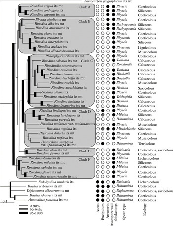
The species analysed are split into two groups, which correspond to the previously recognized Buellia and Physcia groups (Fig. 1, Grube & Arup Reference Grube and Arup2001; Helms et al. Reference Helms, Friedl and Rambold2003; Kaschik Reference Kaschik2006). Genetic variation is poorly resolved in the Buellia-group with our markers, therefore we concentrate on the Physcia group in the following discussion. Our combined analysis shows that there are several more or less well supported units of Rinodina species within the Physcia group. These units of Rinodina species also include other genera such as Physcia, Phaeophyscia, Physconia, Phaeorrhiza H. Mayrhofer & Poelt and Rinodinella H. Mayrhofer & Poelt. Except for the last two genera, these are characterized by a more complex morphological organization, such as foliose thalli with more phenotypic characters of taxonomic value than found in the crustose groups.
Bayesian consensus nrITS and mtSSU rDNA phylogeny of selected Physiaceae compared with phenotypical and ecological characters. Ascus type: • Bacidia-type, ○ Lecanora-type; excipulum: •lecideine, ○ lecanorine; atronorin and ascospore thickenings: • present, ○ absent.

The monophyletic and well-supported Clade A includes only the three species Rinodina exigua (Ach.) Gray, R. confragosa (Ach.) Körb. and R. capensis Hampe. All these species contain atranorin and have ascospores of the Physcia-type.
The moderately supported Clade B is nesting Physcia aipolia (Ehrh. ex Humb.) Fürnr. basal with Rinodina alba Metzler ex Arnold and R. atrocinerea (Hook.) Körb. The last two are related silicicolous species with ascospores of the Pachysporaria-type. The other Rinodina species in Clade B are corticolous, except for the siliceous R. interpolata (Stirt.) Sheard and the muscicolous R. olivaceobrunnea C. W. Dodge & G. E. Baker, and most have Physcia-type ascospores, except for R. orculata Poelt & M. Steiner and R. archaea (Ach.) Arnold which possess the related Physconia-type ascospores. A highly supported monophyletic subclade includes R. orculata, R. interpolata, R. archaea and R. olivaceobrunnea. These are species with varying ecology.
The monophyletic and highly supported Clade C includes Phaeophyscia ciliata (Hoffm.) Moberg and Rinodinella controversa (A. Massal.) H. Mayrhofer & Poelt. Three subclades can be recognized, the first of these consists of calcareous taxa such as Rinodinella controversa, Rinodina tunicata H. Mayrhofer & Poelt, R. immersa (Körb.) Arnold, R. bischoffii (Hepp) A. Massal., R. zwackhiana (Kremp.) Körb. and interestingly the muscicolous R. roscida (Sommerf.) Arnold. The second subclade includes species with Teichophila-type ascospores (R. teichophila (Nyl.) Arnold and R. albana (A. Massal.) A. Massal.). The third subclade includes species occurring on calcareous rocks with Bicincta-type ascospores, such as R. luridata (Körb.) H. Mayrhofer et al. and R. lecanorina (A. Massal.) A. Massal.
Clade D is distinguished here for pragmatic reasons although it is not monophyletic. It includes species of Rinodina with ambiguous relationships as well as Physconia distorta (With.) J. R. Laundon and Phaeorrhiza sareptana (Tomin) H. Mayrhofer & Poelt. A well-supported monophyletic subclade comprises R. luridescens (Anzi) Arnold together with the morphologically peculiar species Rinodina lindingeri (Erichsen) Giralt & van den Boom and R. parvula H. Mayrhofer & Poelt. All species in this clade have different types of excipulum and ascospores and occur on different substrata. However, the relationships of this clade to other lineages in Rinodina including Clade C remain poorly resolved.
Clade E consists of the two species, Rinodina oleae Bagl. with Dirinaria-type ascospores and R. pyrina (Ach.) Arnold with Physconia-type ascospores.
The monophyletic Clade F includes species with Milvina-type ascospores such as Rinodina obnascens (Nyl.) H. Olivier, R. milvina (Wahlenb.) Th. Fr., and the type species R. sophodes (Ach.) A. Massal., as well as those with Physcia-type ascospores of the related corticolous species pair R. glauca Ropin and R. septentrionalis Malme.
The species analysed of the Buellia group belong to the genera Amandinea M. Choisy, Buellia, Diplotomma Flot. and Endohyalina Marbach. The phenotypically unusual Endohyalina insularis, previously suggested to belong to Rinodina, is distinct from the latter genus and is clearly a member of the Buellia group (Fig. 1).
Taxonomy
Endohyalina insularis (Arnold) Giralt, van den Boom & Elix
Mycological Progress 9: 44 (2010).—Buellia saxatilis f. insularis Arnold, Verh. zool.-bot. Ges. Wein 46: 119 (1896).—Rinodina insularis (Arnold) Hafellner, Beih. Nova Hedwigia 62: 87 (1979); type: South Tirol, Plan (Südtirol, Gröden, Augitporphyr oder plan gegen das Sellajoch), Aug. 1895, leg. Arnold (M—holotype).
The phylogenetic analysis based on nrITS and mtSSU sequence data indicates that Endohyalina insularis is not monophyletic with other species of Rinodina, but groups in Buellia s. lat. (Fig.1), as already shown by Kaschik (Reference Kaschik2006 as ‘Rinodina’ insularis). Morphologically, Endohyalina insularis has lecideine apothecia, Dirinaria-type ascospores and Bacidia-type asci similar to other representatives of Buellia s. lat. As a lichenicolous lichen, the species grows on lichens belonging to the Lecanora rupicola (L.) Zahlbr. group (Rambold et al. Reference Rambold, Mayrhofer and Matzer1994). The species was described as Buellia saxatilis f. insularis by Arnold (Reference Arnold1896). The combination with Rinodina was mainly based on the ascospore wall thickenings (Hafellner Reference Hafellner1979), while it was speculated that lichenicolous Rinodina species might easily loose algae in their excipula. Supported by ascus and excipulum structure, ascospore characters, as well as molecular data, we accept this species as Endohyalina insularis. The genus Endohyalina was described by Marbach (Reference Marbach2000).
The species is widely distributed. In Europe it has been found in the British Isles, France, Greece, Italy, Austria, Romania (e.g., Hafellner Reference Hafellner1979; Mayrhofer & Poelt Reference Mayrhofer and Poelt1979; Mayrhofer Reference Mayrhofer1984a; Triebel et al. Reference Triebel, Rambold and Nash1991; Calatayud et al. Reference Calatayud, Atienza and Barreno1995; Giralt Reference Giralt2001; Hitch Reference Hitch2006), in Macaronesia (e.g. Hafellner Reference Hafellner1995), and in Turkey (Halici et al. Reference Halici, John and Aksoy2005). Samples from North America thus far originate from Arizona and British Columbia (Triebel et al. Reference Triebel, Rambold and Nash1991; Sheard Reference Sheard, Nash, Ryan, Diederich, Gries and Bungartz2004) and the species is also found in the Southern Hemisphere in Australia, New Zealand and Chile (Triebel et al. Reference Triebel, Rambold and Nash1991; Mayrhofer & Lambauer Reference Mayrhofer and Lambauer2004; Kaschik Reference Kaschik2006; Hafellner & Mayrhofer Reference Hafellner and Mayrhofer2007).
Selected specimen examined. Greece: Crete: Monophatsi, Asterusi Gebirge, bei Kapetaniana, 1942, Rechinger (M, associated with Tephromela atra).
Endohyalina brandii Giralt, van den Boom & Elix
Mycological Progress 9: 41 (2010).
This species is similar to Endohyalina insularis and obligately lichenicolous on Aspicilia intermutans (Nyl.) Arnold. Apart from growing on Lecanora rupicola, E. insularis is distinguished by the larger, initially immersed apothecia, the larger ascospores and the darker hypothecium. The new species was previously known only from Tenerife and El Hierro in the Canary Islands (Giralt et al. Reference Giralt, van den Boom and Elix2010b). We here report the first specimens from Europe (Iberian Peninsula: Spain) and La Palma, another island in the Canary Islands.
Other specimens examined. Spain: Prov. Barcelona: Sierra de Montseny, Hänge NE der Paßhöhe des Coll Formich, 1983, Hafellner 17322 (GZU). Prov. Tarragona: Sierra de Prades, NW-geneigte Hänge SW ober dem Monasterio de Poblet, an der Straße nach Prades, 1983, Hafellner 17489 (GZU). Canary Islands: La Palma: Caldera de Taburiente, Taburiente, am Westrand des Schotterfeldes “La Playa”, 1991, Mayrhofer 10643 & Matzer (GZU).
Endohyalina diederichii Giralt, van den Boom & Elix
Mycological Progress 9: 42 (2010).
This species is obligately lichenicolous on the genus Ramalina Ach. It is characterized by a densely inspersed hymenium with abundant oil droplets and fusiform ascospores. It is closely related to E. ericina (Nyl.) Giralt et al. but differs in its lichenicolous habit and its smaller apothecia. It is known only from Lanzarote, Fuerteventura, El Hierro and Tenerife in the Canary Islands (Giralt et al. Reference Giralt, van den Boom and Elix2010b).
Other specimen examined. Canary Islands: La Gomera: La Fortalezza, 1983, Rambold 1709 (M).
Rinodina albana (A. Massal.) A. Massal
Ric. Auton. Lich. Crost.: 15 (1852).—Hagenia albana A. Massal., Verh. zool.-bot. Ges. Wien 1: 221 (1851); type: locality not indicated, hb. Massalongo (VER—lectotype).
Rinodina albana is characterized by a cortex consisting of hyaline hyphae with globose lumina, apothecia with a brown disc, distinct proper margin visible as a ring within the thick thalline margin, and large ascospores of the Teichophila-type (Sheard & Mayrhofer Reference Sheard and Mayrhofer2002) with lumina, if well developed, mainly of the Physcia-type. A torus is not visible. The surface of the ascospores is microrugulate and already visible at magnification ×400. The hymenia often do not include any good ascospores, and where ascospores are present, the asci usually do not contain a complement of eight fully developed ascospores. Ropin & Mayrhofer (Reference Ropin and Mayrhofer1993), Giralt & Mayrhofer (Reference Giralt and Mayrhofer1995) and Giralt (Reference Giralt2001) referred the ascospores to the Physcia-type. There are frequent conidiomata around the base of the apothecia containing bacilliform conidia. Rinodina albana resembles R. trevisanii (Hepp) Körb. and R. exigua. Rinodina trevisanii is characterized by ascospores of the Physconia-type with a distinct torus (Mayrhofer & Sheard Reference Mayrhofer and Sheard2007). Rinodina exigua differs in containing at least traces of atranorin in a much weaker developed apothecial cortex and in having Physcia-type ascospores with a distinct torus (Ropin & Mayrhofer Reference Ropin and Mayrhofer1993). The phylogenetic analysis supports the close relationship of the corticolous R. albana to the saxicolous R. teichophila. The ascospores of the latter species and its relatives in the Southern Hemisphere were discussed by Matzer & Mayrhofer (Reference Matzer and Mayrhofer1994). Rinodina albana is a rare species occurring mainly on the bark of deciduous trees. It is reported from Central Europe (Austria, Czech Republic, France, Germany, Romania, Slovakia, Slovenia, Switzerland) and southern Europe (Italy, Montenegro, Spain) by Ropin & Mayrhofer (Reference Ropin and Mayrhofer1993) and Giralt & Mayrhofer (Reference Giralt and Mayrhofer1995).
Selected specimens examined. Austria: Kärnten: Gailtaler Alpen, Gemeinde Paternion, Anwesen Farchen nördlich Kreuzen, Fagus sylvatica, 2000, Mayrhofer 13976 & Pichorner (GZU). Salzburg: Tennengau, Vorderwiestal, alte Wiestalstraße, bei Almdurchbruch, 550 m, 1985, Blieberger & Türk (SZU). Vorarlberg: Bregenzerwald, Hittisau, von Hinterberg nach Gfreren, Fraxinus excelsior, 1992, Pfefferkorn 8525 (SZU).—Montenegro: Lovćen: Ivanova Korita, on bark of Fagus sylvatica, 2004, Mayrhofer 18277, Nedović, Knežević & Jakić (GZU).—Slovenia: Triglav National Park: Bohinj area, Voje valley, c. 3·5 km N from Stara Fužina, Juglans regia, 2002, Batič & Mrak (GZU).— Switzerland: Bern: Gemeinde Hasliberg, Acer platanoides, 1998, Dietrich 12082 (G).— Glarus: Gemeinde Elm, Acer pseudoplatanus, 1997, Roth 11707 (G). Graubünden: Gemeinde Rueun, Fraxinus excelsior, 1998, Dietrich 12090 (G). Schwyz: Muotatal, Mittst Weid, Waldrand unterhalb Pragelstraße, Fraxinus excelsior, 1997, Groner 1843 (Groner). St. Gallen: Gemeinde Walenstadt, Walenstadtberg, Ulmus glabra, Roth 11705 (G). Tessin: Gemeinde Malvaglia, Caslou, Juglans regia, 1995, Keller 2108 (G). Waadt: Gemeinde Chateau-d'Oex, Lac de l'Hongrin, Fraxinus excelsior, 1996, Groner 11697 (G). Wallis: Gemeinde Kippel, Haslä, Fraxinus excelsior, 1997, Scheidegger 11695 (G).
Rinodina lindingeri (Erichsen) Giralt & van den Boom
Bryologist 113: 101 (2010).—Buellia lindingeri Erichsen, Hedwigia 66: 281 (1926); type: Macaronesia, Canary Islands, Tenerife, Mesa Gallardina near Laguna, on Agave americana, 750 m, 1917, L. Lindinger (HB—holotype).
In the present phylogenetic study Rinodina lindingeri belongs in the Physcia-group, and is closely related to R. luridescens and R. parvula (Fig. 1). This confirms previous ITS data, where Rinodina lindingeri was not supported in Buellia s. lat. (Grube & Arup Reference Grube and Arup2001; Molina et al. Reference Molina, Crespo, Blanco, Hladun and Hawksworth2002; Wedin et al. Reference Wedin, Baloch and Grube2002; Helms et al. Reference Helms, Friedl and Rambold2003; Kaschik Reference Kaschik2006, all as Buellia lindingeri). The species has lecideine to pseudolecanorine apothecia, Physcia-type ascospores and Lecanora-type asci (Giralt & Matzer Reference Giralt and Matzer1994; Rambold et al. Reference Rambold, Mayrhofer and Matzer1994; Giralt et al. Reference Giralt, van den Boom and Elix2010a). The species was described as Buellia lindingeri by Erichsen (Reference Erichsen1926) with material from Macaronesia and all subsequent records have been from this region (Giralt & Matzer Reference Giralt and Matzer1994; Giralt et al. Reference Giralt, van den Boom and Elix2010a).
Selected specimen examined. Canary Islands: Fuerteventura: Morro de la Cruz, c. 600 m, 2006, Zimmermann (hb. Zimmermann).
Rinodina parvula H. Mayrhofer & Poelt
Bibl. Lich. 12: 138 (1979); type: Macedonia, Sar planina, Rudoka, Popova sapka W Tetovo, nge W der Bergstation der Bergbahn, ± 2000 m alt., 8 July 1977, Poelt (GZU—holotype).
In the phylogenetic study Rinodina parvula is nested within the Physcia group, close to R. luridescens (Fig. 1), confirming previously published ITS trees (Grube & Arup Reference Grube and Arup2001; Helms et al. Reference Helms, Friedl and Rambold2003; Kaschik Reference Kaschik2006). Despite the general similarity of thalline and apothecial characters and substratum of R. parvula and R. lecanorina (Fig. 1, Clade C), these two species are clearly distinguished by their types of ascospores, i.e. Buellia- and Bicincta-type, respectively. Their superficial morphological similarity may be the reason for numerous misidentifications of R. parvula as R. lecanorina. A specimen of R. lecanorina from GZU (Mayrhofer 13120, GenBank AF540545, Helms et al. Reference Helms, Friedl and Rambold2003) is here identified as R. parvula. We also suspect that the R. lecanorina specimen used in Grube & Arup (Reference Grube and Arup2001; M283, GenBank AF250810) belongs to R. parvula, due to the phylogenetic position close to R. lindingeri (as Buellia lindingeri) on their respective phylogenetic tree.
Rinodina parvula is characterized by cryptolecanorine to lecanorine apothecia, Beltraminea-type ascospores and Lecanora-type asci (Rambold et al. Reference Rambold, Mayrhofer and Matzer1994). The species was described as Rinodina parvula by Mayrhofer & Poelt (Reference Mayrhofer and Poelt1979) because of its type of apothecium. The authors emphasized that this species may be more closely related to Buellia according to the type of ascospores, and it was noted that this species deserves comparative study with other Buellia species (Mayrhofer Reference Mayrhofer1984a). Scheidegger (Reference Scheidegger1993) transferred it to Buellia and hypothesized a relationship of this species with Buellia aethalea (Ach.) Th. Fr. The relationship with the Buellia group is not supported at all by our phylogenetic data and we therefore retain this taxon as Rinodina parvula.
The species was described from Macedonia, but is also known from Austria, Switzerland (Mayrhofer & Poelt Reference Mayrhofer and Poelt1979; Mayrhofer Reference Mayrhofer1984a; Hofmann et al. Reference Hofmann, Wittmann, Türk and Breuss1993; Hafellner et al. Reference Hafellner, Petutschnig, Taurer-Zeiner and Mayrhofer2005) and Spain (Navarro-Rosinés & Hladun Reference Navarro-Rosinés and Hladun1990; Giralt Reference Giralt2001).
Other specimens examined. Austria: Styria: Grazer Bergland, Jungfernsprung bei der Ruine Gösting, 1996, Mayrhofer 13120 (GZU).—Italy: Südtirol: Gröden, Unterkofel über St. Ulrich, 1899, Arnold (M).—Switzerland: Tessin: Adula Alpen, Val Piora, NE-exponierte Abhänge des Föisc über dem Ritomsee, 1984, Mayrhofer 4418 (GZU).—Slovakia: Tatra Magna: pars Belanské Tatry. in monte Bujačic, 1955, Vězda [hb. Vězda, associated with Rinodina bischoffii and R. castanomela (Nyl.) Arnold].
Discussion
In the present study we have examined the phylogenetic position of the phenotypically peculiar species Buellia lindingeri, B. parvula and Rinodina insularis, whose systematic positions are controversial (Scheidegger Reference Scheidegger1993; Helms et al. Reference Helms, Friedl and Rambold2003; Kaschik Reference Kaschik2006; Giralt et al. Reference Giralt, van den Boom and Elix2010a, Reference Giralt, van den Boom and Elixb). Our analysis shows that Buellia lindingeri and B. parvula belong to the genus Rinodina, although the first species has a lecideine excipulum and the second is characterized by Buellia-type ascospores. Both taxa are phylogenetically close to Rinodina luridescens (Fig. 1, Clade D), which differs in exciple character and spore-type. This clade is particularly interesting as it shows an impressive phenotypic variation among crustose species. Apparently morphological evolution of taxonomically important traits is more dynamic in clades of the Physcia-group, which can clearly complicate the consistent application of characters traditionally used for classification. Analogously, Rinodina insularis was transferred to the genus Endohyalina by Giralt et al. (Reference Giralt, van den Boom and Elix2010b). It is placed with species of the Buellia-group in spite of spore wall thickenings, which were previously regarded as characteristic for Rinodina.
The combined analysis of the nrITS and mtSSU rDNA confirms the monophyly of Buellia and Physcia groups as well as the heterogeneity of Rinodina (Fig. 1, Grube & Arup Reference Grube and Arup2001; Helms et al. Reference Helms, Friedl and Rambold2003; Kaschik Reference Kaschik2006). We tried to assess correlations of some anatomical, morphological, chemical and ecological characters with highly supported monophyletic groups within Rinodina. Our analysis shows that there are still no distinctly diagnostic criteria that can be used for clear delimitation of groups within Rinodina or Buellia. While the ascus type consistently distinguishes the Buellia and Physcia groups, the excipulum type is highly variable in the Physcia group where some of the Rinodina species have either lecideine, pseudolecanorine or lecanorine apothecia (Fig. 1; see also Rinodinella in Grube & Arup Reference Grube and Arup2001). The presence of a hypothecium pigmentation is a constant character in the Buellia group, but is present only in the phylogenetically close Rinodina lindingeri and R. luridescens from the Physcia group (Fig. 1, Clade D). The presence of atranorin is correlated with one Clade A (Fig. 1) which joins three Rinodina species with Physcia-type ascospores, R. exigua, R. confragosa and R. capensis. Atranorin was also found in two other clades (Fig. 1). Ascospore wall thickenings varied considerably among the species studied. The Physcia group may also include species without spore wall thickenings such as Rinodinella controversa, Rinodina parvula and Phaeorrhiza sareptana, whereas the Buellia group can include species with spore wall thickening, such as Endohyalina insularis (and also species traditionally assigned to Hafellia spp.). Therefore, spore wall thickenings are not suitable for the delimitation within the Buellia and Physcia groups. The type of ascospores did not correlate with any of the subclades in the phylogenetic tree presented here, but it was in fact more or less constant in smaller monophyletic groups. They are only useful in providing distinct diagnostic characters. The substratum preferences of the investigated species were also variable in each group (Fig. 1). However, it should be pointed out that the different clades show at least some substratum preferences: Clade A consists of mainly corticolous species, Clades B and F contain mainly corticolous and siliceous and Clade C includes calcareous species. Preliminary results of our ecological analysis are not presented here, but they suggest that other ecological preferences may also be useful in distinguishing distinct clades within Rinodina and Buellia. Indeed some mycobiont species appear to demonstrate preferences for particular strains of trebouxioid photobionts, which appear to be better adapted to the habitats where the lichen grows. Classical morphological and anatomical characters should thus be re-evaluated and augmented by other parameters that may eventually prove more useful to arrive at a new generic delimitation within the Physciaceae.
This study was partially supported by an INTAS Young Scientist Grant (Ref. Nr. 05-109-4864) granted to Olga Nadyeina. Financial support from the Austrian Science Foundation (P20842-BIO) and from SYNTHESYS made available by the European Community – Research Infrastructure Action under the FP6 Structuring the European Research Area Programme (project DE-TAF 5385) is acknowledged by Helmut Mayrhofer. The curators of the following herbaria supplied loan material used in this study: GZU, M, and SZU. Thanks are also due to the late Antonin Vězda, Urs Groner and Erich Zimmermann for specimen loans from their private herbaria. Olga Nadyeina is grateful to Barbara Fetz, Sigrun Kraker and Lucia Muggia for their support and advice in the molecular laboratory at Graz University. The authors appreciated comments and suggestions made by Frank Bungartz, Mireia Giralt, John Sheard, Harrie Sipman and Toby Spribille that helped to improve this publication. Olga Nadyeina is much indebted to Sergey Kondratyuk for his continued support.