Case report
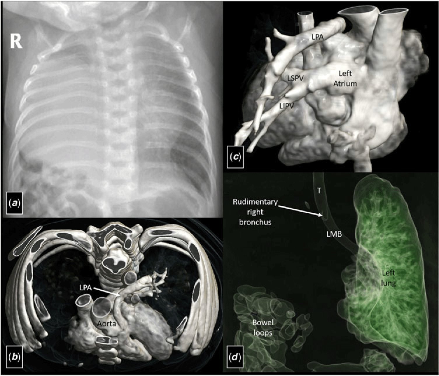
A 7-month-old boy presented with respiratory distress, cyanosis, and feeding difficulties. Oxygen saturation on room air was 78%, which improved to 83% on oxygen supplementation. Chest radiograph revealed near-complete opacification of the right hemithorax with herniation of liver and small bowel loops; however, no apparent tracheo-mediastinal shift was noted (Fig 1a). Transthoracic echocardiography established a diagnosis of tetralogy of Fallot with non-visualisation of right pulmonary artery. Patient was subsequently referred for computed tomography angiography for evaluation of the pulmonary vasculature and airways. Computed tomography angiography confirmed the diagnosis of tetralogy of Fallot with complete absence of right lung, right pulmonary artery, and right pulmonary veins. A rudimentary right-sided bronchus was, however, present (Fig 1b–d). Associated herniation of liver and small bowel loops in the right hemithorax was noted.
Chest radiograph (a) reveals near-complete opacification of the right hemithorax with herniation of the liver and small bowel loops without any apparent tracheo-mediastinal shift. Volume-rendered images (b–d) of computed tomography angiography show the absence of the right pulmonary artery and the right pulmonary veins with a rudimentary right-sided bronchus. LIPV = left inferior pulmonary vein; LMB = left main bronchus; LPA = left pulmonary artery; LSPV = left superior pulmonary vein; T = trachea.

Unilateral pulmonary agenesis is an exceedingly rare anomaly and is associated with other congenital anomalies in ~50% cases. Pulmonary agenesis is classified into three types depending on the degree of arrest of development. Type I (pulmonary agenesis) is characterised by complete absence of a lung along with its bronchus and vessels. Type II (pulmonary aplasia) shows complete absence of a lung albeit with a rudimentary bronchus present. In type III (pulmonary hypoplasia), there is partial presence of the bronchial tree along with parts of unilateral lung and its vasculature.Reference Boyden1 As development of the lung occurs in the 4th–5th week of gestation around the same time the migration of heart occurs, pulmonary agenesis is frequently associated with CHDs. To the best of our knowledge, this unique combination of anomalies in association with tetralogy of Fallot has been reported only once previously.Reference Labovsky, Hoffman and Scott2
Acknowledgements
None.
Financial Support
This research received no specific grant from any funding agency, commercial, or not-for-profit sectors.
Conflicts of Interest
None.