Introduction
Human lymphatic filariasis is a tropical disease, caused by the nematodes Wuchereria bancrofti and Brugia malayi, that infects about 128 million people worldwide (Hotez, Reference Hotez2009) and is a major cause of clinical suffering, disability and handicap (World Health Organization, 2006). The parasites are transmitted via mosquitoes and severely damage the lymphatic system. When an infective mosquito bites, L3 larvae of parasites enter the body through the skin, subsequently moult to the L4 stage within 9–14 days post infection and become adult worms in the lymphatic vessels after a period of 6–12 months.
Studies have established that immunity is operative during early infection, when the infective stage L3 larvae first encounter the host (Ramachandran et al., Reference Ramachandran, Kumar, Rami, Chinnaiah, Nutman, Kaliraj and McCarthy2004). ALT-2 represents >3% of all L3 expressed sequence tags (ESTs) in filarial genome analysis. Alt-2 transcripts are first in abundance among all L3-expressed cDNAs in the EST dataset (Gregory et al., Reference Gregory, Atmadja, Allen and Maizels2000). Abundant larval transcript (ALT) proteins are synthesized in the later phase of growth in the mosquito. The conservation of alt genes in all filarial nematodes studied so far and the very weak similarity to a single Caenorhabditis elegans locus imply that ALT products have a critical filaria-specific role.
ALT proteins have been reported to show highest reactivity against putatively immune sera and are strongly implicated in protective immunity (Ramachandran et al., Reference Ramachandran, Kumar, Rami, Chinnaiah, Nutman, Kaliraj and McCarthy2004). Their highly regulated expression, abundance and presence in excreted–secreted products of mammalian-stage nematodes have been implicated in establishing and maintaining infection (Allen et al., Reference Allen, Daub, Guilliano, McDonnell, Lizotte-Waniewski, Taylor and Blaxter2000). Further, ALT proteins have immunomodulatory function (Hoerauf et al., Reference Hoerauf, Satoguina, Saeftel and Specht2005) and their role in the evasion strategy of filarial nematodes is established (Gomez-Escobar et al., Reference Gomez-Escobar, Bennett, Prieto-Lafuente, Aebischer, Blackburn and Maizels2005).
Immunization with recombinant ALT-1 conferred 76% protection (Gregory et al., Reference Gregory, Atmadja, Allen and Maizels2000) while ALT-2 showed over 75% protection against a challenge infection in the jird model (Thirugnanam et al., Reference Thirugnanam, Pandiaraja, Ramaswamy, Murugan, Gnanasekar, Nandakumar, Reddy and Kaliraj2007) and 74% protection in a murine model (Ramachandran et al., Reference Ramachandran, Kumar, Rami, Chinnaiah, Nutman, Kaliraj and McCarthy2004). About 78% protection was observed when immunized along with thioredoxin peroxidase (TPX) as a multiple DNA vaccine (Anand et al., Reference Anand, Murugan, Prabhu, Anandharaman, Reddy and Kaliraj2008). These studies indicate that ALT is an important filarial target that needs to be fully explored. Since ALT protein has not yet been thoroughly characterized, we attempted to investigate the role of this crucial antigen by a detailed structural and epitope analysis.
We modelled the structure of B. malayi ALT-2 (BmALT-2) and analysed its motifs compared to other homologues. Further, we mapped the B epitopes of BmALT-2 to locate the potential domains contributing to its superior protective efficacy, using various immunological assays. A recombinant gene construct was engineered, containing the putative immunodominant epitopic domains of BmALT-2 while eliminating the immunosuppressive region. The prophylactic potential of the recombinant ALT epitope protein (AEP) was evaluated in Mastomys coucha, a widely used filarial animal model for the development of B. malayi (Sänger et al., Reference Sänger, Lämmler and Kimmig1981).
It is generally believed that epitope-based vaccines would be more effective if multiple epitopes covering a region of immunological hot spots are simultaneously delivered as a single chimeric construct. Identification of immunological hot spots and formulation of vaccine targets have become increasingly popular with advances in immunological and molecular biology techniques (Srinivasan et al., Reference Srinivasan, Brusic and August2004). Peptides derived from the 28-kDa glutathione S-transferase of Schistosoma mansoni showed best results with mono-epitopic tetravalent multiple antigen peptides (MAPs) (Niederhafner et al., Reference Niederhafner, Sebestík and Jezek2005). Epitope-based vaccines have been shown extensively to be promising in various diseases, capable of inducing protective immunity (Ben-Yedidia & Arnon, Reference Ben-Yedidia and Arnon1997; Srinivasan et al., Reference Srinivasan, Brusic and August2004). Peptides containing protective epitopes of malarial antigens have shown more promise than the whole recombinant antigenic proteins (Chatterjee et al., Reference Chatterjee, Wery, Sharma and Chauhan1995). Since the whole ALT protein has immunosuppressive domains, targeting this antigen using an immunodominant multi-epitope vaccine would be beneficial in filarial management.
Materials and methods
Structure analysis and modelling of B. malayi ALT-2
To analyse the structural complexity and globularity of ALT-2, the server GlobPlot (Linding et al., Reference Linding, Russell, Neduva and Gibson2003) was used. The secondary structure of ALT-2 was predicted using the PDBsum (Laskowski, Reference Laskowski2001) and PSIPRED server (McGuffin et al., Reference McGuffin, Bryson and Jones2000). The probability of crystallizing the protein was predicted by XtalPred prediction server, which compares nine of the protein's biochemical and biophysical features and gives a crystallization score (Slabinski et al., Reference Slabinski, Jaroszewski, Rychlewski, Wilson, Lesley and Godzik2007).
The model was built using HHpred (Söding, Reference Söding2005), a web server for automated comparative modelling of proteins based on the program Modeller (Fiser & Sali, Reference Fiser and Sali2003). HHpred builds a profile hidden Markov model (HMM) from a query sequence and compares it with a database of HMMs representing annotated protein families or domains with known structure (Protein Data Bank (PDB), Structural Classification of Proteins (SCOP)) used especially for those interesting groups of proteins for which no orthologue has yet been studied.
The templates Drosophila melanogaster protein giant lens (PDB ID: 3C9A), Lycopersicon esculentum fruit-specific protein (PDB ID: 2HLG_A) and yeast oligosaccharyl transferase subunit (PDB ID: 1RKL_A) were selected for building the model. This model was further refined with the graphics program Coot (Emsley & Cowtan, Reference Emsley and Cowtan2004). The geometric quality of the modelled structure was assessed by Ramachandran plot occupancy of residues using MOLPROBITY (Lovell et al., Reference Lovell, Davis, Arendall, de Bakker, Word, Prisant, Richardson and Richardson2003), PROCHECK server (Laskowski et al., Reference Laskowski, MacArthur, Moss and Thorton1993) and the structure validation tools errat (Colovos & Yeates, Reference Colovos and Yeates1993) and whatif (Vriend & Sander, Reference Vriend and Sander1993).
Epitope prediction and peptide synthesis
The bioinformatics tools BcePred (Saha & Raghava, Reference Saha and Raghava2004) and IEDB (Peters et al., Reference Peters, Sidney, Bourne, Bui, Buus, Doh, Fleri, Kronenberg, Kubo, Lund, Nemazee, Ponomarenko, Sathiamurthy, Schoenberger, Stewart, Surko, Way, Wilson and Sette2005) were used for B-epitope prediction, which utilizes the physico-chemical properties of proteins, such as hydrophilicity, flexibility and surface probability. Also, the antigenicity index predicted in the DNAstar software, which combines the other existing methods (Jameson & Wolf, Reference Jameson and Wolf1988), was used. Eight peptide regions were identified in the BmALT-2 protein sequence, predicted to carry linear B epitopes. Peptides were synthesized by solid-phase technique, using f-moc chemistry and assembled on Wang resin. Peptides were purified and the purity was analysed by high-performance liquid chromatography.
Recombinant protein purification and circular dichroism spectra
The BmALT-2 gene in the T7 expression vector pRSET B (Invitrogen, Carlsbad, California, USA) was expressed in Escherichia coli (BL21plysS). Bacterial cultures were induced with 1 mm isopropyl β-d-thiogalactoside at optical density (OD) 0.6 for 3 h. The protein expression profile was analysed on 12% sodium dodecyl sulphate–polyacrylamide gel electrophoresis (SDS–PAGE) and the recombinant protein was purified to homogeneity by hydrophobic interaction chromatography using phenyl sepharose fast flow (GE Healthcare Life Sciences, Chennai, India).
ALT-2 proteins were characterized by circular dichroism (CD) spectra in the far-UV region, which reflects the secondary structure content of the protein, such as helical structures, folded or unordered conformations. CD measurements were performed using a Jasco-600 spectropolarimeter (JASCO International Co. Ltd., Tokyo, 192-0046 Japan). Samples were scanned at a rate of 10 nm/min and were recorded at ambient temperature (~25°C) between 190 and 260 nm, using a 1-mm optical path. Spectra were corrected for background scattering. Proteins (~0.5 mg/ml) were analysed in 10 mm phosphate-buffered saline (PBS), pH 7.2. The secondary structure content of the protein was analysed using the K2D2 server (Perez-Iratxeta & Andrade-Navarro, Reference Perez-Iratxeta and Andrade-Navarro2008).
Western blotting
The expressed and purified protein was confirmed by Western blotting. Five micrograms of purified proteins were resolved on 12% SDS–PAGE and electroblotted on to nitrocellulose membrane. The membrane was blocked in 5% skimmed milk powder in PBS at 4°C overnight. After three washings with PBST (phosphate-buffered saline with Tween 20), it was incubated with mouse anti-histidine (Invitrogen, San Diego, California, USA) for 1 h at 37°C. After washing, bound antibodies were detected with alkaline phosphatase (ALP)-conjugated goat anti-mouse IgG and nitro-blue tetrazolium and 5-bromo-4-chloro-3′-indolyl-phosphate (NBT-BCIP) substrate (Sigma, St. Louis, Missouri, USA).
Mice, immunization and sera collection
Six- to eight-week-old female BALB/c (H-2d) mice were procured from TamilNadu Veterinary and Animal Sciences University, Chennai. Groups of six mice were injected via the intramuscular route with either 50 μg of peptides or 25 μg of protein, in 50 μl of PBS mixed with 50 μl of alum. The same dose of booster was given on days 7, 14 and 21. Blood was collected on days 0, 14, 28, 35 and 42. The sera were separated and stored at − 80°C.
Measurement of total IgG and IgG isotypes
Peptide-specific IgG levels in the sera were determined by enzyme-linked immunosorbent assay (ELISA). Briefly, 96-well microtitre plates (Immunlon2, Dynatech, Virginia, USA) were coated with 100 μl of peptide (500 ng/well) or protein (100 ng/well). After washing and blocking with 5% skimmed milk powder, a serial two-fold dilution (1:500–1:1,28000) of antisera was used. After three washings with PBST, ALP-conjugated anti-mouse IgG was incubated for 1 h at 37°C. The plate was developed using p-nitrophenyl phosphate substrate (1 mg/ml) (Sigma) in substrate buffer (100 mm Tris-Cl, pH 9.5, 100 mm NaCl, 5 mm MgCl2) and absorbance was read at 405 nm.
For estimating IgG isotypes, mice sera (1:100) from different immunization groups were incubated for 1 h at 37°C, with respective peptides/protein coated on ELISA plates. IgG isotype binding was detected using secondary rabbit anti-mouse IgG specific for each subclass (Pierce Isotyping kit, Pierce, Rockford, Illinois, 61105 USA) as instructed by the manufacturer. The absorbance was read at 405 nm.
Reactivity with anti-ALT sera
Reactivity of each peptide antiserum with ALT-2 protein was studied by direct binding assay. ELISA plates were coated with ALT-2 protein and optimal dilutions of peptide antisera were incubated at 37°C for 2 h. Serum from mice immunized with alum alone was used as the negative control and the mean absorbance was used as the cut-off value. Alternatively, the reactivity of ALT-2 antisera with different peptides was also performed, where ELISA plates were coated with peptides and incubated with ALT-2 antisera at 1:1000 dilution. Binding of ALT-2 with anti-ALT sera was used as the positive control in both the assays.
Establishment of ALT-2 monoclonal antibodies
Generation of hybridomas was carried out as described previously (Gangatirkar et al., Reference Gangatirkar, Gangadharan, Narendranath, Nagpal, Salunke and Karande2002). Briefly, 6- to 8-week-old female BALB/c mice were immunized subcutaneously with 50 μg of purified rALT-2 protein emulsified in Freund's complete adjuvant, followed by administration of 50 μg of the antigen in Freund's incomplete adjuvant. After 30 days, and 4 days prior to fusion, mice were injected intraperitoneally with 250 μg of antigen in saline. Spleens were excised from the mice, splenocytes were prepared as a single-cell suspension and fused with the myeloma line SP2/O-Ag14 at a ratio of 5:1 using polyethylene glycol 4000 (PEG4000) (Merck, Rahway, New Jersey, USA). Cells were then dispensed into 96-well plates in Iscove's Modified Dulbecco's Medium (IMDM) supplemented with 20% fetal bovine serum, 50 μg β-mercaptoethanol (βME) and HAT (10 mm hypoxanthine, 4 × 10− 5 m aminopterin, 1.6 mm thymidine). When hybridoma cells reached >50% confluence, the supernatant from each well was tested by ELISA for the presence of antigen-specific antibodies.
Reactivity of ALT-2 peptides and recombinant protein with three different monoclonal antibodies (Mabs) were tested. ELISA plates were coated with peptides and were incubated with different Mabs for 1 h at 37°C after blocking. Plates were developed as described above. An unrelated protein was used as a negative control and its mean absorbance was taken as the cut-off value.
Epitope gene construction and protein purification
DNA sequences of epitope domains 1–30 (EDI) and 89–128 (EDII) of ALT-2 were polymerase chain reaction (PCR) amplified using specific primers, and ligated using the BglII restriction site in between them. The ligated product was further amplified using the forward primer of EDI and the reverse primer of EDII. The amplified gene was cloned in the pRSET B vector in BamHI and EcoRI sites. The clone was confirmed by PCR and restriction analysis. The construct designated as ALT epitope protein (AEP) was sequenced and submitted to GenBank (GU596489). AEP was expressed in the E. coli salt-inducible strain (GJ1158) induced with 50 mm NaCl at OD 0.6 for 3 h. The 12-kDa fusion protein with histidine tag was analysed in a 12% SDS–Tricine gel and confirmed by Western blotting using anti-histidine antibodies. The recombinant protein was purified using Ni2+ affinity column chromatography.
Immunoreactivity and dot-blot analysis of AEP
Reactivity of anti-ALT sera with rAEP was studied by ELISA to confirm the recognition of the epitope protein by antisera raised against the whole antigen. Plates were coated with AEP and incubated with ALT antiserum (1:1000) at 37°C for 1 h following blocking and washing. After three washings with PBST, ALP conjugated anti-mouse IgG was incubated for 1 h at 37°C. Alternatively, reactivity of AEP antisera with rALT was also performed – ELISA plates were coated with ALT and incubated with AEP antisera (1:1000). Binding of proteins with respective antisera was considered as a positive control in both the assays.
To test the reactivity of antisera raised against AEP with native parasitic ALT, a dot-blot assay was performed using 1 μg of crude protein extract from B. malayi microfilariae (mf), L3 or adult parasites. Five hundred nanograms of purified recombinant protein was used as the control. After subsequent blocking and washing of the blot, antisera at appropriate dilutions were incubated for 1 h at 37°C. Anti-ALT-2 serum was used as the positive control and alum immunized serum as the negative control. After washing, bound antibodies were detected with ALP conjugated goat anti-mouse IgG and NBT-BCIP substrate (Sigma).
Mastomys for protection study, immunization and parasites
Four- to 6-week-old, male Mastomys coucha used in this study (Lok & Abraham, Reference Lok and Abraham1992) were obtained from Mahatma Gandhi Institute of Medical Sciences, India. Six mastomys were used per group. Mastomys were immunized with 50 μg of rALT-2 or rAEP protein in alum. Four doses at weekly intervals were administered intraperitoneally. The control group received PBS alone in alum. Sera, collected periodically after immunization, were used to check the antibody titre by ELISA. For B. malayi infection, mosquito colony and parasites were maintained as described previously (Thirugnanam et al., Reference Thirugnanam, Pandiaraja, Ramaswamy, Murugan, Gnanasekar, Nandakumar, Reddy and Kaliraj2007). Twelve days after infection, B. malayi L3 larvae were obtained by crushing the insects (Suzuki & Seregeg, Reference Suzuki and Seregeg1979) and carefully removing the L3 larvae. Larvae were counted by microscopy and used for the protection study.
Protection study in Mastomys coucha
The protection study was done using a micropore chamber method, as described previously (Abraham et al., Reference Abraham, Lange, Yutanawiboonchai, Trpis, Dickerson, Swenson and Eberhard1993; Chenthamarakhshan et al., Reference Chenthamarakshan, Reddy and Harinath1995). After the final booster immunization, mastomys were challenged by intraperitoneal implantation of a micropore chamber containing ten live B. malayi L3 larvae in RPMI 1640 medium (Gnanasekar et al., Reference Gnanasekar, Rao, He, Mishra, Nutman, Kaliraj and Ramaswamy2004). After 48 h the micropore chamber was harvested; the contents were removed on to a glass slide and examined microscopically for cytotoxicity. The numbers of live and dead L3 were enumerated by observing the movement of L3 under a microscope. Percentage protection was expressed as: [(average number of worms recovered from the control mastomys − average number of worms recovered from the vaccinated mastomys)/average number of worms recovered from the control mastomys] × 100.
Data analysis
All statistical analysis was done using graphpad prism software version 5 (GraphPad Software, La Jolla, California, USA). Comparisons between two individual data points were made using a Student's t-test. For multiple comparisons, the non-parametric Kruskal–Wallis test was used along with the Bonferroni's post test. A probability (P) value ≤ 0.05 was considered statistically significant.
Results
Structural analysis and modelling
Secondary-structure prediction analysis of ALT-2 showed an N-terminal α-helix (1–21), a 310 helix (68–72), two β-sheets (100–117) and random coils (see supplementary fig. S1a). Further, CD spectra of ALT-2 in the far-UV region (240–190 nm) revealed that it comprises about 10.45% α-helix and 29.09% β-strand (see supplementary fig. S1d). This is also in close agreement with the secondary structure predicted by PSIPRED (10.2% α-helix, 28.1% β-strand and 61.7% coils).
Disorder propensity plot using GlobPlot showed a highly disordered region in the variable acidic domain (27–50) of ALT-2 (see supplementary fig. S1c). Prediction of the probability of crystallization showed the highest score (class 5) for ALT-2, which indicates that ALT-2 is very difficult to crystallize due to the longest disorder region and high percentage of coil structures (60%). This led to the construction of an in silico theoretical model of ALT-2 structure.
Since there are no protein homologues with known structure available for ALT-2, homology modelling using a single template is not possible. The target–template sequence similarity is less than 30% and hence multiple templates were used to improve the model accuracy. The quality check of modelled structure by Ramachandran plot analysis showed that 85.7% (108/126) of all residues were in favoured regions and 96.0% (121/126) of all residues were in allowed regions, with five outliers (see supplementary fig. S2a).
Intriguingly, ALT-2 shows 28% sequence identity with bee venom phospholipase A2 (PDB ID: 1POC). The region 87–99 shows 50% identity with snake curaremimetic toxin (PDB ID: 1CMR) with almost complete structural similarity (see supplementary fig. S2b). This nuclear magnetic resonance (NMR) structure carries an alpha/beta scorpion fold which is shared by scorpion toxins and insect defensins. Also, the region 66–84 has 26% identity with HPTX2, a toxin from Heteropoda venatoria spider venom that blocks the kv4.2 potassium channel (PDB ID: 1EMX). Although the exact significance and function of ALT protein has not yet been elucidated, structural homology with other toxins indicates that ALT proteins are important in parasite defence and might have originally been a toxin that has been mutated.
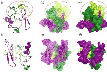
The model of ALT-2 confirms the random-coiled (green coloured), highly-disordered structure in the region 21–90, which encompasses the variable acidic domain (fig. 1a–c). The regions 1–21 and 90–128 (purple coloured) has secondary structural features as predicted and the protein is folded in such a way that these regions lie almost parallel and closer to each other, possibly forming a conformational epitope (fig. 1d–f).
Homology models of ALT-2 using HHPred, based on the program Modeller and refined using the program Coot. Different views of the model, with the predicted highly-disordered structure circled in red in the region 21–90 encompassing a variable acidic domain. Residues 22–60 have been indicated as dark green and residues 61–90 as light green. (a) and (d) Secondary structure view of the model as ribbons; (b) and (e) mesh view of the protein; (c) and (f) space-filling view of the protein. The epitopic domains of ALT-2 that form a possible conformational epitope are shown in purple (residues 1–20 and residues 91–128).

Epitope prediction, peptide and protein synthesis
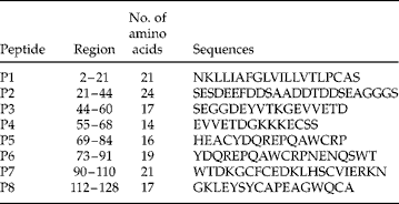
For epitope mapping, the ALT protein sequence was analysed using epitope prediction tools. Eight peptide sequences containing putative linear B epitopes predicted by three tools, i.e. BcePred, IEDB and DNAstar, were chosen for the study (table 1). Peptides were synthesized and purified to >90%. The recombinant protein ALT-2 was expressed in E. coli (BL21plysS) and purified by hydrophobic interaction chromatography to yield ~3–4 mg/l (see supplementary fig. S3a). The protein was further confirmed by Western blotting using anti-His antibody (see supplementary fig. S3b).
Sequences of selected ALT-2 peptides based on in silico epitope prediction and their corresponding position.

Antibody titre and isotypes
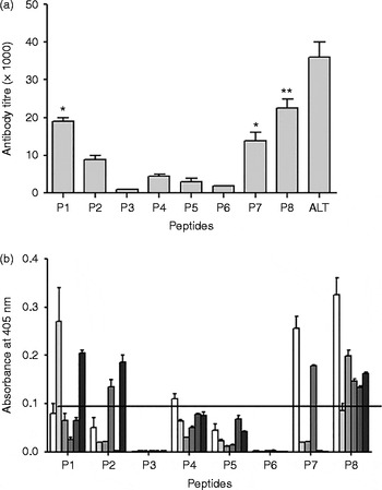
In order to determine the antigenicity of peptides, the antibody response was measured in terms of peak titres. Peptides P8, P1 and P7 induced high peak titres of 22,000, 18,000 and 14,000, respectively, in mice at the 35th day after immunization (fig. 2a). P2 showed a peak titre of ~8000–10,000, while the other peptides did not show significant antibody levels. ALT-2 protein elicited a high antibody titre of 36,000. This suggests that peptides P8, P1 and P7 are highly immunogenic and contain dominant linear B epitopes, while P2 may carry a sub-dominant epitope. Also the C-terminal peptide, P8, is capable of eliciting a potent antibody response.
(a) Peak antibody titres induced by ALT-2 peptides and whole protein in mice on the 35th day post immunization, determined by ELISA. Data represent the mean titre of six mice ± SEM. The asterisks on top of the bars indicate a significantly high antibody titre compared to control sera (P < 0.05). (b) Antibody isotype profile of ALT-2 peptides in mice sera analysed by isotype ELISA (from left to right: IgG1 (white bars), IgG2a (light-grey bars), IgG2b (mid-grey bars), IgG3 (grey bars), IgA (dark-grey bars) and IgN (black bars)). The horizontal line represents the cut-off value derived from the mean absorbance of sera from alum-immunized mice.

To analyse the type of antibody response induced by individual peptides, IgG isotypes were assayed. P8 and P7 showed a predominant IgG1 response while P1 induced high levels of IgG2a. P2 induced IgM antibodies predominantly (fig. 2b).
Reactivity with anti-ALT sera
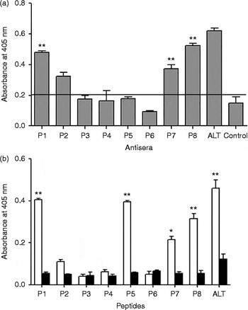
To check the binding of peptide-specific antibodies against whole protein, the reactivity of recombinant ALT-2 protein with peptide antisera was tested by direct binding assay. Antisera raised against P1, P8 and P7 showed the highest reactivity with ALT-2, in correlation with antibody titre (fig. 3a). Also, the ability of synthetic peptides to bind antisera raised against ALT-2 was assessed. ALT-2 antisera showed significantly high reactivity (P < 0.001) against P1, P5, P8 and P7 compared to alum control sera (fig. 3b). This shows that these peptides carry B epitopes recognized by ALT-2 polyclonal sera. Although P5 was recognized by polyclonal sera, it was not capable of eliciting an immune response as a linear peptide.
(a) Reactivity of recombinant ALT-2 protein with antisera raised against ALT-2 peptides, determined by direct ELISA. Anti-ALT serum was used as the positive control. The horizontal line represents the cut-off value derived from the mean absorbance of sera from alum-immunized mice. (b) The reactivity of ALT-2 peptides and protein with anti-ALT-2 sera (white bars) and alum control sera (black bars). The asterisks on top of the bars indicate a significantly high absorbance compared to control sera: *, P < 0.01; **, P < 0.001.

Reactivity of peptides with ALT monoclonal antibodies
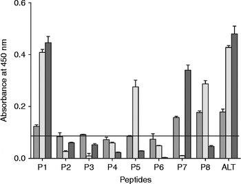
To further confirm the identified epitopes, reactivity of peptides was tested with three Mabs developed against BmALT-2 (fig. 4). Peptides P1, P7 and P8 were reactive with two of the Mabs, while peptide P5 was detected by only one Mab. This confirms that P1, P7 and P8 carry dominant epitopes while P5 may carry a less dominant epitope.
Reactivity of ALT-2 peptides and whole protein with three different ALT-2 mouse monoclonal antibodies: Mab1 (mid-grey bars), Mab2 (light-grey bars) and Mab3 (dark-grey bars). The horizontal line represents the cut-off value derived from the mean absorbance of an unrelated protein with ALT-2 Mabs.

Based on these observations, peptides P1 (2–21), P7 (90–110) and P8 (112–128) were found to carry dominant B epitopes (see supplementary fig. S4a, b). Our previous studies also showed the presence of T-cell epitopes in P1, P7 and P8 (Madhumathi et al., Reference Madhumathi, Prince, Nageswara Rao and Kaliraj2010b). The N-terminal region 1–30 and the C-terminal region 90–128 of ALT-2 carried both B and T epitopes. Hence, these two regions (1–30 and 89–128) of ALT-2 were found to be the hot spots rich in overlapping epitope clusters.
We have shown earlier that the region 55–91 of ALT-2 induces interleukin (IL)-10 secretion and thus contributes to T-cell immunosuppression in the host (Madhumathi et al., Reference Madhumathi, Pradiba, Prince, Jeyaprita, Rao and Kaliraj2010a, b). Eliminating this region may inhibit the immunomodulatory behaviour of ALT-2. Thus, we designed a recombinant multi-epitopic protein, termed ALT epitope protein (rAEP), carrying the N-terminal 1–30 amino acids, designated as epitopic domain I (EDI) and the C-terminal end 89–128, named epitopic domain II (EDII) (see supplementary fig. S4b).
rAEP construction and protein synthesis
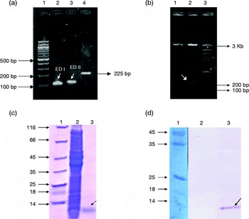
The two epitopic domains (EDI and EDII) identified in ALT-2 were cloned as a single gene to rAEP. The AEP construct carrying DNA sequences of EDI and EDII was amplified and cloned in pRSET B vector. PCR and restriction analysis showed an insert size of ~225 bp, which was confirmed by DNA sequencing (fig. 5a, b). The 12-kDa His-tagged fusion protein was expressed in E. coli (GJ1158) and purified (fig. 5c). The protein was confirmed by Western blotting using anti-His antibody (fig. 5d).
(a) Construction of the AEP gene by PCR amplification of epitopic domains using specific primers. Lanes: 1, 100-bp DNA ladder; 2, amplified product of EDI; 3, amplified product of EDII; 4, AEP gene of 225 bp, amplified from the ligation product of EDI and EDII using EDI forward and EDII reverse primers. (b) Restriction analysis using BamHI and EcoRI double digestion at 37°C for 1 h. Lanes: 1, pRSET B vector; 2, pRSET B–AEP clone, showing the insert of 225 bp and vector backbone; 3, 100-bp DNA ladder. (c) Expression of 12-kDa rAEP protein in E. coli (GJ1158) and purification analysed by 12% SDS–Tricine PAGE. Lanes: 1, protein molecular marker; 2, pRSET B–AEP in E. coli (GJ1158) cells induced with 50 mm NaCl; 3, purified AEP protein. (d) Reactivity of purified rAEP with mouse anti-His confirmed by immunoblotting. Lanes: 1, protein molecular marker; 2,3, induced culture of E. coli (GJ1158) with pRSET B vector (2) or with pRSET B–AEP clone (3).

Immunoreactivity and dot-blot analysis
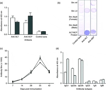
Reactivities of AEP with anti-ALT-2 sera and ALT with anti-AEP sera were assayed. Reactivities of the antigens with their respective sera were used as positive controls. Both ALT-2 and AEP proteins showed significant reactivity (P < 0.05) with anti-AEP and anti-ALT sera, respectively, compared to alum control sera (fig. 6a). Thus, the epitopic domains of AEP are recognized by ALT-2 antisera and anti-AEP sera binds the ALT-2 whole protein.
(a) Reactivity of ALT-2 (white bars) and AEP proteins (black bars) with anti-ALT-2 and anti-AEP sera, determined by direct ELISA. Alum-immunized mouse serum was used as the negative control. (b) Dot-blot of the crude extract of the filarial antigens from different stages of the parasite (mf, L3, adult (male) and adult (female)) with antisera raised against ALT-2 or AEP (1:1000) in mice. Reactivity with alum-immunized mouse serum was used as the negative control. (c) Peak antibody titres of ALT-2 (black circles) and AEP (black squares) in Mastomys coucha at the 35th day post immunization. Data represent mean peak titres of six mastomys ± SEM. (d) Antibody isotype profiles of ALT-2 (white bars) and AEP (grey bars) in mastomys.

In order to analyse the reactivity of anti-AEP sera with native parasitic proteins, dot-blot analysis was performed with crude extracts of all stages of the parasite (fig. 6b). Reactivity with anti-ALT-2 sera was used as the positive control and alum-immunized mouse sera as the negative control. Both anti-ALT-2 and anti-AEP sera showed high reactivity with crude extracts of L3 antigens but not with any other stages, as expected, since ALT protein is highly L3-stage specific. This shows the ability of antibodies raised against AEP to recognize the native protein, which is crucial for parasite clearance.
Immune response and protection study in mastomys
Mastomys were immunized with ALT-2 and AEP to compare the immunogenicity by measuring antibody titres. Both the antigens showed a peak titre of ~32,000–38,000 on day 35 (fig. 6c). Isotype analysis showed variation in the type of antibody subtypes induced by both the antigens. Interestingly, AEP showed a predominant IgG1 antibody while ALT-2 induced higher levels of IgG2b and IgG2a (fig. 6d).
Parasite clearance by ALT-2 and AEP in the immunized mastomys was evaluated by in situ micropore chamber studies. Interestingly, the epitope construct AEP showed 74.59% protection, which was significantly higher (P < 0.001) than that of ALT-2 protein, which showed 61.95% protection. The mean worm count of live L3 parasites after removal of the chambers was 2.33 ± 0.18 for AEP and 3.5 ± 0.17 for ALT, whereas the alum control showed 9.2 ± 0.20 live L3. Although ALT is known to be a potential vaccine candidate, the ability of AEP to clear parasites shows that elimination of region 31–111, which includes the immunomodulatory domain of ALT, does not affect its protective efficacy. Our study also proves that the N- and C-terminal regions of ALT-2 carry protective epitopes that are sufficient and more efficient in clearing the parasites.
Discussion
Filariasis is a disease resulting from host immune dysfunction or an erratic immune regulation triggered by the parasites residing in the lymphatics. ALT proteins are stockpiled in the oesophageal glands of infective larvae (Wu et al., Reference Wu, Egerton, Pappins, Harrison, Wilkinson, Underwood and Bianco2004) and are secreted by the parasites when they encounter mammalian culture conditions. Although their function is unknown, it is presumed that ALT may help in promoting parasite survival within the host immunological environment. ALT protein was thought to interfere with the critical first interaction between the innate immune system and the nematode invaders.
alt-like genes are present in other filarial nematode species and the proteins have unique structural features characterized by a highly variable acidic domain (9.4% D and 12.5% E residues) (22–60) and a conserved cysteine-rich domain (61–128) (Gregory et al., Reference Gregory, Atmadja, Allen and Maizels2000). It is interesting to note that this acidic domain is highly disordered and does not have intact secondary structure conformation. This structural complexity may contribute to making ALT-2 a very difficult protein to crystallize and thus the lack of any structural information. The unavailability of homologues with known structure has additionally hampered any attempt to build theoretical structure models for ALT-2. However, recent reports on the use of multiple template-based homology modelling (Sokkar et al., Reference Sokkar, Mohandass and Ramachandran2011) led us to identify such templates and to utilize the same strategy for ALT-2. The utility of multiple templates for comparative modelling combines information from many template structures and relies on the accuracy of their multiple structure alignment (Fernandez-Fuentes et al., Reference Fernandez-Fuentes, Rai, Madrid-Aliste, Fajardo and Fiser2007).
The putative ALT model revealed that the random coils in the variable acidic domain are highly disordered. A homology analysis showed that ALT-2 has 28% sequence identity with bee venom phospholipase A2. The regions 87–99 and 66–84 show ~50% and ~26% identity with snake curaremimetic toxin and spider venom toxin, respectively, although the relevance of this is not understood. The HPTX2 spider toxin belongs to the structural protein family called knottins, which are small, disulphide-rich proteins with a characteristic knot motif, and include protease inhibitors, toxins from cone snail, spider, bug, horseshoe crab and scorpion, gurmarin-like peptides and anti-microbial peptides. The knottin fold is an attractive scaffold for drug design. The regions 55–68 and 73–91 of ALT-2 were found to carry immunomodulatory domains that induced high IL-10 cytokine levels in BALB/c mice in our previous study (Madhumathi et al., Reference Madhumathi, Prince, Nageswara Rao and Kaliraj2010b). Since these immunomodulatory regions encompass sequences homologous to toxins and knottins, it could be speculated that they might have evolved from the toxin family and could be partly responsible for parasitic defence induced by ALT-2 proteins.
To characterize ALT-2 immunologically and probe its epitopic regions, we mapped the linear B epitopes of BmALT-2, using a series of peptides with overlapping sequences. Immunoinformatic analysis was carried out initially to screen the putative epitope peptides. Eight such peptides predicted to carry epitope sequences, spanning the entire protein, were chosen and synthesized.
Peptides P1 (2–21), P7 (90–110) and P8 (112–128) were found to be highly immunogenic, capable of inducing high titres in mice. P7 and P8 induced a Th2 type of response, with high levels of IgG1 subtype, and thus could facilitate parasite clearance (Lawrence, Reference Lawrence2001). It has been established that larval stages that express ALT transcripts abundantly elicit a strong Th2 response in mice (Osborne & Devaney, Reference Osborne and Devaney1998) and thus the epitopic region 90–128 of ALT is responsible for such a heightened Th2 response.
Structural analysis of ALT revealed that the regions 1–21 and 90–128 (purple coloured in fig. 1) are in proximity and might possibly form conformational epitopes. Since conformational epitopes could be identified by Mabs, we developed three ALT-2-specific Mabs. To confirm the specific binding of epitope regions, peptides were assayed with three Mabs developed against BmALT-2. Peptides P1, P7 and P8, which induced high antibody titres, were distinctly recognized by the ALT-2 monoclonal antibodies, proving the presence of dominant epitopes. Moreover, these peptides also showed high reactivity with the polyclonal anti-ALT-2 sera. Reactivity of anti-peptide sera with recombinant ALT-2 also showed similar results, which prove that the anti-peptide antibodies are able to recognize the surface epitopes of whole protein (Dong et al., Reference Dong, Qi, Ying, Chen and Chen2006) that was purified in non-denaturing conditions to avoid any adverse structural changes (Van Regenmortel et al., 2006).
In order to study if these domains are protective against the parasite, we made a gene construct carrying the putative epitopic domains (EDI: 1–30 and EDII: 89–128) and devoid of region 30–89, to eliminate the variable acidic domain and immunomodulatory regions (55–89). The immunoprophylactic efficacy of the multi-epitope protein, termed ALT epitope protein (AEP), was analysed against the infective L3 stage of the parasite in a permissive animal model for filariasis, M. coucha (Sänger et al., Reference Sänger, Lämmler and Kimmig1981, Khan et al., Reference Khan, Gaur, Dixit, Saleemuddin and Murthy2004).
Recombinant AEP induced antibody titres equal to ALT-2 protein, which proves that the dominant B epitopes alone were sufficient for immunogenicity and that elimination of other regions does not affect the antigenic potential of ALT-2. Antibody isotype analysis indicates a shift from type 1 response by ALT-2 to type 2 response by AEP. This could be due to the elimination of an immunomodulatory domain of ALT-2 that contributes to type 1 immune responses. Since parasite clearance is attributed mainly to type 2 responses in filariasis (Lawrence, Reference Lawrence2001), this could be expected to enhance the protection against challenge infection.
Further, reactivity of AEP with ALT-2 antisera and AEP antisera with ALT-2 protein shows that epitopic domains form the exposed surface or solvent-accessible regions (Khan et al., Reference Khan, Babu, Gupta and Rao2008). Although antibodies against AEP recognize the recombinant protein, it may not bind to native ALT-2 of the parasite due to changes in folding or glycosylation. Hence, the specific reactivity of L3 antigens with AEP antisera was tested by dot-blot analysis, which showed recognition of native ALT protein by anti-AEP sera.
The protective efficacy of the epitopic construct was determined by a micropore chamber experiment, which is a robust (Abraham et al., Reference Abraham, Lange, Yutanawiboonchai, Trpis, Dickerson, Swenson and Eberhard1993; Chenthamarakshan et al., Reference Chenthamarakshan, Reddy and Harinath1995) and widely used method for immunoprophylactic studies (Gnanasekar et al., Reference Gnanasekar, Rao, He, Mishra, Nutman, Kaliraj and Ramaswamy2004; Dabir et al., Reference Dabir, Dabir, Goswami and Reddy2008; Vanam et al., Reference Vanam, Pandey, Prabhu, Dakshinamurthy, Reddy and Kaliraj2009). Interestingly, AEP showed higher protective efficacy (74.59%), compared to ALT-2 (61.95%), indicating that these epitopes are protective in nature and are responsible for the protective nature of ALT-2. This encourages the use of new-generation vaccines for specific targeting in parasitic infections where the whole protein could not be used. Epitopic vaccines carrying dominant epitopes of the antigen and devoid of domains triggering immunosuppression are not only effective in protecting the host but also have an added benefit of preventing immune dysregulation. This is especially beneficial in diseases caused by malfunction of the immune system, such as lymphatic filariasis, for regulating immune function.
Peptide vaccines have become extremely popular recently, giving promising results for almost all types of diseases. B–T epitope-based vaccines containing immunodominant regions from antigens have been developed for malaria (Nardin et al., Reference Nardin, Calvo-Calle, Oliveira, Nussenzweig, Schneider, Tiercy, Loutan, Hochstrasser and Rose2001), schistosomiasis (Yang et al., Reference Yang, Jackson, Zeng and McManus2000) and many other parasitic diseases (Nardin et al., Reference Nardin, Oliveira, Calvo-Calle and Nussenzweig1995; Arnon et al., Reference Arnon, Tarrab-Hazdai and Steward2000). In addition to the insights into ALT-2 structure and identification of its epitome, our study also proves the potential of epitope-based vaccines in filariasis, which could be exploited in rational vaccine design for other parasitic diseases.
Supplementary material
To view supplementary material for this article, please visit http://dx.doi.org/10.1017/S0022149X16000055
Financial support
The authors are thankful to the Department of Biotechnology (DBT) and University Grants Commission (UGC), Government of India, for their financial assistance.
Conflict of interest
None.
Ethical standards
All experiments were performed in accordance with Indian Animal Ethics Committee regulations. Mastomys were handled in accordance with the institutional guidelines, approved by the institutional animal care committee.