Introduction
Congenital anomalies are also identified as birth defects, congenital disorders, or congenital malformations.1 Although they have accompanied man throughout his history, congenital anomalies have currently acquired an important role in morbidity and mortality since they can provoke spontaneous abortions, stillbirths, and long-term disability, thus affecting not only individuals and their families but also national health systems.1
The prevalence of congenital anomalies varies globally, ranging from approximately 3% to 6% of all live births.Reference Hobbs, Chowdhury and Cleves2 According to World Health Organization, the most common severe congenital anomalies are heart defects, neural tube defects, and Down syndrome.1 Most congenital anomalies occur during the 3rd to 8th weeks post-fertilization, but the period of greatest susceptibility is the first 2 weeks.Reference Sadler3 There are multiple mechanisms, causes, and factors involved in the development of congenital anomalies, including physical, chemical, genetic, environmental, and other unknown potential agents.Reference Ujházy, Mach, Navarová, Brucknerová and Dubovický4
Ancestrally, humans have consumed medicinal plants to cure various afflictions. It is estimated that 80% of the world population trusts traditional medicine and plant-based drug compounds to improve health.5 However, numerous phytochemical studies have shown the presence of secondary metabolites in commonly used medicinal plants, which can become pharmacologically active and may be associated with cytotoxic and mutagenic properties involved at the origin of various carcinogenic processes and the development of congenital malformations because they are easily disseminated through the placenta and fetal metabolism.Reference Macías-Peacok, Pérez-Jackson, Suárez-Crespo, Fong-Domínguez and Pupo-Perera6
Bocconia frutescens L. (1753) is a plant native to tropical America, from México to South America, and the infusion of its leaves has been traditionally used in extensive parts of the American continent for treatment of gastrointestinal disorders, skin ulcers, dermatitis, some respiratory tract infections, as well as tuberculosis.Reference Velázquez, Calzada, Torres, Gonzáles and Ceballos7,Reference Yu, Gao and Zhu8 In vitro, extract of B. frutescens L. has shown anti-malarian,Reference Chinchilla, Valerio and Sánchez9 anti-mycobacterium tuberculosis,Reference Cruz-Vega, Verde-Star and Salinas-González10 anti-leishmanial,Reference Chinchilla-Carmona, Valerio-Campos and Sánchez-Porras11 and anti-trichomonal activity.Reference Calzada, Yépez-Mulia and Tapia-Contreras12 In a rat model of high intestinal secretion induced by cholera toxin, B. frutescens L. extract exhibited potent antidiarrheal activity.Reference Velázquez, Calzada, Torres, Gonzáles and Ceballos7 Additionally, B. frutescens L. reduces peristalsis activity.Reference Calzada, Arista and Pérez13
Phytochemical analyses of B. frutescens L. have detected high levels of saponins and coumarins, followed by flavonoids, sesquiterpenlactones, alkaloids,Reference Yu, Gao and Zhu8 and cardiotonic in chloroformic, ketonic, and ethanolic extracts, whereas tannins and sterols were not detected. The effects of consuming metabolites such as alkaloids during pregnancy must be evaluated since exposure to these compounds may cause defects in developing embryos and fetuses.Reference Green, Lee, Panter and Brown14–Reference Panter, Welch, Gardner and Green16 Therefore, this study investigated the possible association between the consumption of B. frutescens L. at a dose equivalent to that consumed by humans and the development of teratogenesis in rat progeny.
Method
Collection of plant material
B. frutescens L. is usually found in the ecotonia between tropical and temperate zones. Bocconia frutescens L. (Papaveraceae family) leaves were collected in Ixhuatlancillo Veracruz (18° 53′ 32″N, 97° 8′ 51″W). Plant material was dried in an oven at 35–40° for a period of 72 h and ground in a conventional mechanical mill until a fine powder was obtained. This powder was macerated with 300 ml of methanol at room temperature, covered to protect it from light for a week and vacuum concentrated at 35–40° in a rotary evaporator (Heidolph Laborota 4001. Heidolph Instruments GmbH CO. KG, Germany). Subsequently, the product was lyophilized (lyophilizer; Labconco, Kansas City, MO, USA) at –50ºC, pressure 0.002 bar, to obtain the plant extract. For administration to the animals, the extract was diluted in canola oil (Capullo®. © 2015 ACH Foods, Guatemala) heated at 50°C as vehicle.
Animals and experimental design
The experiments were carried out with Wistar rats from the Animal Facility in the Health Sciences Unit, Universidad Veracruzana. All procedures described here fully complied with the National Guide for the Production, Care and Use of Laboratory Animals (Norma Oficial Mexicana NOM-062-ZOO-1999).
Virgin female Wistar rats, weighing 200 ± 10 g, were housed in a temperature-controlled room (22 ± 2ºC) in a 12/12-h light/dark cycle and had free access to food (Rodent Lab Chow 5001®; Purina. © 2019 Lab Diet. St. Louis, MO, USA) and water. After a habituation period of 2 weeks, rats were mated with a sexually experienced male. We considered day 0 of gestation the day that sperm cells appeared in a vaginal smear. Pregnant animals were housed individually and randomly assigned to the control group (Ctrl, n = 12) or the B. frutescens L. group (Bocf, n = 11). Bocf females were administered lyophilized extract of B. frutescens L. in a single orogastric dose of 300 mg/kg/day, using an adapted intravenous catheter of 16G × 47 mm. Ctrl rats received only orogastric-administered canola oil via. Bocconia frutescens L. extract or vehicle was administered from day 7 to day 13 of gestation, which constitutes the period of organogenesis. The body weight of each mother was recorded on gestational days 0 and 20.
Dams were euthanized by an overdose of sodium pentobarbital (120 mg/kg, i.p.) on day 20 of pregnancy (the entire gestation period in the Wistar rat is 22 ± 1 days). The two uterine horns of each rat were immediately dissected in a Petri dish and fetuses, and investing membranes were teased apart with fine jewelers’ forceps using a dissecting stereoscope (Fisher Scientific Stereomaster®. © 2016 Thermo Fisher Scientific Inc., Massachusetts, USA.). The yolk sac was isolated from the surrounding deciduas and the fetuses were removed. All fetuses were inspected to determine malformations. Half of each litter, randomly assigned, was analyzed using the Wilson technique, which allows determining internal malformations through soft tissue analysis, and the remaining fetuses were treated by the Peters technique to identify external malformations through skeletal abnormalities. In addition to malformations in the fetuses, implantation and resorption sites in the uterine horns were recorded. Growth development was obtained by measuring pup weight immediately after caesarean procedure.
Wilson technique
This technique allows the analysis of soft tissues using the technique of anomalies of cross sections by Wilson and Warkany.Reference Wilson and Warkany17 The internal malformations searched were hydrocephalus, craneomeningocele, rhinocephaly, cyclopia, exophthalmia, agnatia, micrognathia, protruding tongue, craneoschisis, gastroschisis, umbilical hernia, union of twin fetuses (Thoracopagus twins), spina bifid oculi, polydactyly, ectrodactyly, focomelia, syndactyly, coiled tail, vestigial tail, tail omission, and retarded growth. Fetuses assigned for this technique were placed in a falcon tube with 6 ml of Bouin fixative for 24–48 h. Afterwards, fetuses were washed in ethyl alcohol 70% until the yellow color was removed to ensure total elimination of picric acid. The Bouin fixative consisted of 25-ml concentrated formaldehyde, 5-ml concentrated acetic acid, and 75-ml aqueous solution of 1% picric acid diluted with distilled water. This fixative was prepared fresh every day.
Peters technique
This technique allows the observation of both cartilage and bone tissue in the same specimen.Reference Peters, Neubert, Merker and Kwasigroch18 We searched for the external malformations, extra front suture, open and elongated fontanelle, rib fusion, wavy ribs, extra ossification of thoracic centers, rib central ossification, double form of ossified thoracic centers, hypoplasia of sternebrae, centers of sternebrae, omission of sternebrae, omission of the fibula, hypoplastic fibula, bent fibula, and short tibia. Fetuses were placed in a water bath for 40 s at 56°C and then were shallow cut on the back of the shoulder blades to separate the skin. Subsequently, the abdominal cavity was opened, and fetuses were eviscerated with fine forceps and their internal organs removed. They were fixed in 96% ethanol for 48 h and then stained for 3 days in Alciano’s blue solution; after that, they were dehydrated for 5 days in absolute alcohol. The soft tissue was clarified for 1 to 2 days in a 1% solution of potassium hydroxide. The tissue was then stained for 1 day by adding Alizarina red solution. Subsequently, fetuses remained for 1 day in a 25% glycerin solution in distilled water (this step was repeated using 50% then 80% glycerin). All the fetuses were kept stained in pure glycerin for evaluation. Alciano’s blue solution, which stains cartilage tissue, consisted of mixing 80 ml of 96% ethanol, 20 ml of picric acid, and 15-mg alciano blue. Alizarina red solution consisted of 10-g potassium hydroxide and 0.1-g red alizarin in 1-l distilled water. This dye stains fetus bones for evaluation of possible congenital malformations.
Statistical analysis
The response variables were analyzed with a generalized linear model, applying a unifactorial ANOVA design with adjustment of the Poisson error distribution (variance is equal to the mean) because the data refer to counts, and we use log link to compare the control (Ctrl) and B. frutescens L. groups (Bocf).Reference Crawley19,Reference Bolker, Brooks and Clark20
The frequency of external and internal malformations was obtained from the number of events per litter. Differences between groups were considered statistically significant at α = 0.05. All analyses were performed in JMP 6.0 (SAS Institute, Inc. Cary, NC, USA, 1989–2007).
Results
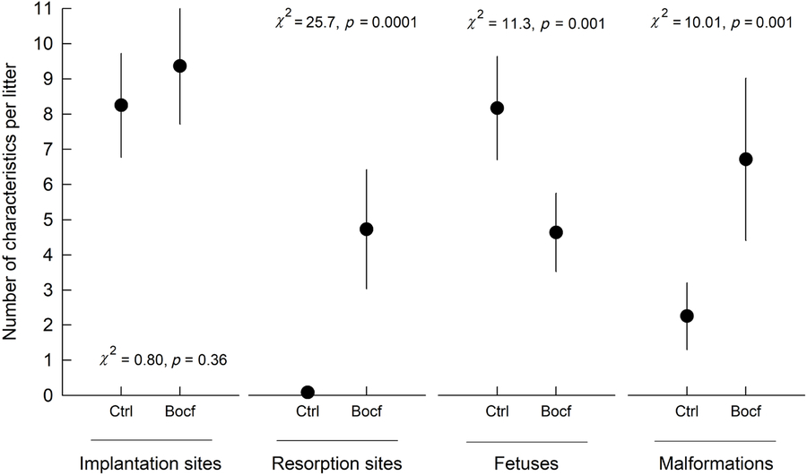
The treatment with B. frutescens L. had no effect on weight of the pregnant mothers since no difference in body weight gain from gestational day 0 to day 20 was found between the groups (p > 0.05). At gestational day 20, the number of implantation sites did not differ between Ctrl and Bocf dams (Fig. 1). However, we found that animals treated with B. frutescens L. had a lower number of fetuses, a higher number of resorption sites, and more fetal malformations per mother (Fig. 1).
Progeny features on gestational day 20. Effect of the administration of Bocconia frutescens L. 300 mg/kg/day (Bocf) during gestational days 7 to 13 on the number of implantations, resorption sites, fetuses, and malformations on the fetuses, compared to control (Ctrl) group. Data are presented as mean ± standard error of the mean.

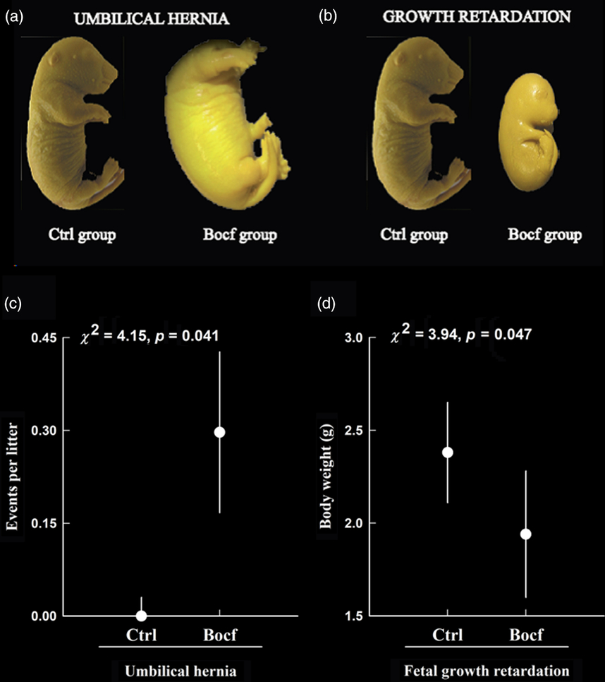
In the case of external malformations, we did not find that the B. frutescens L. treatment increased the number of damaged fetuses. However, among the internal malformations studied, we found differences in the number of fetuses with umbilical hernias (χ2 = 4.15, p = 0.041 per litter; Fig. 2a and 2c) and retarded growth (χ2 = 3.94, p = 0.047 per litter; Fig. 2b and 2d).
Main congenital malformations induced by maternal Bocconia frutescens L. orogastric administration (300 mg/kg/day; Bocf group) during pregnancy. The internal malformations found on day 20 of gestation in the Bocf group were umbilical hernia (2a and 2c) and growth retardation (2b and 2d). Data are presented as mean ± standard error of the mean.

Discussion
To our knowledge, this is the first report showing that the orogastric intake of B. frutescens L. at concentrations of human consumption during pregnancy produces teratogenic effects. Our findings may be more relevant given that women tend to consume medicinal plants more frequently during the first trimester than during the rest of the pregnancy, increasing the risk of their offspring manifesting some malformation.Reference Macías-Peacok, Pérez-Jackson, Suárez-Crespo, Fong-Domínguez and Pupo-Perera6 Furthermore, the dose of 300 mg/kg/day used in our study is closely equivalent to one cup of B. frutescens L. used by Mexican and Central American people in infusions or decoctions taken three times/day for the treatment of gastrointestinal disorder such as diarrhea.Reference Calzada, Arista and Pérez13
We showed that the administration of B. frutescens L. during the organogenesis period caused internal alterations such as umbilical hernia and growth retardation. Similar results have been described in relation to the development of internal congenital defects (neural tube defects) caused by consuming caffeine during pregnancy.Reference Ma, Qin and Wang21
The exposure of a developing embryo or fetus to alkaloids from plants, plant products, or plant extracts has the potential to cause developmental defects in humans and animals. For example, piperidine and quinolizidine alkaloids induce the inhibition of fetal movement, generally referred to as multiple congenital contracture-type deformities.Reference Green, Lee, Welch and Panter15 It has been demonstrated that the administration of rue (Ruta chalepensis L.) in pregnant albino mice causes exocephaly and spina bifida in the progeny, possibly linked to its alkaloid content.Reference Gonzáles, Benavides, Rojas and Pino22
The Bocconieae family has been described in the literature to contain more than 75 alkaloids, such as morphine, codeine, protopine, isocorydine, and tetrahydropalmatine,Reference Yu, Gao and Zhu8 but only a few have been studied for their possible teratogenic effects. Some alkaloids isolated from B. frutescens L. are also found in other medicinal plants used in China, such as Berberis vulgaris L., which has been described as a potent maternal toxin with teratogenic effects.Reference Rad, Rameshrad and Hosseinzadeh23 Berberine and sanguinarine are alkaloids present in B. frutescens.Reference Yu, Gao and Zhu8 The former has been reported to cause cytotoxicity and defects in the synthesis of DNA, and its administration can cause uterine contraction and teratogénesis.Reference Singh and Sharma24 Sanguinarine, on the other hand, even at physiological doses, can adversely influence embryonic development in rats during pre-implantation and post-implantation.Reference Vrba, Dolezel, Vicar and Ulrichová25 More studies are needed to determine whether other alkaloids contained in B. frutescens have intrauterine effects.
In summary, the results obtained in this study give scientific support to the effect of a selected medicinal plant widely used in Mesoamerican tradition. Traditional medicine is a fundamental part of developing countries, where the population still has many restrictions in terms of access to medical care, motivating the use of medicinal plants, which may increase the risk of adverse effects on health, such as those associated with teratogenesis. For this reason, there is an urgent need to test the safety of traditional medicines before recommendation and commercialization.
Acknowledgements
We are grateful to María Elizabeth Márquez López from the Faculty of Chemical Sciences in Orizaba (Universidad Veracruzana) for collecting B. frutescens L. and obtaining the extract and to José Antonio Palma Jacinto for his technical assistance in care of the animals. The authors thank to Diane Fumiko Miyoshi for her assistance in copyediting the article. The authors are entirely responsible for the scientific content of the paper.
Financial Support
This research was supported by grant PRODEP PTC-812 (VEBG). SLL was supported by CONACyT-México297211 scholarship. These funding sources were not involved in the study design, data collection, analysis or interpretation, writing the manuscript, or decision to submit it for publication.
Conflicts of Interest
None.
Ethical standards
The authors assert that all procedures contributing to this work comply with the ethical standards of the relevant national guides on the care and use of laboratory animals (Norma Oficial Mexicana NOM-062-ZOO-1999) and the work has been approved by the institutional committee (Comité Interno para el Cuidado y Uso de Animales de Laboratorio del Instituto de Ciencias de la Salud CICUAL-ICS: 2015-0017).