Introduction
“Lepraria is an important lichen genus because its species are usually dominant on shaded rock, bark, and, to a lesser extent, earth in the temperate regions of the world. Unlike most lichen genera it never produces ascomata or conidiomata and its specialized unorganized thallus is deficient in morphological characters. On the other hand, most specimens contain lichen substances, and a study of their chemistry is therefore essential in providing an understanding of the species. The genus has been little studied by modern taxonomic methods.”
Laundon (1992: 315)The repeated and independent evolution of asexual reproduction is one of the most interesting aspects of lichen biology. Although asexual reproduction via lichenized diaspores occurs throughout the diverse lineages of lichens, the genus Lepraria represents a unique group (albeit presently polyphyletic) that has lost the ability to reproduce sexually yet continues to speciate (Myllys et al. Reference Myllys, Hognabba, Lohtander, Thell, Stenroos and Hyvonen2005; Fehrer et al. Reference Fehrer, Slavíkova-Bayerová and Orange2008). The genus currently includes more than 70 species representing a heterogeneous assemblage of asexually reproducing lichenized fungi (Nelsen et al. Reference Nelsen, Lumbsch, Lücking and Elix2008; Saag et al. Reference Saag, Saag and Randlane2009; Lendemer Reference Lendemer2010a).
Historically, Lepraria has been used in an extremely broad sense as a catchall for all normally sterile lichens with leprose thalli (Laundon Reference Laundon1992), a circumscription clearly based on superficial similarity rather than shared evolutionary history. The first monograph of the group was published by Hue (Reference Hue1924), who described more than 100 new taxa based on specimens in the herbarium of Bouly de Lesdain. The destruction of de Lesdain's herbarium during World War II, coupled with the cryptic nature of Lepraria species, created an impasse in taxonomic progress that was only overcome when Laundon (Reference Laundon1992) reconstructed Hue's work using duplicates of cited specimens that survived in other herbaria. Laundon's revision of Lepraria was the first to utilize chemotaxonomy in concert with morphological characters and thus made Lepraria accessible to present-day lichenologists. My current studies of the genus draw considerable inspiration from this seminal work.
Recently, Lepraria has become the subject of considerable attention (see summary in Saag et al. Reference Saag, Saag and Randlane2009), mostly falling into one of two categories. The first being attempts to accurately circumscribe and place Lepraria s. str. within the larger framework of the Lecanoromycetes (Ekman & Tønsberg Reference Ekman and Tønsberg2002; Myllys et al. Reference Myllys, Hognabba, Lohtander, Thell, Stenroos and Hyvonen2005; Nelsen et al. Reference Nelsen, Lumbsch, Lücking and Elix2008). The second being descriptions of new taxa and documentation of range extensions (e.g., Flakus & Kukwa Reference Flakus and Kukwa2007; Knudsen & Elix Reference Knudsen and Elix2007; Lendemer & Harris Reference Lendemer and Harris2007; Saag et al. Reference Saag, Hansen, Saag and Randlane2007; Tønsberg Reference Tønsberg2007; Lendemer et al. Reference Lendemer, Knudsen and Elix2008; Lendemer Reference Lendemer2010a).
Unfortunately progress in Lepraria taxonomy following Laundon's revision has occurred in a highly haphazard and uneven fashion, resulting in a systematic framework plagued by a fundamental lack of consistency in taxonomic concepts, as well as a lack of standardized descriptive morphological terminology (Lendemer Reference Lendemer2010a). The chaotic state of present-day Lepraria taxonomy can be attributed to a diverse array of factors. Foremost among these is that due to the lack of apothecia and pycnidia, the genus is regarded as having few, if any, reliable morphological characters that can be used to differentiate taxa. With this preconception in mind, taxonomists working on Lepraria have almost universally relied upon the diversity of its secondary metabolites, above morphology and ecology, when delineating taxa, despite ample evidence that chemotaxonomy does not necessarily result in the circumscription of monophyletic entities (e.g., Ekman & Tønsberg Reference Ekman and Tønsberg2002; Tretiach et al. Reference Tretiach, Muggia and Baruffo2009). There is also mounting evidence that, contrary to popular belief, morphology, ecology, and biogeography are salient taxonomic characters in this group (e.g., Lendemer & Harris Reference Lendemer and Harris2007; Lendemer et al. Reference Lendemer, Knudsen and Elix2008; Lendemer Reference Lendemer2010a).
After several years of initial study I began work on a revision of Lepraria s. lat. in North America as the subject of my dissertation. In addition to the discovery of numerous apparently undescribed and overlooked taxa (e.g., Lendemer Reference Lendemer2010a) these studies revealed that it was almost impossible to reconcile accounts of species in the published literature. The primary reason for this was the lack of a standardized morphological terminology and descriptive scheme. This led me to develop such a system through extensive morphological studies conducted in conjunction with my revision. Though the modern roots of this system lie in the descriptions of Laundon (Reference Laundon1992), in reality they draw from the excellent historical studies of Lepraria and other sterile lichens produced by a number of lichenologists (e.g., Hue Reference Hue1924; Ozenda Reference Ozenda1963). This system was first presented by Lendemer et al. (Reference Lendemer, Knudsen and Elix2008) in the context of an extended discussion of why the term ‘soredia’ should be rejected from use in Lepraria descriptions. An improved version was then published by Lendemer (Reference Lendemer2010a). Subsequent to that publication, substantive additional morphological studies have been carried out to arrive at the treatment presented here. Many aspects of this system, however, remain theoretical because they are based on observations of character states and discrete stages of developmental processes rather than continuous laboratory growth studies. That said, my goal is to provide a conceptual framework through which the non-lichenized secondary structures in any given species can be described and placed in the broader context of tertiary thallus structure. It is hoped that the present contribution provides a simple system to facilitate dialogue in the Lepraria community, and renders this difficult genus more accessible to non-specialists.
Materials and Methods
This study is based primarily on material of Lepraria s. lat. in the herbarium of The New York Botanical Garden (NY), supplemented by material received on loan from the following herbaria: BG, CANL, COLO, CONN, DUKE, FH, GZU, H, KANU, MIN, MSC, SBBG, and UCR. Included in the holdings of this group at NY are the >1000 specimens collected by the author during his extensive field studies of Lepraria throughout North America. To date these total 1·5 years in the field and span the period August 2007 to December 2010. Through these field studies the author has been able to gather first-hand observations of ecology and morphology for all the species known to occur in North America. The chemistry and morphology of all specimens was studied using the methodology outlined in detail by Lendemer et al. (Reference Lendemer, Knudsen and Elix2008) with the SEM methods as corrected by Lendemer & Elix (Reference Lendemer and Knudsen2010).
Results: A Standardized Morphological Terminology
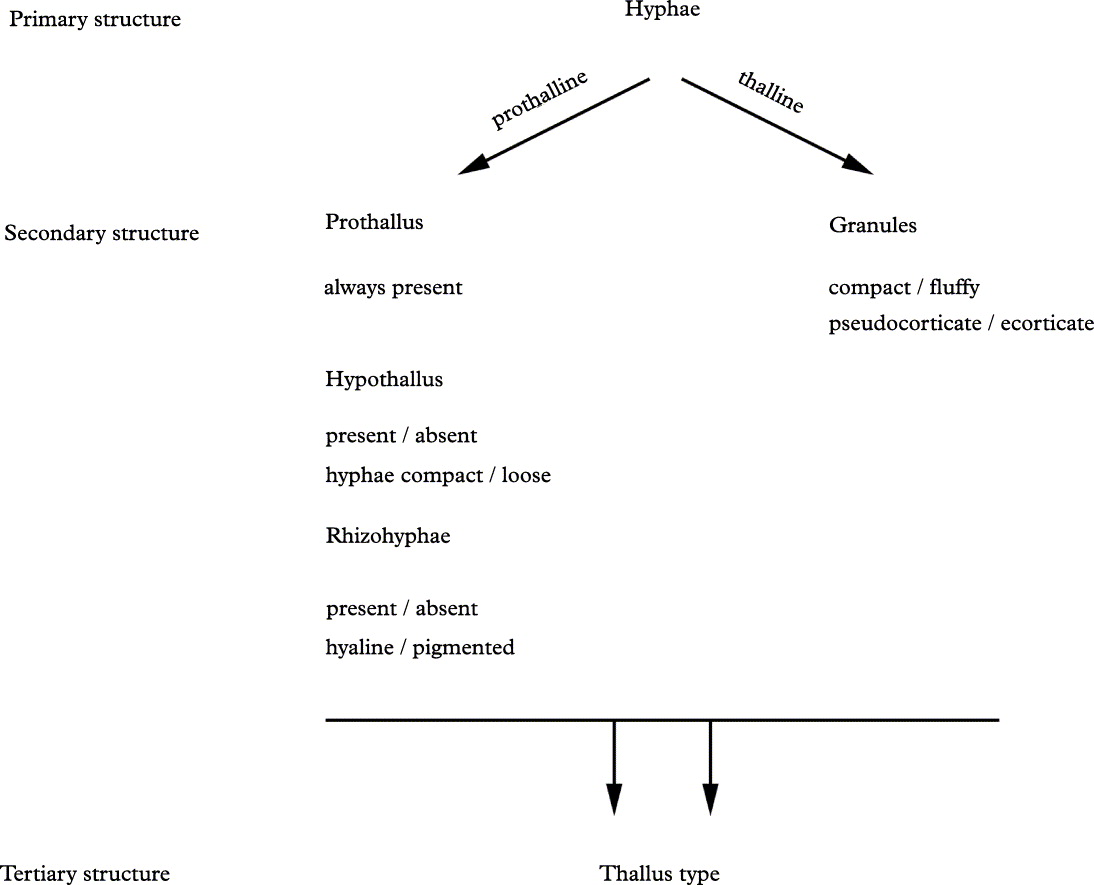
This section aims to elucidate a standardized morphological terminology for Lepraria by building the thallus from the ground up. It is divided into subsections that correspond to different scales of structural complexity similar to those used to describe biomolecular structure (i.e., primary, secondary, and tertiary structures; see Fig. 1). Employing this system of classification is advantageous because it forces anyone attempting to describe Lepraria thalli to contemplate ontogeny in addition to the morphological character states observed in any given specimen.
Schematic diagram of hierarchy of morphological terminology employed here.
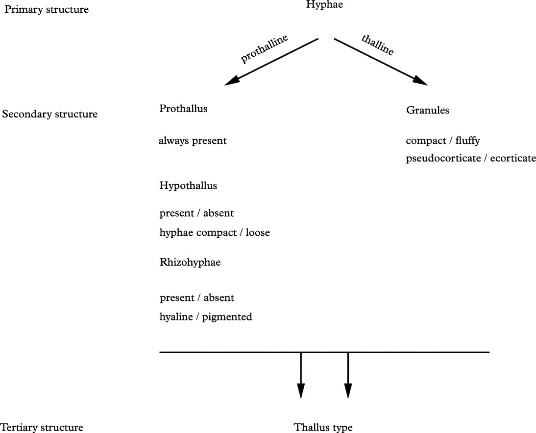
The discussion begins with the description of the smallest identifiable unit comprising the thallus, the hyphae. It continues with the description of the micromorphological structures (e.g., granules) formed by these units, and their known character states. Finally the section concludes with a survey of the thallus types displayed across Lepraria s. lat. and how these macromorphologies are derived from differing combinations of character states of a set of shared micromorphological structures.
Primary structure
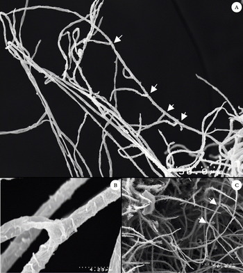
Fungal hyphae (Fig. 2) are the smallest identifiable unit of the Lepraria thallus. These are the primary structural units whose differing arrangements form all the component parts of the thallus and are integral to its ultimate macromorphology. In all the species that have been studied to date the morphology of the hyphae is uniform. To some degree this is remarkable considering the wildly polyphyletic nature of Lepraria as presently defined. There are no significant differences between the hyphae of different species within Lepraria s. str., or for that matter within Lepraria s. lat. However, differing hyphal characteristics, particularly differences in thickness, have been noted between Lepraria and members of the Roccellaceae with byssoid or leprarioid thalli (Jørgensen & Tønsberg Reference Jørgensen and Tønsberg1988; Lendemer & Knudsen Reference Lendemer and Knudsen2010).
Hyphal branching in Lepraria. A, typical secund branching as seen when isolated from the hypothallus of L. cryophila (arrows point to branches); B, detail of branch attachment from A; C, typical secund branching in the context of the hypothallus in L. finkii (arrows point to secund branching in one hypha). (A & B, Lendemer 18702; C, Lendemer s. n.).

Hyphae in Lepraria species are 3–5 µm in diameter, hyaline, septate, and have a secund branching pattern. New branches form at the septa, consistently diverging in the same direction away from the primary branch (Fig. 2). Within any given Lepraria thallus, the hyphae can be subdivided into two groups based on whether they are involved in the formation of lichenized or non-lichenized secondary structures. These subdivisions are artificial and are not derived from differences in the hyphae themselves but rather from the fundamental differences between the structures they eventually form. Those that form lichenized secondary structures (i.e., granules) are hereafter referred to as thalline hyphae. The hyphae that form non-lichenized secondary structures (i.e., the prothallus, hypothallus, and rhizohyphae) are referred to as prothalline hyphae.
The terms prothalline and thalline hyphae are introduced here to distinguish between hyphae that comprise non-lichenized and lichenized secondary structures, respectively. Although both groups of hyphae are morphologically identical, they form two fundamentally different sets of secondary structures. Thalline hyphae form lichenized secondary structures, which are those that incorporate photobiont cells. In all the species of Lepraria, granules represent the only lichenized secondary structures that have been observed. All other structures (i.e., the prothallus, hypothallus, and rhizohyphae) are formed from prothalline hyphae and do not incorporate photobiont cells.
Whether new Lepraria thalli are established through prothalline or thalline hyphae is not clear at present. It is also unknown whether prothalline hyphae are always derived from thalline hyphae, or vice-versa. Throughout this study prothalline hyphae were routinely observed extending from the thalline hyphae of granules (Fig. 6). Conversely, prothalline hyphae were neither observed anastomosing photobiont cells nor forming granules. The possibility that new Lepraria thalli can form from prothalline hyphae rather than from a lichenized diaspore cannot, however, be excluded. Each secondary structure is described below.
Secondary structure
Secondary structures represent the next level of morphological complexity in the thallus in that their form is derived directly from the patterns of organization of the primary hyphal structure. These secondary structures are the basic parts of the thallus (e.g., granules, rhizohyphae, etc.). It is important to recognize that these structures are not the smallest divisible unit of the thallus, but rather are derived from different arrangements of the primary units (i.e., hyphae).
Lichenized secondary structures
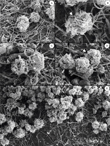
Granule division in Lepraria friabilis Lendemer. A, typical spherical granule and associated prothalline hyphae; B, initiation of vegetation division (arrow points to cleavage point); C, later stage of division; D, aggregation of divided granules (arrow points to granule undergoing further division); E, resultant tertiary structure of the thallus. (A, C & D, from Lendemer 9063; B & E, from Lendemer 8308).

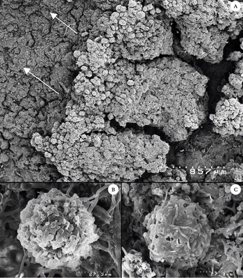
Granules are the only secondary structures in Lepraria thalli that are lichenized. These structures are always spherical or globose in shape and are composed of one or more layers of fungal hyphae that form a layer encapsulating a core of photobiont cells. In the vast majority of species studied, the granules vary from 20–50 µm in diameter. Lepraria alpina (de Lesd.) Tretiach & Barffuo, L. gelida Tønsberg & Zhurb., and L. lanata Tønsberg are notable exceptions in having enormous granules ∼1·0 mm in diameter. It is important to describe the character states of the granules observed in a single thallus as well as across different populations. In most taxa the granules can be classified either as fluffy or compact, visual features that correlate to the thickness and degree of formation of a gelatinous layer around the outer layer of hyphae (Fig. 4C vs. 5D). These characters vary between species and can vary depending on the age of the granule (Figs 4 & 5). It is tempting to refer to the outer layer of hyphae in the granules of some taxa (e.g., the L. caesioalba-neglecta group) as a cortex because it is thick and gelatinized, causing the granule to appear compact and corticate (Fig. 4). The use of the term ‘cortex’ is inappropriate, however, because the granules in all species are initially ecorticate (i.e., the photobiont core is incompletely surrounded by a network of hyphae). Instead the term pseudocortex should be employed to describe cases in which the outer layer of hyphae appears to form a cortex-like layer (Fig. 4C).
Developmental progression of the granules in Lepraria caesioalba/neglecta. A, macroscopic view of thallus illustrating progressive development of the pseudocortex and gelatinization with age, arrows indicate direction of new growth (L. caesioalba, Lendemer 17098); B, young granule with a poorly developed pseudocortex (L. neglecta, Lendemer 14020); C, senescent granule with fully developed strongly gelatinized pseudocortex (L. caesioalba, Harris 55753).

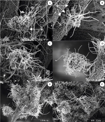
Development of granules in Lepraria rigidula. A, young granule establishing prothallus (p) on substratum, note absence of projecting hyphae; B & C, further development of prothallus (p) including some projecting hyphae not attached to the substratum; D, mature granule nearly covered in projecting hyphae; E, projecting hyphae anchor granules together to form an aggregation; F, mature granules with (right) and without (left) projecting hyphae.

Rather than growing exponentially in size the granules undergo a process of regular vegetative division (Fig. 3). It appears this process is initiated when the algal core reaches a critical size that can no longer be contained within a single continuous layer of hyphae. As the photobiont cells divide and the alga increases pressure on the wall of fungal hyphae, the hyphae begin to grow into the core and surround individual photobiont cells. Through this process the granule is cleaved apart to form a variable number of granules that begin the process anew.
Many previous workers have used differences in granule morphology to justify the circumscription of taxa. Unfortunately this has taken place while concurrently overlooking the fact that granules undergo a developmental series during which the granules of one taxon may resemble those of another. That this has led to considerable confusion in the descriptive literature is perhaps best illustrated by the use of the term ‘projecting hyphae’. This term was first introduced to describe the conspicuous hyphae that extend outward from the hyphal layer in the mature granules of Lepraria rigidula (de Lesd.)Tønsberg (Tønsberg Reference Tønsberg1992; Fig. 5 herein). As subsequent workers studied other species the term began to be employed for any hyphae extending outward from the hyphal layer of a granule, and thus was included in the descriptions of more and more taxa. Eventually the taxonomic value of the character was obscured as its usage, and concept, expanded through the literature. Through careful study of granule development across different members of Lepraria s. lat. it eventually became clear that the granules in all species produce some form of projecting hyphae at some stage of their development (Fig. 3E). It also became clear that few species retain these hyphae in the final stages of development in the manner of L. rigidula. Clearly an understanding of the developmental stages of the granules, not just the final state, in a given species should be undertaken when available material and resources permit. Similarly, this type of problem will only be avoided when descriptions include this information, regardless of its hypothetical nature, rather than simply describing the granules in an idealized state.
Previous studies have referred to the granule as the basic structural unit of Lepraria thalli (Lendemer et al. Reference Lendemer, Knudsen and Elix2008; Lendemer Reference Lendemer2010a). To some degree this is misleading because, as has been discussed above, fungal hyphae form the primary structure of the thallus. Nevertheless, the importance of the granule in Lepraria thalli cannot be underestimated because, in fact, it represents the only portion of the thallus that is actually involved in the process of lichenization. Assuming that Lepraria species reproduce through the dispersal of lichenized diaspores, then granules also represent the only means through which new thalli can be established. Thus granules have an unusual dual nature as both lichenized thallus and lichenized diaspore. Granules have also frequently been referred to as ‘soredia’ by some workers. Detailed critiques of why this is both conceptually dubious and historically divergent from descriptive morphology in lichenized fungi have been published elsewhere (Lendemer et al. Reference Lendemer, Knudsen and Elix2008; Lendemer Reference Lendemer2010a) and will not be repeated here.
Non-lichenized secondary structures
Most early morphological work in Lepraria focused on the lichenized portion of the thallus which is comprised of granules. Focusing on these structures overlooks the fact that the gross morphology, or the aspect, of the thallus is owed entirely to development of the non-lichenized portions of the thallus. The non-lichenized secondary structures in Lepraria collectively form the support structure for the granules. These structures unite the granules into a single thallus through a network of interconnecting hyphae (i.e., a prothallus or hypothallus) and serve to anchor/buffer them from the substratum through specialized attachment hyphae (i.e., rhizohyphae).
I – The prothallus
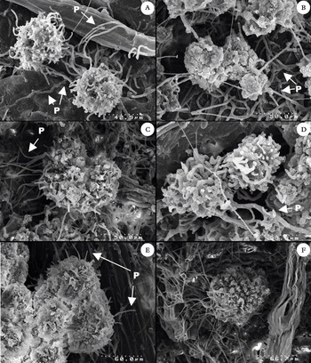
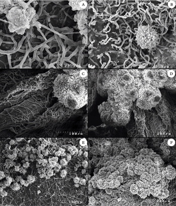
The first non-lichenized secondary structure to form during the establishment of a new thallus from a lichenized diaspore (i.e., granule) is the prothallus (Fig. 6). All species of Lepraria produce a prothallus, at least initially. The prothallus is here defined as a network comprising one or more hyphae that extend outward from the granule, attaching it directly to the substratum and to the prothalli produced by other granules. In species with an aggregate thallus (see tertiary structure below) the hyphae eventually coalesce as the granules aggregate to form piles, at which time the prothallus is only visible as a carpet of hyphae extending outward from the edge of the piles (Fig. 7). The prothallus in such species remains distinct (Fig. 7E) and can be observed throughout all stages of thallus development. In species of Lepraria with thick placodioid thalli (see tertiary structure below) the prothallus either develops into, or is replaced by, a thick layer of interconnecting hyphae (i.e., hypothallus) in which the granules are embedded (Fig. 8).
Development of the prothallus in different species of Lepraria (‘p’ points to the prothallus). A & D, L. pacifica (Lendemer 19325); B, L. caesiella (Lendemer 18773). C & F, L. caesioalba (Harris 55753); E, L. neglecta (Lendemer 14020).

Prothalli of different Lepraria species (‘p’ indicates location of prothallus). A, L. caesiella (Lendemer 18773); B, L. pacifica (Lendemer 19325); C & D, L. borealis (Hassellrot s.n.); E, L. frabilis (Lendemer 8308); F, L. neglecta (Harris 52596).

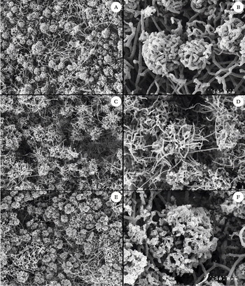
Hypothallus (left) and granules in the context of the hypothallus (right). A & B, Lepraria cryophila (Lendemer 18702); C & D, L. rigidula (Lendemer 14812); E & F, L. squamatica (Elix 37755).

II – The hypothallus
Here the term hypothallus is used to refer to the network of fungal hyphae in which the granules of species that have thick thalli are embedded (Fig. 8). As the thallus ages, the thickness of the hypothallus increases as older granules senesce and are overgrown by newly formed granules. Descriptions of the hypothallus should refer to the degree of development (thin to thick) and to the hyphal arrangement (compact or loose). The development of a hypothallus follows that of the prothallus in species of Lepraria with placodioid thalli (see tertiary structure below). Species of Lepraria with aggregate thalli (see tertiary structure below) do not produce a hypothallus.
The term ‘medulla’ has incorrectly been applied to this structure by some workers, presumably because it appears as a whitish layer just below the granules. Although it is unclear how and when the term was first used in the genus, it should be abandoned. This usage is in direct conflict with the traditional definition of the medulla as the layer of fungal hyphae below the cortex and algal layer (Kirk et al. Reference Kirk, Cannon, David and Stalpers2001). Lepraria thalli are neither corticate nor produce an algal layer, therefore they cannot have a medulla. The usage of the term hypothallus for this structure is clearly more appropriate as it linguistically and conceptually links it to the prothallus while concurrently preserving the meaning as applied in the Pannariaceae.
III – Rhizohyphae
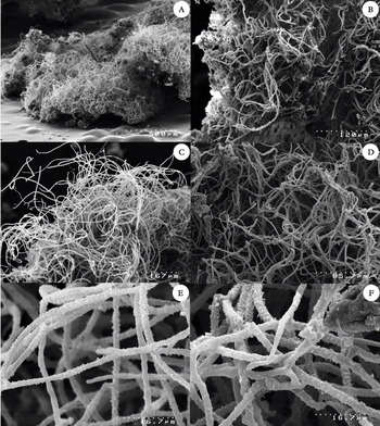
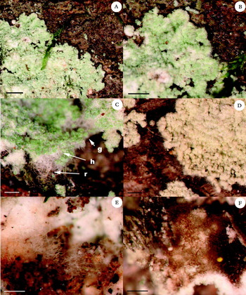
The term rhizohyphae is here applied to the fungal hyphae that extend vertically downward from the hypothallus to anchor the thallus to the substratum (Fig. 9). These structures are present only in the species that produce thick thalli with a well-developed hypothallus. They should not be confused with the hyphae of the prothallus that extend from the granules in the early stages of thallus formation (Fig. 6). In some species (e.g., Lepraria normandinoides Lendemer & R. C. Harris), the walls of the cells in the rhizohyphae are melanized, giving them a distinctive dark colour. The presence or absence of pigmentation in the rhizohyphae has been used as a character in delineating species when correlated with other traditional characters.
Rhizohyphae, gross aspect on overturned thallus (A), macromorphology (B–D), detail (E & F). A, B, D & E, Lepraria cryophila (A–D, Lendemer 11961 and E, Lendemer 18702). C & F, L. normandinoides (Lendemer 16746).

Tertiary structure
Tertiary structure represents the largest morphological scale in the descriptive terminology outlined here and is best understood as the gross morphology, or aspect, of the thallus. It is defined by the arrangement, combination, and character states expressed in the secondary structures of the thallus. Based on the species of Lepraria studied to date, there are seven basic types of tertiary structure, each of which is outlined below. These types can be classified into two broad categories based on whether a hypothallus is present or absent. Species that lack a hypothallus have discontinuous thalli comprised of granules that are dispersed on a prothallus and aggregate into piles. Conversely species that produce a hypothallus have continuous thalli comprised of granules embedded in a well-developed network of prothalline hyphae. The former group is referred to as having ‘aggregate’ thalli while the latter group is referred to as having ‘placodioid’ thalli.
Any given specimen or species of Lepraria should be able to be classified using the scheme presented here, but it should be recognized that this scheme is preliminary. It should also be noted that shared tertiary structure does not a priori indicate a close evolutionary relationship. Such theoretical relationships can only be established using a combination of biogeographical, chemical,ecological, and morphological characters thatare correlated with molecular characters.
Type 1 – Aggregate thalli
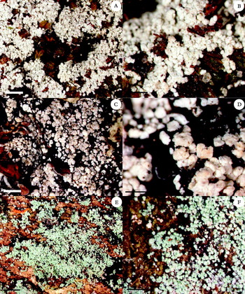
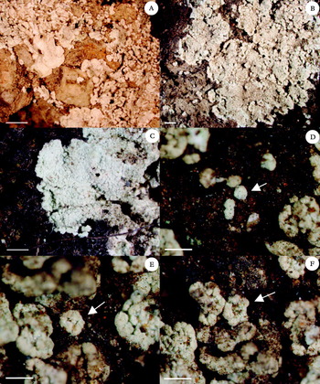
Species of Lepraria with aggregate thalli are characterized by a thin thallus that begins as isolated, dispersed granules that form piles, or aggregations, as they divide (Figs 10B, 11). Aggregate thalli are best recognized by examining the thallus margins where the dispersion of the young granules is most readily observed (Figs 11E–F). In this thallus type the piles of granules eventually overlap and become confluent, giving the false appearance of a continuous thallus in areas of older growth (Fig. 11A). Such areas can become thick and then misinterpreted as a hypothallus. This feature is best described as a necrotic layer that develops through the build-up of dead granules underlying theuppermost layer of lichenized granules. So far three subtypes of aggregate thalli have been observed. These subtypes are distinguished primarily by the degree of development of a pseudocortex and the size of granules.
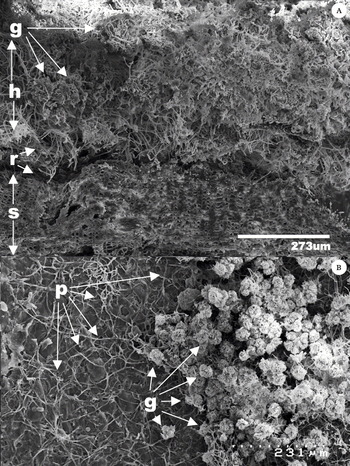
Tertiary structure in Lepraria. A, section of thallus of L. finkii (Lendemer 22897); B, thallus of L. friabilis (Lendemer 8308) (g = granules, h = hypothallus, p = prothallus, r = rhizohyphae, s = substratum).

Subtypes of aggregate thalli. A & B, caesioalba-type exemplified by Lepraria borealis (holotype, Tønsberg 1519, BG); C & D, alpina-type exemplified by L. alpina (isoneotype, Willard s.n., NY); E & F, caesiella-type exemplified by L. caesiella (Lendemer 18767, NY). Scales: A, C & E = 1·0 mm; B, D & F = 0·5 mm.

Subtype 1 – Caesioalba-type (Fig. 11A–B). Typified by Lepraria caesioalba (de Lesd.) J. R. Laundon.
This type of aggregate thallus is found in members of the Lepraria caesioalba/ L. neglecta group and is characterized by the dense, compact granules with a well-developed pseudocortex. When growing in exposed habitats these species produce rosette-forming thalli that grow in a radial fashion and appear placodioid. The absence of a conspicuous hypothallus in such cases indicates that the thallus is not truly placodioid (see Type 2 below).
Subtype 1a – Alpina-type (Fig. 11C–D). Typified by Lepraria alpina (de Lesd.) Tretiach & Baruffo.
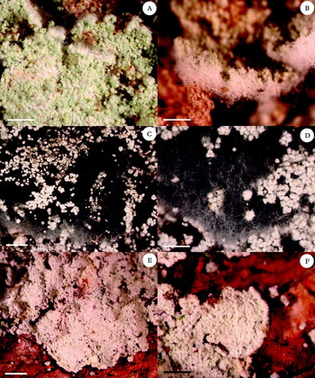
Subtypes of placodioid thalli. A & B, cryophila-type exemplified by L. cryophila (Lendemer 11961, NY); C & D, cryophila-type exemplified by L. yunnaniana (Goffinet 10077, NY); E & F, normandinoides-type exemplified by L. normandinoides (Lendemer 18674, NY). Scales: A, C & E = 1·0 mm; B, D & F = 0·5 mm.

Subtypes of placodioid thalli in Lepraria, finkii-type (g = granules, h = hypothallus, r = rhizohyphae). A–C, finkii-type exemplified by L. finkii (Lendemer 25433, NY); D, finkii-type exemplified by L. membranacea (Lewis 450, CANL); E, pale rhizohyphae of L. finkii (Lendemer 25433, NY); F, dark rhizohyphae of L. membranacea (Lewis 450, CANL). Scales: A = 1·0 mm; B = 0·5 mm; C–F = 0·25 mm.

Subtypes of placodioid thalli in Lepraria, xerophila-type. A, xerophila-type exemplified by Lepraria xerophila (Knudsen 7624, NY); B & C, xerophila-type exemplified by L. larrainiana (Lendemer 15936, NY); D–F, development of thallus through the marginal expansion of the granules, arrows point to developing granule (Lendemer 15936, NY). Scales: A–C = 1·0 mm; D–F = 0·5 mm.

Lepraria alpina and L. gelida are two species with caesioalba-type thalli that differ from other members of the L. caesioalba/ neglecta group in having larger granules (≥ 100 µm vs. 30–50 µm diameter).
Subtype 2 – Caesiella-type (Fig. 11E–F). Typified by Lepraria caesiella R. C. Harris.
This type of aggregate thallus is found in many members of Lepraria s. str. and is characterized by loose, ± fluffy granules that do not develop a distinct pseudocortex with age. The type species of Lepraria, L. incana (L.) Ach., displays this thallus type, as does the common North American species L. caesiella.
Type 2 – Placodioid thalli
Species of Lepraria with placodioid thalli are characterized by a thick thallus with discrete margins and a well-developed hypothallus in which the lichenized granules are embedded (Fig. 10A). Placodioid thalli are best recognized by examining the thallus margins to determine whether they are discrete and distinctly separated from the substratum, indicating the presence of a hypothallus (Figs 12F, 13). In many species with placodioid thalli a distinct fuzz of hyphae projects outward from the growing edge (Figs 12F, 13D). The majority of species with placodioid thalli also produce rhizohyphae which are best observed by overturning a portion of the thallus with a razor blade (Fig. 13E & F) or by cutting a section through the thallus with a fresh razor blade (Fig. 10A). So far four subtypes of placodioid thalli have been observed (Figs 12–14). These subtypes are distinguished primarily by characteristics of the hypothallus (loose vs. compact) and the development of a pseudocortex on the granules.
Subtype 1 – Cryophila-type. Typified by Lepraria cryophila Lendemer (Fig. 12A–D).
This type of placodioid thallus is characterized by a hypothallus composed of loosely intertwined hyphae, granules that do not develop a pseudocortex, the production of a weft of rhizohyphae on the lower surface, and thallus margins that are not distinctly ‘crisped’ with a lip.
Subtype 2 – Finkii-type (Fig. 13). Typified by Lepraria finkii (Hue) R. C. Harris (= L. lobificans auct.).
This type of placodioid thallus is characterized by a hypothallus composed of closely intertwined hyphae, granules that do not develop a pseudocortex, the sparse production of rhizohyphae on the lower surface, and thallus margins that are not distinctly ‘crisped’ with a lip.
Subtype 3 – Normandinoides-type (Fig. 12E & F). Typified by Lepraria normandinoides Lendemer & R. C. Harris.
This thallus type is characterized by a compact hypothallus composed of closely intertwined hyphae, granules that develop a poorly defined pseudocortex, the production of abundant rhizohyphae on the lower surface, and thallus margins are distinctly ‘crisped’ with a lip.
Subtype 4 – Xerophila-type (Fig. 14). Typified by Lepraria xerophila Tønsberg.
This thallus type is characterized by a compact hypothallus composed of tightly intertwined hyphae, granules that form a well-developed pseudocortex, the absence of rhizohyphae on the lower surface, and thallus margins that are distinctly ‘crisped’ with a lip. Notably this thallus type has only been found in species that occur on stabilized soil in regions with a Mediterranean climate. It is represented by L. xerophila in North America, L. isidiata (Llimona) Llimona & A. Crespo and L. santosii Argüello & A. Crespo (= L. lobificans Nyl. s.str. ?) in Europe, and L. larrainiana Lendemer in South America. Species with a Xerophila-type thallus appear to have a unique ontogeny in which the thallus initially forms through the apical unfolding of a granule (Fig. 14D–F; Tretiach et al. Reference Tretiach, Muggia and Baruffo2009; Lendemer Reference Lendemer2010b). Molecular studies are currently underway to determine whether these taxa form a monophyletic group.
Key to the known thallus types in Lepraria s. lat
1 Thallus aggregate-type [hypothallus absent; granules dispersed on a prothallus, eventually aggregating to form piles] ... 2
Thallus placodioid-type [hypothallus present; granules embedded in a hypothallus, always connected and forming a continuous thallus] ... 4
2(1) Granules ecorticate, appearing fluffy ... caesiella-type
Granules with a well-developed pseudocortex, appearing compact ... 3
3(2) Granules <50 µm diameter ... caesioalba-type
Granules >100 µm diameter ... alpina-type
4(1) Thallus margins with a discrete ‘crisped’ lip ... 5
Thallus margins without a discrete ‘crisped’ lip ... 6
5(4) Rhizohyphae present; granules ecorticate, appearing fluffy ... ... normandinoides-type
Rhizohyphae absent; granules with a well-developed pseudocortex, appearing compact ... xerophila-type
6(5) Hypothallus composed of loosely arranged hyphae; rhizohyphae forming a well-developed waft ... cryophila-type
Hypothallus composed of tightly arranged hyphae; rhizohyphae not forming a well-developed waft ... finkii-type
Richard Harris and Kerry Knudsen are thanked for their numerous discussions and extensive commentary on the manuscript. Martin Kukwa and Tor Tønsberg reviewed the manuscript. Mike Baxter is thanked for his help preparing samples for SEM studies. Unlimited access to the SEM was made possible through Lehman College of the City University of New York (CUNY). This study was made possible in part by funding from the following sources: California Lichen Society (CALS), City University of New York (CUNY), New York Botanical Garden (NYBG), Southern Appalachian Botanical Society (SABS), Torrey Botanical Society (TBS), and Western Pennsylvania Conservancy (WPC).