Introduction
Vocal cord palsy (VCP) can be the initial and sole symptom of head and neck cancer (HNC).Reference Chen, Jen, Wang, Lee and Lin1 Since time to treatment plays a crucial role in cancer survival,Reference Chen, King, Pearcey, Kerba and Mackillop2 an efficient diagnostic workup is essential. Malignancies have been reported to account for as much as 57 per cent of VCP aetiologies.Reference Chen, Jen, Wang, Lee and Lin1, Reference El Badawey, Punekar and Zammit-Maempel3–Reference Mehlum, Faber, Grøntved and Andersen5 However, a broad range of neurological, traumatic and other benign conditions may also cause VCP.Reference Chen, Jen, Wang, Lee and Lin1 Considering the variety of causes of VCP, various imaging modalities are used in the diagnostic investigation. Additionally, even after a comprehensive examination, the aetiology might remain unknown, categorizing the VCP as idiopathic.Reference Chen, Jen, Wang, Lee and Lin1
An optimal protocol for investigation and follow-up of VCP has been discussed for decades without a consensus on the initial diagnostic approach or the extent and duration of the follow-up in cases of indeterminate aetiology.Reference Bilici, Yildiz, Yigit and Misir6–Reference MacGregor, Roberts, Howard and Phelps9 Despite this apparent lack of agreement upon practices, it is of great importance to identify malignancies or other severe, treatable causes of VCP.
The aim of this retrospective study was to assess the diagnostic yield of imaging modalities utilized in probing the causes of VCP with initially unknown aetiology and to evaluate the performance of an extensive follow-up protocol in order to improve future investigation and optimize the use of resources.
Methods
The patients
In 2007, the Danish National Board of Health, in collaboration with national multidisciplinary cancer groups and the Danish Regions, introduced fast-track clinical pathway solutions to reduce unnecessary waiting times. This initiative was founded on so-called “package solutions”, which incorporated pre-scheduled slots for outpatient assessments, imaging and diagnostic surgical procedures.Reference Roennegaard, Rosenberg, Bjørndal, Sørensen, Johansen and Godballe10, Reference Sorensen, Johansen, Gano, Sørensen, Larsen and Andersen11 Based on the well-known risk of underlying malignancy, all patients with a newly diagnosed VCP are referred to this fast-track programme. Patients with VCP are diagnosed by flexible fiberoptic laryngoscopy or videostroboscopy. These examinations are supplemented with a chest X-ray (CXR) or computed tomography (CT) of the neck and chest, ultrasound (US) of the neck and magnetic resonance imaging (MRI) of the neck and brainstem. If no pathology is identified, the patient is categorized with idiopathic VCP. In our department, the CT of the neck and chest was replaced by full-body positron emission tomography (PET-CT) in 2012.
If no cause was determined during the initial investigation and the VCP persisted, patients underwent repeated clinical phoniatric follow-ups and after six months new imaging; CT of the neck and chest and/or MRI of neck and brainstem to detect potential cases of late-manifesting extra-laryngeal disease.Reference Mehlum, Faber, Grøntved and Andersen5
In 2020, the guidelines for the fast-track program for HNC were revised by the Danish Health Authority. Since 1 October 2020, it has been mandatory to conduct a US of the neck and either a CT of the neck and chest or a PET-CT for all patients entering the fast-track program if no other obvious causes of VCP are found during the patient´s initial consultation.
Study design
In this retrospective study, we reviewed the records of all patients diagnosed with VCP at the Department of Otorhinolaryngology - Head and Neck Surgery, Odense University Hospital (OUH), Denmark, over a 12-year period from 1 January 2005, to 31 December 2016.
The study received approval from the Regional Scientific Ethical Committee for Southern Denmark (17/21199) and the National Committee on Health Research Ethics (3-3013-2237/1).
Data sources
The primary data source was patient medical records. All relevant records were identified and accessed on-site at OUH using the International Classification of Diseases, version 10 (ICD-10) codes for VCP (J38.0, J38.01 and J38.02).
Data collection
Patients’ medical records were retrospectively reviewed by two members of the research team. Data were extracted on the following variables: age at diagnosis, gender, side of VCP, symptoms, date of the initial consultation regarding VCP, imaging modalities utilized, diagnostic findings, follow-up setup and final conclusion regarding cause of VCP. All data were compiled into a secure, access-logged database using Research Electronic Data Capture (REDCap) tools, hosted at Odense Patient Explorative Network (OPEN) at OUH.
Study population
During the study period, we identified 678 patients diagnosed with VCP. Of these, 173 (26 per cent) were eligible patients with an initially undetermined aetiology. The diagnosis of VCP was established based on findings from flexible laryngoscopy or rigid videostroboscopy.
Inclusion criteria
Patients diagnosed with VCP where on evident cause was discerned following the initial clinical assessment.
Exclusion criteria
Patients aged below 16 years, cases with direct association between surgical trauma and the onset of VCP and patients previously diagnosed with cancer invading structures along the course of the affected recurrent nerve. In case an obvious relation between surgical iatrogenic trauma and VCP was apparent, further diagnostic imaging was deemed redundant, leading to the exclusion of such cases from the study.
Statistical analysis
The diagnostic yield was calculated as the proportion of procedures with pathological findings to the total number of procedures performed. Descriptive statistics were utilized to describe demographic data. The Mann–Whitney U test and chi-squared test were employed for the comparison of continuous and categorical variables, respectively. The statistical tests were two-sided, with a p value of less than 0.05 considered statistically significant. All statistical analyses were conducted using Stata 17 (StataCorp. 2021. College Station, TX: StataCorp LLC).
Results
Out of 173 eligible patients, 156 (90 per cent) presented with unilateral VCP, with 111 (71 per cent) being left-sided and 45 (29 per cent) right-sided. The remaining 17 patients (10 per cent) had bilateral VCP. The mean age was 62 (range 16–92) years. On average, patients were followed for seven months (range 0–36). Patient characteristics are presented in Table 1.
Characteristics of patients with VCP of unknown aetiology after initial clinical examination

Note: Percentages in the benign and malignant columns should be interpreted horizontally.
* Specific benign diagnosis or idiopathic after investigation.
mo. = month; n = number; VCP = vocal cord palsy.
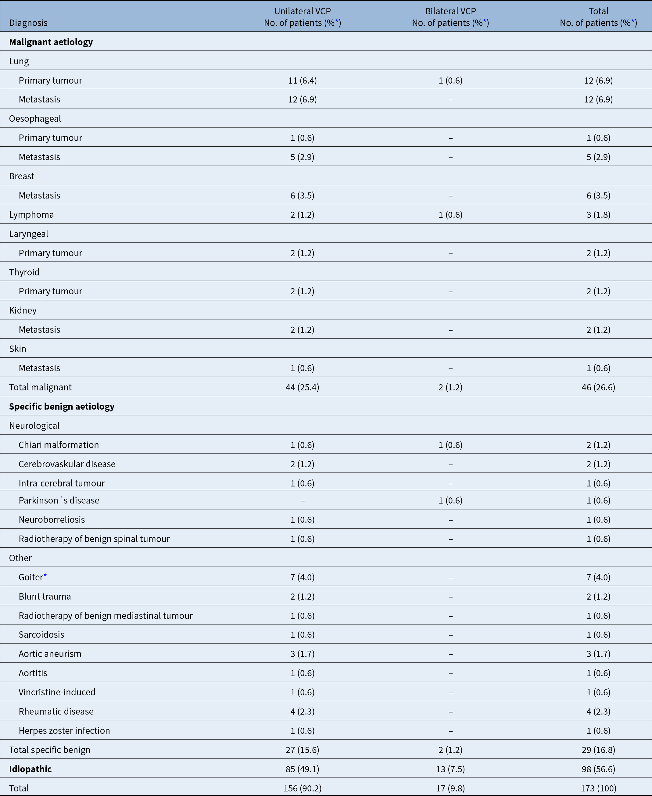
After completing imaging examinations, both initial and follow-up, 46 of the 173 patients (27 per cent) were diagnosed with malignant disease. Men had a one-in-three probability of a malignancy, while women had a one-in-five probability. The aetiologies of VCP are detailed in Table 2.
Aetiology of VCP

Note: Percentage of the 173 patients with VCP.
* Subsequent hemithyroidectomy in 6/7 cases, all with benign histology.
VCP = vocal cord palsy.
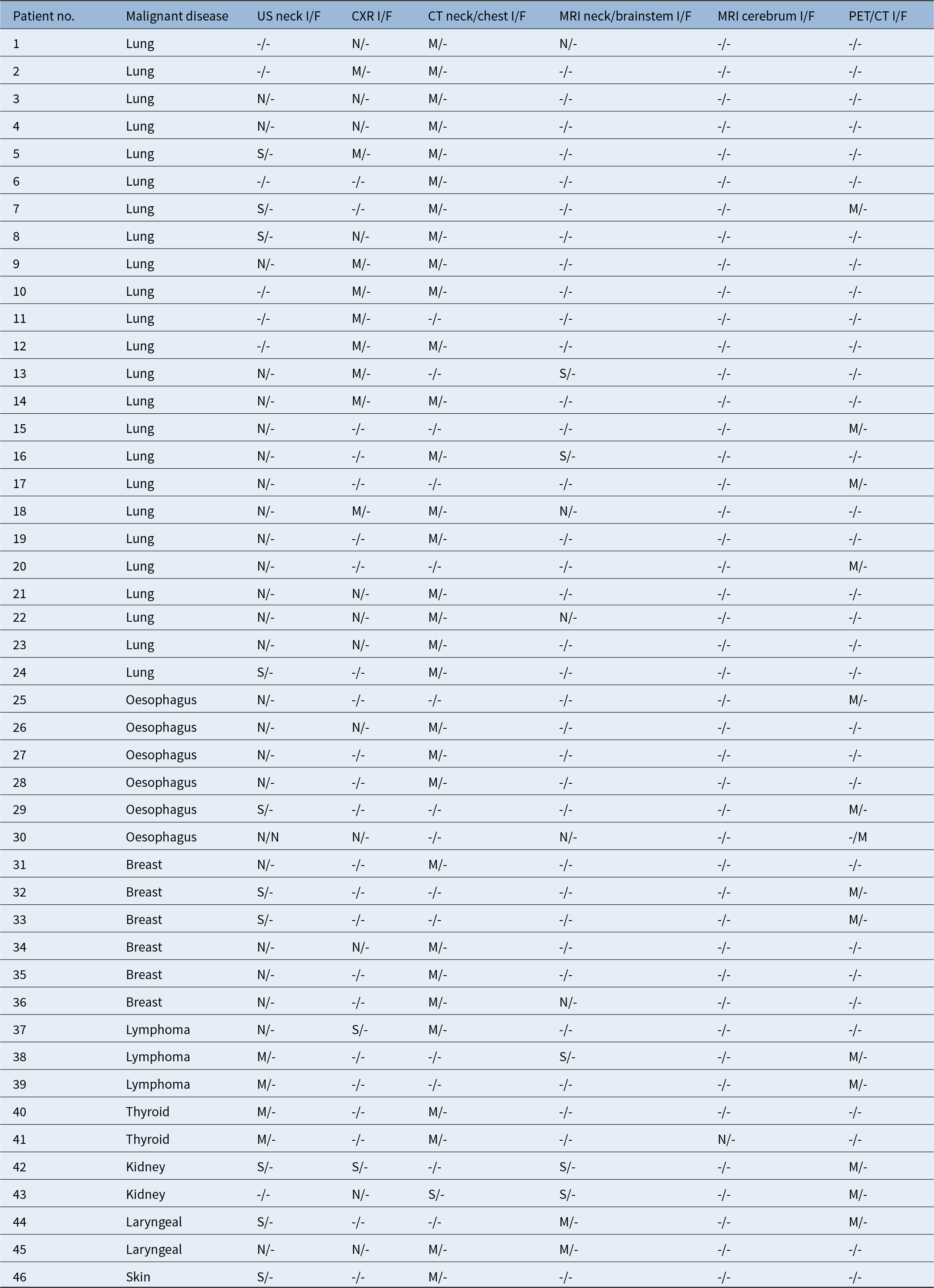
The majority of the malignancies originated from the lungs, 24/46 (52 per cent), followed by the breast, 6/46 (13 per cent) and the oesophagus, 6/46 (13 per cent), while the remaining 10/46 (22 per cent) patients had miscellaneous cancers. Disseminated disease to the neck and/or mediastinal lymph nodes were identified in 14/24 patients with lung cancer, 6/6 patients with breast cancer, 4/6 patients with oesophageal cancer, 2/2 patients with kidney cancer, 1/2 with thyroid cancer and 1/1 with skin cancer. Table 3 provides a comprehensive outline of the imaging modalities used to diagnose malignancy in each patient.
Diagnostic imaging results at initial and follow-up examinations in patients with malignant aetiology of VCP

- = imaging modality not applied; CT = computed tomography; CXR = chest x-ray; F = follow-up examination; I = initial examination; M = malignant diagnosis found; MRI = magnetic resonance imaging; N = no malignant diagnosis found; PET/CT = positron emission tomography–computed tomography; S = suspicious findings; US = ultrasonography; VCP = vocal cord palsy.
A total of 127 (73 per cent) patients either had a specific benign aetiology or remained idiopathic without signs of malignancy during follow-up. In 16/173 (9 per cent) patients with VCP, diagnostic imaging found a specific causative structural benign disorder: 5 with intra-cerebral disease (2 patients with Chiari malformation, 2 with cerebral apoplexy and 1 with a meningioma), 4 patients with aortic disease and 7 with goiter, of which 6 patients underwent hemithyroidectomy. In 13/173 (8 per cent) patients with normal imaging findings, VCP was assumed to be caused by medical conditions comprising infectious disorders, Parkinson’s disease, radiotherapy complications, neck trauma and vocal fold fixation due to rheumatoid arthritis. The cause of VCP was classified as idiopathic in the remaining 98/173 (57 per cent) patients.
In patients with unilateral VCP, malignancies were detected in 32 per cent of those with left-sided and 18 per cent with right-sided VCP.
For the subgroup with bilateral VCP, 2 out of 17 patients had malignant diseases (lymphoma and lung cancer), 2 had specific benign diseases (1 Chiari malformation and 1 Parkinson’s disease) and 13 were idiopathic.
In our study, the majority (89 per cent) underwent neck US, followed by CT of the neck and chest (68 per cent), MRI of the neck and brainstem (57 per cent), CXR (47 per cent), PET-CT (23 per cent) and cerebral MRI (8 per cent). Diagnostic imaging resulted in diagnoses in 62/173 (36 per cent) patients. The CT of the neck and upper chest, as well as the PET-CT, demonstrated the highest overall diagnostic yield at 36 per cent and 35 per cent, respectively. In the patients who underwent PET-CT, no pathological aetiologies underlying VCP were detected that were not identified on CT of the neck and upper chest. For patients presenting with specific benign disorders, the CT of the neck and upper chest and MRI of the neck and brainstem outperformed other modalities with a diagnostic yield of 14 per cent and 9 per cent, respectively. While the MRI proved pivotal in identifying cerebral lesions, such findings were infrequent. In the subset of patients diagnosed with malignant conditions, the CT of the neck and upper chest and the PET-CT achieved the highest diagnostic yield at 97 per cent and 100 per cent, respectively. Notably, in a case involving a patient with disseminated kidney cancer (patient no. 43, Table 3), both the CT and MRI revealed potential metastatic lesions in the left lung and right jugular foramen. However, the subsequent PET-CT was instrumental in pinpointing the primary kidney lesion. The remaining imaging modalities rendered considerably lower diagnostic yield when identifying underlying malignancies. Table 4 delineates the diagnostic yields associated with each employed imaging modality.
Diagnostic yield of the imaging modalities

CT = computed tomography; CXR = chest x-ray; MRI = magnetic resonance imaging; PET/CT = positron emission tomography–computed tomography; US = ultrasonography.
Following the initial imaging, 100/173 (58 per cent) patients remained diagnostically unresolved. Of these patients, 42 patients regained normal vocal fold mobility upon follow-up, 11 patients declined further examination, 4 patients failed to appear and 1 patient had died due to heart disease. Of the remaining 42 patients, 10 showed partial improvement in their VCP, while 32 persisted with immobility. Follow-up imaging for these 42 patients identified extra-laryngeal pathology in two cases. For one patient (no. 30, Table 3), the initial imaging examination included neck US, CXR and MRI of the neck and brainstem. However, a follow-up CT of neck and upper chest detected a proximal oesophageal carcinoma. The second patient initially underwent US and CT of neck and upper chest. On follow-up, an MRI of the cerebrum revealed a cerebellopontine angle meningioma. Notably, these two patients were diagnosed in 2005 and 2007, a time when PET-CT and MRI of the neck and brainstem were not yet incorporated into the standard preliminary assessment.
Upon reviewing the medical records of patients with VCP categorized as idiopathic, we found no indications of late-emerging conditions that could be associated with the onset of VCP.
Discussion
We retrospectively evaluated a cohort of 173 patients with VCP of initially unknown aetiology and evaluated the use and diagnostic yield of the imaging modalities applied at initial assessment and during follow-up.
Our cohort consisted of 90 per cent unilateral VCP (64 per cent left-sided and 26 per cent right-sided) and 10 per cent bilateral VCP, a distribution comparable to other studies.Reference Bilici, Yildiz, Yigit and Misir6, Reference Paddle, Mansor, Song and RA8, Reference Terris, Arnstein and Nguyen12, Reference Seyed Toutounchi, Eydi, Golzari, Ghaffari and Parvizian13 Malignant aetiology was diagnosed in 27 per cent of our cases, a specific benign aetiology in 16 per cent, leaving 57 per cent of our cohort with idiopathic VCP.
The most frequently reported malignancies causing VCP are thyroid, pulmonary and oesophageal carcinomas, as well as mediastinal metastasisReference Seyed Toutounchi, Eydi, Golzari, Ghaffari and Parvizian13–Reference Wang, Chen, Lou, Yang, Hu and Shieh16, consistent with our findings. The reported proportion of idiopathic VCP varies from 3 to 76 per cent,Reference Mehlum, Faber, Grøntved and Andersen5, Reference Terris, Arnstein and Nguyen12, Reference Kang, Roh, Lee, Jung, Choi and Nam14, Reference Wang, Chen, Lou, Yang, Hu and Shieh16 reflecting the discrepancies in investigation approaches. Diagnostic tools should possess high sensitivity for detecting occult diseases, but cost-effectiveness must also be considered. CXR, CT, MRI and neck US have been applied in the investigation of VCP for decades.Reference MacGregor, Roberts, Howard and Phelps9, Reference Kang, Roh, Lee, Jung, Choi and Nam14
CT of neck and upper chest is considered standard care when evaluating patients with VCP.Reference Kang, Roh, Lee, Jung, Choi and Nam14 In our study, we observed a total diagnostic yield of CT of the neck and upper chest of 36 per cent, compared to 97 per cent in the subgroup diagnosed with malignancy. Comparable findings have been observed in several other studies. In a prospective study, Gupta et al.Reference Gupta, Varshney, Bist and Bhagat4 reported on 120 patients with vocal fold paralysis, wherein a CT from the base of skull to the chest revealed malignancy in 34 per cent of the patients. Bilici et al.Reference Bilici, Yildiz, Yigit and Misir6 reported diagnostic yields for CT of the neck and chest of 24.5 per cent and 30.9 per cent, respectively. However, Paddle et al.Reference Paddle, Mansor, Song and RA8 presented a case series with chart review determining the diagnostic yield of CT in patients with presumed idiopathic unilateral vocal fold paresis, reporting a diagnostic yield of just 2.9 per cent in a study of 174 patients, thus arguing against routine CT. This opinion was supported by the retrospective study of Badia et al.,Reference Badia, Hillel, Shah, MM and Klein17 who reported a diagnostic yield of CT of neck and chest of a mere 1.7 per cent in 60 patients with presumed idiopathic, unilateral vocal fold paresis.
Bilateral VCP is more commonly attributed to lesions in the central nervous system, especially in the medulla oblongata, such as demyelinating diseases, infections and both benign and malignant intra-axial tumours.Reference Dankbaar and Pameijer18 MRI provides superior resolution, especially in the brainstem and skull base compared to CT,Reference Bilici, Yildiz, Yigit and Misir6, Reference Kang, Roh, Lee, Jung, Choi and Nam14 making it ideal for detecting lesions in the central nervous system causing VCP. In our study, an MRI scan of the neck and brainstem was conducted in all patients with bilateral VCP, while 84/156 (54 per cent) patients with unilateral VCP underwent an MRI scan of the neck and brainstem. We observed a total diagnostic yield for MRI of the neck and brainstem of 10 per cent (10/99). Additionally, MRI contributed to the detection of metastatic neck disease in five patients. In our cohort, an MRI of the cerebrum was performed only in 14 patients, with a diagnostic yield of 7 per cent (1/14). Between 2012 and 2016, an MRI scan of the neck and brainstem was conducted in 35/41 (85 per cent) patients.
PET-CT has been established as an effective and sensitive diagnostic tool for detection for malignancies, demonstrating a significant capability to rule out malignancy in patients with VCP.Reference Rohde, Nielsen, Pareek, Johansen, Sørensen and Diaz19, Reference Thomassen, Nielsen, Lauridsen, Blomberg, Hess and Petersen20 However, in certain lesions, it can be challenging to differentiate between benign and malignant aetiologies.Reference Heller, Meltzer, Fukui, Rosen, Chander and Martinelli21, Reference Minamimoto, Kubota, Morooka, Ito, Mitsumoto and Okasaki22 Asymmetrical fluorodeoxyglucose uptake in vocal folds is a well-known finding in patients with VCP.Reference Badia, Hillel, Shah, MM and Klein17 Increased uptake in the contralateral, unaffected vocal fold is often observed due to heightened physiological activity serving as a compensatory mechanism.Reference Dankbaar and Pameijer18
In a systematic review by Rohde et al.Reference Rohde, Dyrvig, Johansen, Sørensen, Gerke and Nielsen23 that compared the diagnostic accuracy of PET-CT to standard conventional imaging for patients with head and neck squamous cell carcinoma, the meta-analysis showed a pooled sensitivity and specificity for PET-CT of 89.3 per cent and 89.5 per cent, respectively. In contrast, standard conventional imaging displayed a sensitivity of 71.6 per cent and specificity of 78.0 per cent. Thomassen et al.Reference Thomassen, Nielsen, Lauridsen, Blomberg, Hess and Petersen20 conducted a retrospective study on 65 patients referred for PET-CT due to paresis or paralysis of one or both vocal folds. The results of the PET-CT were compared against clinical evaluations and histopathological findings. Their findings underscored the utility of PET-CT, showing a sensitivity of 100 per cent and highlighting its efficacy in excluding malignancy. Importantly, biopsy-confirmed malignancy (either newly diagnosed or relapsed) was identified as the cause of VCP in 42 per cent of these patients. However, it is worth noting that, in 12 per cent of cases, the PET-CT results were false positive, leading to a series of potentially unnecessary investigations.
During our study period, new guidelines for the diagnostic evaluation of VCP were implemented in 2012, leading to a varied imaging approaches within our cohort. From January 2012 to December 2016, 38/41 (93 per cent) patients with VCP of undetermined aetiology underwent PET-CT examinations during this timeframe (data not presented). In our analysis, the full-body PET-CT showed the highest diagnostic yield of 100 per cent in the 46 patients with malignant diseases, though the overall yield was 35 per cent.
In Denmark, neck US is a routine procedure in all ENT departments and is incorporated into the fast-track evaluation during the initial examination.Reference Mehlum, Faber, Grøntved and Andersen5 US offers high sensitivity and serves as a precise imaging tool for differential diagnosis of neck pathologies, especially thyroid pathologies.Reference Wang, Chen, Lou, Yang, Hu and Shieh16 In cases where thyroid lesions are located deep within in the posterior part of the thyroid lobe, VCP can be the primary manifestation. The strength of US lies in its accessibility, cost-effectiveness and lack of radiation exposure. Nevertheless, potential inter-observer variability should be acknowledged.Reference Wang, Chen, Lou, Yang, Hu and Shieh16 Our study found the diagnostic yield of neck US to be 7 per cent (11/155) in all patients. Still, it was 24 per cent (10/41) in patients with malignant neck and/or mediastinum conditions, 67 per cent for lymphoma cases (2/3) and 100 per cent for all thyroid diseases (9/9). This suggests that patients with thyroid lesions might not require additional imaging. Bilici et al.Reference Bilici, Yildiz, Yigit and Misir6 reported a diagnostic yield of neck US at 26.2 per cent, advocating for neck US and CT of the chest as initial assessment tools for unilateral vocal fold paralysis. Wang et al.Reference Wang, Chen, Lou, Yang, Hu and Shieh16 reported the significance of neck US, including the thyroid gland, as the primary imaging approach for unilateral vocal fold paralysis.
As for follow-up protocols, no consensus has been reached and various strategies have been suggested throughout the years. El Badaway et al.Reference El Badawey, Punekar and Zammit-Maempel3 followed 62 patients, all of whom underwent initial CT of the neck and chest without discernible pathological findings. Clinical evaluations were performed with six-month intervals, and no additional positive findings were observed during the follow-up period spanning 18 to 66 months. In a review, Tsikoudas et al.Reference Tsikoudas, Paleri, El-Badawey and Zammit-Maempel24 argued that patients with initial CT scans without pathological findings should receive subsequent follow-up without additional imaging, unless specific symptoms emerged. In a retrospective study, Noel et al.Reference Noel, Jeffery and Damrose7 found that 90 per cent of 207 patients with idiopathic VCP maintained their diagnosis after 60 months with 42 per cent of the cohort monitored beyond a year. They suggested annual clinical evaluation, resorting to imaging only when new symptoms manifest. In our study, 42 of 173 patients (24 per cent) were qualified for imaging follow-up. Among these, two with persistent symptoms were diagnosed with disease during follow-up. One patient’s CT of the neck and chest unveiled an oesophageal carcinoma, which had not been initially CT-scanned. For another patient, an MRI of the cerebrum found a benign cerebral tumour during the follow-up. In summary, the diagnostic yield at six months follow-up was below 5 per cent. Importantly, we did not discover any late-emerging malignancies disease in the idiopathic group. This insight is pivotal as it spares patients from extensive examinations, associated concerns and can help curtail healthcare expenditures.
In 2021, as a result of this current study, our regional guideline for follow-up underwent a revision. Now, CT scans of the neck and chest and MRIs of neck and brainstem are not routinely performed at the six-month follow-up but only when clinically indicated.
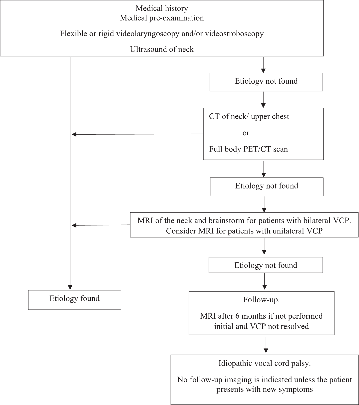
Based on our results, we suggest the workup of patients with unilateral and bilateral vocal fold palsy as illustrated in Figure 1.
Our recommended assessment for a patient with newly diagnosed VCP includes the following steps. PET/CT = positron emission tomography–computed tomography; VCP = vocal cord palsy.

• The optimal protocol for investigation and follow-up on vocal cord palsy (VCP) with undetermined aetiology has been discussed for decades.
• Of the 173 patients with VCP in our study, 27 per cent were diagnosed with malignancy.
• In 45 out of 46 cases, malignancies were detected during the initial imaging investigation.
• Post-initial imaging, 58 per cent of patients had an unresolved diagnosis.
• A total of 42 patients underwent follow-up imaging, identifying 2 additional patients with extra-laryngeal pathology, in both cases, the initial imaging procedure had been incomplete.
• CT of the neck and upper chest, along with full-body PET-CT, demonstrated similarly high diagnostic yields for identifying underlying extra-laryngeal pathology.
Strengths and limitations
During our study period, revised guidelines for the diagnostic evaluation of VCP were implemented, leading to varied imaging examinations within our cohort.
Upon reviewing the medical records of patients in the idiopathic group, we did not identify any additional cases of late-onset disease that could have precipitated VCP. We consider the extended five-year follow-up period to be one of the strengths of our study.
Conclusion
In our study, CT of the neck and upper chest and PET-CT demonstrated comparable diagnostic yields in detecting underlying extra-laryngeal pathology. While our data did not reveal an advantage, MRI remains a suitable modality for identifying lesions within the central nervous system responsible for VCP. We suggest that patients with initial normal findings on CT, PET-CT or MRI be monitored without subsequent imaging unless new relevant symptoms manifest.
Acknowledgements
None.
Financial support
This research received no specific grant from any funding agency, commercial or not-for-profit sectors