Introduction
Non-spicular taxa constitute a significant portion of the biodiversity of living sponges. The non-spicular demosponges (subclasses Verongimorpha and Keratosa, sensu Morrow and Cárdenas, Reference Morrow and Cárdenas2015) are ~8.2% of known sponge species, equaling the contribution of the class Calcarea (8.4%), and surpassing that of the classes Hexactinellida (7.2%) and Homoscleromorpha (1.25%) (van Soest et al., Reference van Soest, Boury-Esnault, Hooper, Rützler, de Voogd, Alvarez, Hajdu, Pisera, Manconi, Schönberg, Klautau, Picton, Kelly, Vacelet, Dohrmann, Díaz, Cárdenas, Carballo, Ríos and Downey2018). In addition, some homoscleromorphs are also non-spicular (e.g., Gazave et al., Reference Gazave, Lavrov, Cabrol, Renard, Rocher, Vacelet, Adamska, Borchiellini and Ereskovsky2013; Domingos et al., Reference Domingos, Lage and Muricy2016). Although the absence of mineral skeletons in living demosponges and homoscleromorphs is probably apomorphic (Maldonado, Reference Maldonado2009; Philippe et al., Reference Philippe, Derelle, Lopez, Pick, Borchiellini, Boury-Esnault, Vacelet, Renard, Houliston, Quéinnec, Da Silva, Wincker, Le Guyader, Leys, Jackson, Schreiber, Erpenbeck, Morgenstern, Wörheide and Manuel2009; Botting et al., Reference Botting, Cárdenas and Peel2014), it is still in debate whether the common ancestor of sponges was spicular (e.g., Botting and Muir, Reference Botting and Muir2018) or non-spicular (e.g., Wood, Reference Wood2011; Luo, Reference Luo2015). However, due to the low fossilization potential of non-biomineralized tissues, the evolutionary history of non-spicular sponges is still largely unexplored. Thus, any new discovery may influence our understanding of the evolutionary history of these clades.
The several species of Vauxia (family Vauxiidae, order Verongiida) from the middle Cambrian of Laurentia have been regarded as the earliest record of non-spicular demosponges (Rigby, Reference Rigby1986; Rigby and Collins, Reference Rigby and Collins2004; Botting and Muir, Reference Botting and Muir2018), along with later Phanerozoic fossils preserved in carbonates (e.g., Luo and Reitner, Reference Luo and Reitner2014, Reference Luo and Reitner2016; Lee et al., Reference Lee, Chen and Chough2015; Park et al., Reference Park, Lee, Hong, Choh, Lee and Lee2015; Friesenbichler et al., Reference Friesenbichler, Richoz, Baud, Krystyn, Sahakyan, Vardanyan, Peckmann, Reitner and Heindel2018). Ehrlich et al. (Reference Ehrlich, Rigby, Botting, Tsurkan, Werner, Schwille, Petrášek, Pisera, Simon, Sivkov, Vyalikh, Molodtsov, Kurek, Kammer, Hunoldt, Born, Stawski, Steinhof, Bazhenov and Geisler2013) confirmed that the skeleton of Vauxia gracilenta Walcott, Reference Walcott1920 is composed of chitin, consistent with the skeletal composition of modern verongiids. Recently, a new vauxiid genus, Angulosuspongia Yang et al., Reference Yang, Zhao, Babcock and Peng2017a, was described from the Kaili Biota (Miaolingian, Wuliuan), which is slightly older than the Burgess Shale. These fossils possess an anastomosing, fibrous skeleton, which is claimed to have been exclusively composed of silica (Yang et al., Reference Yang, Zhao, Babcock and Peng2017a, Reference Yang, Zhao, Babcock and Pengb). However, without a phylogenetic framework of vauxiid sponges and other closely related spicular taxa, it is difficult to interpret the evolutionary significance of these fossils.
Here we describe a new vauxiid sponge from the Chengjiang Biota (Cambrian Series 2, Stage 3), with a preserved carbonaceous endosomal skeleton. This material exhibits intermediate morphological characters between other Vauxia species and Angulosuspongia, although the data is still insufficient to establish a phylogenetic framework for these taxa.
Materials and methods
Fossils were collected from the upper part of the Maotianshan Shale Member, Yu'anshan Formation at the Malong Section during a one-month excavation. The location and sedimentary sequence of this site have been described in detail in Zhao et al. (Reference Zhao, Hu, Caron, Zhu, Yin and Lu2012). The age of the fossil layer was correlated to the uppermost part of the Eoredlichia-Wutingaspis Zone (Wang et al., Reference Wang, Zhang, Holmer, Hu, Wang and Li2012), meaning that the investigated materials belong to the Chengjiang Biota, but are slightly younger than the massively occurring, exceptionally preserved fossils in the Haikou and Chengjiang areas. The latest radiometric dates of detrital zircons from the base of the Chengjiang Biota in the Chengjiang area yielded a maximum deposition age of 518.03 ± 0.69/0.71 Ma (Yang et al., Reference Yang, Li, Zhu, Condon and Chen2018).
Fossils were observed and photographed using a Nikon SMZ18 microscope coupled with a Nikon DS-Ri2 camera. Chemical composition of the fossils was investigated using a HITACHI SU3500 scanning electron microscope (SEM) equipped with an energy dispersive X-ray spectrometer (EDS) in the Nanjing Institute of Geology and Palaeontology, Chinese Academy of Sciences (NIGPAS), and a Horiba Jobin-Yvon LabRam-HR 800 UV confocal Raman spectroscopy at the University of Göttingen (excitation wavelength 488 nm).
Repository and institutional abbreviations
Types and figured specimens examined in this study are deposited in the Nanjing Institute of Geology and Palaeontology, Chinese Academy of Sciences (NIGPAS), Nanjing, China.
Systematic paleontology
Class Demospongiae Sollas, Reference Sollas1885
Order Verongiida Bergquist, Reference Bergquist1978
Remarks
The spelling of the order name follows the suggestion of Morrow and Cárdenas (Reference Morrow and Cárdenas2015), which changes “Verongida” to “Verongiida.” This spelling has been widely accepted (van Soest et al., Reference van Soest, Boury-Esnault, Hooper, Rützler, de Voogd, Alvarez, Hajdu, Pisera, Manconi, Schönberg, Klautau, Picton, Kelly, Vacelet, Dohrmann, Díaz, Cárdenas, Carballo, Ríos and Downey2018).
Family Vauxiidae Walcott, Reference Walcott1920
Diagnosis
Thin-walled tubular to conical sponges, with or without branches. Single- or double-layered skeleton composed of fibrous elements, which fuse to produce a net. Discrete spicules absent. The meshes in the single-layered form or the endosomal meshes in the double-layered form are typically hexagonal or polygonal. Dermal layer, if present, is thin, irregularly or regularly reticular, often supported by radial fibers.
Remarks
This family contains two genera: Vauxia Walcott, Reference Walcott1920 and Angulosuspongia Yang et al., Reference Yang, Zhao, Babcock and Peng2017a. The presented diagnosis is emended from that of Yang et al. (Reference Yang, Zhao, Babcock and Peng2017a) for the reasons discussed below. The terms “dermal” and “ectosomal” in this diagnosis and the text below are following paleontologists’ usage since Rigby (Reference Rigby1986), and not necessarily identical to biological terminology.
The family Vauxiidae was initially described as dictyonine hexactinellids with a single genus, Vauxia (Walcott, Reference Walcott1920; de Laubenfels, Reference de Laubenfels and Moore1955), and later re-interpreted as a specialized offshoot of protospongiids (Finks, Reference Finks1960) or as lithisitid demosponges (Rigby, Reference Rigby1980). Rigby (Reference Rigby1986) re-investigated and illustrated the skeletal structures of the species in this family and established that: (1) the skeletal network of these sponges is composed of cored fibers like those in extant verongiids, instead of jointed triactinal or tetraxial spicules as previously described; and (2) the skeletons exhibit two differentiated layers connected by radial fibers—a relatively regular endosomal layer with polygonal meshes and an irregular dermal layer. Since then, “non-spicular” became a diagnostic character of the family Vauxiidae, which distinguishes it from similar taxa with fibrous skeletons (e.g., members of family Hazeliidae de Laubenfels, Reference de Laubenfels and Moore1955).
Botting et al. (Reference Botting, Muir and Lin2013) observed that part of the skeleton of V. bellula Walcott, Reference Walcott1920 is composed of discrete elements and interpreted those as small monaxons. However, the diagnosis of family Vauxiidae was not emended in this study. Due to the suggested similarity between V. bellula and Hazelia conferta Walcott, Reference Walcott1920, even if this observation is confirmed by further paleontological and taphonomical studies, it would still require a phylogenetic analysis to determine whether to keep V. bellula in the family Vauxiidae and change the family definition, or to remove V. bellula from this family.
Yang et al. (Reference Yang, Zhao, Babcock and Peng2017a) established the genus Angulosuspongia for newly discovered sponge fossils which exhibit an anastomosing skeleton composed of siliceous “spicules.” This work modified the family diagnosis of Rigby (Reference Rigby1986) by removing the word “keratose” and adding “spicules” to the description of skeletal components. In spite of the question whether it is appropriate in theory to resolve a “spicular” taxon under the family Vauxiidae, the figures in Yang et al. (Reference Yang, Zhao, Babcock and Peng2017a, Reference Yang, Zhao, Babcock and Pengb) actually did not show any proper structure indicating the existence of discrete skeletal elements (i.e., spicules). Thus, Angulosuspongia does comply with the old diagnosis of family Vauxiidae in exhibiting a fibrous skeleton, although the origin of silica in those fibers remains unknown.
In addition, although Angulosuspongia was described as an organism possessing a double-layered skeleton, the two layers were neither detailed in the text in how they differentiate from each other, nor identifiable from the illustrations. On the contrary, the illustrated specimens seem to show a compacted, single-layered skeleton (Fig. 3.3). The radial fibers of Angulosuspongia seem to have a free end and do not support any skeletal layer, unlike the radial fibers in other Vauxia species.
Therefore, the family diagnosis is modified in this paper from that of Yang et al. (Reference Yang, Zhao, Babcock and Peng2017a) with the following changes: (1) the expression “cored fibers or spicules” (Yang et al., Reference Yang, Zhao, Babcock and Peng2017a, p. 1336) is changed to “fibrous elements;” (2) “single-layered form” is added; and (3) “often” is added in front of “(dermal layer) supported by radial fibers,” because the new species described below does not have the supporting radial fibers.
Genus Vauxia Walcott, Reference Walcott1920
Type species
Vauxia gracilenta Walcott, Reference Walcott1920.
Vauxia leioia new species
Figures 1, 2, 3.2
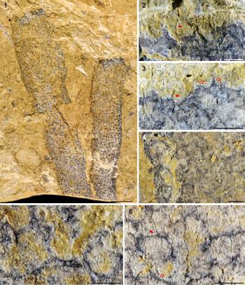
Vauxia leioia n. sp., NIGPAS 169083a and b, holotype: (1) overview of NIGPAS 169083a; areas in rectangles enlarged in (2, 3) and (5, 6); (2, 3) pinnacle-like and fibrous protrusions at the spongocoel margins, indicated by hollow and solid arrows, respectively; (4) a broken edge on NIGPAS 169083b showing that endosomal fibers penetrating into sediments; the well-focused part on the left is on a higher focal plane than the right part; (5, 6) endosomal and dermal skeletons; solid arrow in (6) indicates dermal fibers diverging from an endosomal fiber, hollow arrow indicates where dermal fibers imprints an endosomal fiber. Scale bars = 3 mm (1); 0.5 mm (2–6).

The same area in Vauxia leioia n. sp., NIGPAS 169083a, holotype, observed using (1) stereo microscope and (2) SEM (20.0kV, mode B.3 + UVD); showing the different preservation states of the endosomal and dermal skeletons. Scale bars = 0.3 mm.

Skeleton architecture of three sponge species in the family Vauxiidae: (1) Vauxia gracilenta, USNM 66515, lectotype, drawn after the photo at https://burgess-shale.rom.on.ca, and courtesy of the Smithsonian Institution; indication of colors: red, endosomal skeleton; blue, radial fibers; black, dermal skeleton; (2) Vauxia leioia n. sp., NIGPAS 169083a, holotype; indication of colors: red, endosomal skeleton; black, dermal skeleton; (3) Angulosuspongia sinensis, GTBM17-765a, holotype, drawn after fig. 2b in Yang et al., (Reference Yang, Zhao, Babcock and Peng2017a); not all fibers can be clearly traced in the photo, the blue color indicates fibers that are beneath the others; arrow indicates a node at the edge, from which both blue and black fibers diverge. Scale bars = 1 mm.

Holotype
Sample number: NIGPAS 169083 (including two counterparts NIGPAS 169083a and 169083b), collected from the Maotianshan Shale Member of the Yu'anshan Formation (Cambrian Series 2, Stage 3) at the Malong Section, Yunnan Province, China, now deposited in the Nanjing Institute of Geology and Palaeontology, Chinese Academy of Sciences.
Diagnosis
Thin-walled subcylindrical sponges, branching or not. Surface smooth, with pinnacle-like and fibrous forms of protrusions at spongocoel margin. Double-layered skeleton composed of reticulated fibrous elements. Endosomal skeleton irregular in both fiber diameter and the shape of polygonal meshes. Dermal fibers uniform in diameter, directly diverging from the endosomal skeleton and forming moderately regular, basically hexagonal meshes.
Occurrence
The only known occurrence is from the Maotianshan Shale Member of the Yu'anshan Formation (Cambrian Series 2, Stage 3) at the Malong Section, Yunnan Province, China.
Description
Only one specimen is known. Two subcylindrical fossils without the basal parts are preserved side by side, representing either two unbranched individuals or two branches of a single individual (Fig. 1.1). The larger cylindrical body is ~30 mm long, 6 mm wide at the top, 7 mm across at the widest place, and 4.5 mm at the base. The corresponding data for the smaller one is 22 mm, 4 mm, 5 mm, and 3 mm, respectively. These fossils retain a certain thickness after compaction, and the space among the skeletal fibers is filled with sediments. The shape of the spongocoel is not identifiable.
The endosomal skeleton is preserved as carbonaceous fibers of ~30–50 μm in diameter, which form irregularly polygonal meshes up to 850 μm, but mainly 350–600 μm across. These dark fibers are often thickened at the joints of the network. Viewed on the broken edge of the smaller body on NIGPAS 169083b, part of the endosomal skeletal layer is buried in sediments (Fig. 1.4). At the spongocoel margin, these carbonaceous skeletons extend to form pinnacle-like protrusions, which seem to be soft at some places (Fig. 1.3, hollow arrows).
The dermal skeleton is composed of thin, regular fibers that are ~15 μm in diameter and forming moderately regular hexagonal, pentagonal, and tetragonal meshes that are mainly 100–250 μm and up to 300 μm across. These fibers seem to be distributed only on the sponge surface and do not penetrate into sediments as does the endosomal skeleton. Accordingly, some dermal fibers imprint the underlying endosomal skeleton, while in other places, the former clearly diverge from the latter (Fig. 1.6). Dermal fibers are mainly preserved as impressions. Fibrous protrusions of the same preservation are also observed at the spongocoel margin (Fig. 1.2, 1.3, solid arrows).
Etymology
“leio-,” Greek, means smooth, referring to the smooth surface of the new species.
Remarks
Although only one specimen has been collected so far, the morphological characters are elegantly preserved, and thus provide sufficient information for the taxonomic resolution.
The double-layered, anastomosing fibrous skeleton of the described fossils is in accordance with the definition of genus Vauxia. Six valid species have been described in this genus, including V. bellula Walcott, Reference Walcott1920, V. densa Walcott, Reference Walcott1920, V. gracilenta Walcott, Reference Walcott1920, V. magna Rigby, Reference Rigby1980, V. venata Walcott, Reference Walcott1920, and V. irregulara Rigby and Collins, Reference Rigby and Collins2004.
Most of these previously described species exhibit an endosomal skeleton with more or less regular meshes, different from that of V. leioia n. sp. Specifically, the endosomal meshes of V. densa and V. venata show a ladder-like pattern, those of V. bellula and V. magna are in a regularly hexagonal form, and those of V. gracilenta are subrectangular to hexagonal in shape (Rigby, Reference Rigby1980, Reference Rigby1986). Compared with these five species, the endosomal skeleton of V. irregulara is more irregular, showing circular to hexagonal or rectangular openings. However, the endosomal skeletal fibers of V. irregulara is much thicker (120–150 μm, up to 250 μm in diameter) than that of V. leioia n. sp., and the dermal skeletal fibers of the former species form an irregular network, different from the regular dermal meshes in the latter species (Rigby and Collins, Reference Rigby and Collins2004).
In previously described Vauxia species, the endosomal and dermal skeletal layers are typically connected by radial fibers. This was clearly illustrated in V. bellula and V. gracilenta, in which the radial fibers diverge from the endosomal layer and support the dermal skeleton (Rigby, Reference Rigby1986; Rigby and Collins, Reference Rigby and Collins2004). In the remaining four species, the connection between the dermal and endosomal layers was not described, partly due to unfavorable preservation. In contrast to these fossils, the dermal skeletal fibers of V. leioia n. sp. directly diverge from the endosomal layer.
For these reasons, the specimen described here is assigned to a new species under the genus Vauxia.
Discussion
The endosomal and dermal skeletons in V. leioia n. sp. are preserved in different ways: the former is preserved in dark-colored carbonaceous material, without discernible topographic differences from the host rock under SEM (Figs. 2, 4); while the latter is impressions, generally devoid of the dark carbonaceous material (Fig. 2). The protrusions at the spongocoel margin also exhibit these two different preservation states (Fig. 1.2, 1.3). These facts indicate that these skeletal elements probably had different compositions. Although it is impossible to confirm the original compositions based on the current material, the impressions were probably produced by mineralized tissues. Silica and carbonates, both commonly utilized in sponge skeletal construction, are often diagenetically dissolved in Chengjiang fossils (e.g., Forchielli et al., Reference Forchielli, Steiner, Hu and Keupp2012).
Raman spectra measured from Vauxia leioia n. sp., NIGPAS 169083a, holotype. Colors: black and gray, measured from endosomal skeletal fibers; yellow, from host rock; green, referential spectrum of quartz, from Handbook of Minerals Raman Spectra (http://www/wns-lyon.fr/LST/Raman).

Authigenic crystalline aragonite and amorphous silica have recently been reported from the chitinous skeleton of some extant verongiids (Ehrlich et al., Reference Ehrlich, Simon, Carrillo-Cabrera, Bazhenov, Botting, Ilan, Ereskovsky, Muricy, Worch, Mensch, Born, Springer, Kummer, Vyalikh, Molodtsov, Kurek, Kammer, Paasch and Brunner2010). In light of this fact, it is conceivable that there might have been some fossil keratose sponges that incorporated a higher proportion of minerals in their fibrous skeletons. On the other hand, chitin and spongin, the basic components of the organic skeleton of keratose sponges (e.g., Gross et al., Reference Gross, Sokal and Rougvie1956; Ehrlich et al., Reference Ehrlich, Maldonado, Spindler, Eckert, Hanke, Born, Goebel, Simon, Heinemann and Worch2007), have been used in material science as templates for in vitro mineralization, including calcification and silicification (e.g., Wysokowski et al., Reference Wysokowski, Petrenko, Stelling, Stawski, Jesionowski and Ehrlich2015; Szatkowski and Jesionowski, Reference Szatkowski, Jesionowski and Ehrlich2017). This indicates that some favorable diagenetic physio-chemical conditions also have a potential to trigger mineralization of the originally non-mineralized organic skeletons. However, if further investigations confirmed the mineralization in V. leioia n. sp., it would be more likely a biological feature than a diagenetic product, because the distinct compositional difference between the dermal and endosomal skeletons should invoke some biological regulation.
With the description of V. leioia n. sp. and the discussion above, it is now possible to establish a continuous spectrum of morphological characters in the family Vauxiidae: (1) from double-layered skeleton separated by radial fibers (e.g., V. gracilenta) to a double-layered skeleton without intermediate radial fibers (V. leioia n. sp.), and to single-layered skeleton (Angulosuspongia sinensis Yang et al., Reference Yang, Zhao, Babcock and Peng2017a); and (2) if the mineralization in V. leioia n. sp. and A. sinensis were confirmed by future studies, from organic skeleton (e.g., V. gracilenta) to a partly mineralized (possibly silicified) skeleton (V. leioia n.sp.), and to completely mineralized siliceous skeleton (A. sinensis). In both aspects, V. leioia n. sp. represents an intermediate state between other Vauxia species and Angulosuspongia. Considering the older age of V. leioia n. sp., this species may represent an ancestral form, from which other Vauxia species and Angulosuspongia derived from. However, other possible phylogenetic relationships still cannot be excluded at this stage, such as that V. leioia n. sp., Angulosuspongia, and other Vauxia species were parallel, contemporaneous derivatives of a common ancestor.
Rather than resolving any existing questions, this study reveals unexpected complexity in the early evolutionary history of sponge skeletons. Further data are required to forge a more comprehensive picture.
Acknowledgments
We appreciate discussions with J. Botting and L. Muir (both were visiting scholars at NIGPAS), and the help of N. Schäfer (University of Göttingen) and Y.-Q. Mao (NIGPAS) with the Raman and SEM measurements, respectively. Thanks also go to D.H. Erwin and M.S. Florence for access to the Burgess Shale specimens. The helpful comments from the anonymous reviewers are deeply appreciated as well. This study was supported by the National Natural Science Foundation of China (41602001, 41472012) and the Strategic Priority Research Program (B) of the Chinese Academy of Sciences (XDB26000000).