I. INTRODUCTION
The introduction of bioactive glasses in the use of artificial prosthetic implants has revolutionized the biomedical field since its inception in the late 1970s. Bioactive glasses and glass ceramics have been extensively investigated as bone grafts or fillers due to their ability to form a direct bond with the living bone.Reference Hench and West1–Reference El Batal, Azooz, Khalil, Soltan Monem and Hamdy3 Bioactive glasses are a class of unique synthetic materials that interacts with the living tissue and dissolves relatively quickly compared to calcium phosphate ceramics and enhances the body's ability to heal and regenerate bone tissue. These bioactive glasses function by attracting essential biological elements produced by the body to the healing site and holding them at the defect site while natural healing occurs. Bioactive glasses can make a biological fixation of the implant with bone since they can form an interfacial apatite-like layer. Their bioactivity has been demonstrated in vivo, by accelerating new bone formation.Reference Stanely, Hall, Clark, King, Hench and Berete2,Reference El Batal, Azooz, Khalil, Soltan Monem and Hamdy3
A few methods have been developed to prepare the bioactive glass materials, such as melting, sol–gel process, plasma spraying for coating, etc.Reference Hench and Paschall4–Reference Sanders and Hench9 The melting method is the traditional approach and was the first method used to prepare bioactive glass. Bioactive glass materials have a wide range of compositional variation in terms of constituent materials and percent content. Most bioactive glasses are silica-based materials in which calcium and phosphate are the other two required constituents. The conventional Bioglass® has a nominal composition of around 45% SiO2, 24.5% CaO, 24.5% Na2O, and 6% P2O5 and is melt derived.Reference Sanders and Hench9 In addition, reports are available for different compositional variations with added components with different biocompatibility and material characteristics.Reference El Batal, Azooz, Khalil, Soltan Monem and Hamdy3,Reference Hench and Polak10
Melt-derived bioglass material synthesis is simple and is suitable for mass production. However, the glass powders are obtained by mechanical milling, making it difficult to produce ultrafine particles and nanostructured materials. In recent years, the sol–gel method has attracted much interestReference Verné, Ferraris, Ventrella, Paracchini, Krajewski and Ravaglioli8 for the synthesis of bioactive glass. The sol–gel process has the advantage of a low reaction temperature.Reference Hench and Paschall4 A sol–gel-derived bioglass consisting of SiO2–CaO–P2O5 ternary system has been investigated with variable SiO2 contents containing 55, 58, and 60% (mol%) of SiO2, respectively.Reference Franks, Abrahams, Georgious and Knowles5,Reference Vallet-Regí11 Their excellent bioactivity offers great potential for making implant systems for hard tissue surgery. Moreover, by using this method, it is possible to achieve a nanoporous bioglass system with morphological variations,Reference Li, Clark and Hench6,Reference Pirhonen, Moimas and Väänänen12 which paves the way for many unprecedented applications like drug delivery systems and other specialized applications.Reference Sepulveda, Jones and Hench13,Reference Pereira, Clark and Hench14 Initially P2O5 and Na2O were thought to be essential for the glass composition to be bioactive. Calcium silicate glasses with high SiO2 content were not reported to be produced using the traditional melt-quench method. However, studies have been reported regarding the production of CaO–SiO2 binary glasses via the sol–gel processing methodReference Saravanapavan, Jones, Verrier, Beilby, Shirtliff, Hench and Polak15 and this technique has been used for various applications including drug delivery system, tissue engineering, etc. These bioactive glasses have a high content of SiO2 and yet surprisingly show good biocompatibility and bioactivity. However, expensive organometallic precursors are needed for the synthesis or fabrication of the sol–gel-derived bioactive glass systems, making this option cost intensive. The mechanical properties are also prohibitively low in some cases.Reference Serra, Gonzaalez, Liste, Serra, Chiussi, Peerez-Amor, Ylaanen and Hupa16
The main constituent of bioglass is SiO2 which is in a polysilicate structural form in the bioglass. The bioactivity of bioglass is affected by the chemical composition and structure of the bioglass materials. It was reported that the nonbridging Si–O bond, as opposed to the Si–O–Si bond in the silica structure, has a profound effect on the bioactivity.Reference Angelopoulou, Montouillout, Massiot and Kordas17 However, there are limitations in using ordinary solution chemistry to make the complex structure of the bioglass, which is the key to its bioactivity as some of the constituents are very difficult to precipitate. Moreover, the compositional variation of the bioactive glass system can dramatically change the biocompatibility of the SiO2–CaO–P2O5-based bioactive glass. Based on the ternary phase diagram of the conventional SiO2–CaO–P2O5–Na2O composition, while keeping the P2O5 composition relatively unchanged, an increase in SiO2 content rapidly changes the bioglass from resorbable to highly bioactive to bioinert.Reference Hench18 Phosphorous content also needs to be highly controlled and a higher content of phosphorous is said to impair the bioactivity of the bioglass.
However, in the sol–gel method by omitting the alkaline phase from the composition, it is possible to keep the SiO2 content of the glass system relatively high and phosphorous content low if not nonexistent, while still retaining the exceptional biocompatibility of the material. Bioactive glass materials with a SiO2 content as high as 70% was prepared with good biocompatibility.Reference Lin, Ionescu, Pike, Smith and Jones19
Recently, ultrasonic- and microwave-assisted syntheses have gained attention since these approaches can achieve the appropriate reaction in a short time and can modify the reaction environment to produce amorphous materials.Reference Han, Song, Saito and Lee20–Reference Cao, Zhang and Huang22 Ultrasonic energy can create microbubbles in which the pressure and temperature reach unusually high values in an infinitesimal time. This can produce a reaction environment that is impossible to achieve in a normal aqueous environment. The glass materials are amorphous and this technique can be used for the synthesis of bioactive glass.
In the conventional melt-derived Na2O-based bioactive glass, as the SiO2 content is increased, the bioactivity, in terms of fast dissolution and interfacial bonding ability, decreases.Reference Lin, Ionescu, Pike, Smith and Jones19 However, in the case of sol–gel-derived bioglass the bioactivity and the biodegradation are not essentially affected by the Si content. The presence of the alkaline Na2O phase in the melt-derived bioglass is a key to the chemical environment at the interface which makes the bioglass more prone to dissolve due to the strong ionic nature of the bonding. However, this very presence of Na also makes it difficult for the bioglass to be formed in an aqueous environment. Most sodium compounds are highly soluble and it is hard to obtain stable precipitates of sodium-containing material. In the ultrasonic-assisted method, it is difficult to incorporate the sodium into the precipitated powder using solution chemistry and hence it is obvious that the sodium content will be much less compared with that in the melt-derived bioactive glass composition. But in relation to the sol–gel bioglass it is worth investigating how the bioglass prepared by the solution method having amorphous powder characteristics behaves. The sodium content is inherently low in the bioactive glass prepared using this approach. Hence the bioactivity along with the material’s properties are also expected to be different. The ultrasonic irradiation method is known to synthesize amorphous powders because of the vigorous microenvironment created in the microbubbles during ultrasonication. Moreover, the long-range molecular organization at room temperature also has profound effects on the mechanical and biological or biochemical performance of the bioactive glass. It has to be checked whether this long-range molecular organization is retained in the synthesized bioglass and will preserve the biocompatibility of the materials. Subsequent studies have ascertained that despite the lack of appreciable sodium content, the hydrothermally synthesized bioactive glass powders exhibit superior biocompatibility resembling the bioglass powders prepared by the sol–gel route.
In this study, we synthesized silica-based bioactive glass materials using the ultrasonic energy-assisted hydrothermal method and analyzed the powder's morphological characteristics and biocompatibility. The sintering behavior and material properties of the dense compacts were evaluated for the microwave-sintered samples. The biological compatibility of the prepared bioactive glass in terms of cytocompatibility, in situ apatite formation behavior, cell adhesion, proliferation, and cellular activity and gene expression capability was investigated.
II. EXPERIMENTAL PROCEDURES
A. Powder synthesis and dense body fabrication
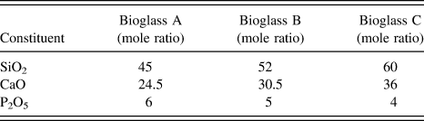
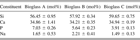
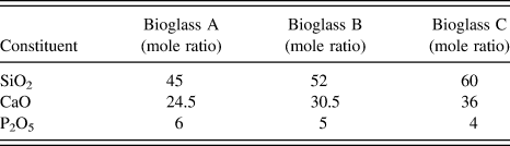
The precursors used in this method were calcium nitrate tetrahydrate (98.5%) (Samchun Pure Chemical, Korea), diammonium hydrogen phosphate (98.5%) (Samchun Pure Chemicals, Korea), and sodium silicate solution as 37% (w/w) SiO2 in 18% (w/w) Na2O (Shwa, Japan). The precursors were dissolved in deionized water, mixed, and transferred to an ultrasonic bath. The slurry suspension was sonicated for 4 h. The precursors were mixed in varying mole ratio as shown in Table I. Three different compositions were taken into consideration and the resulting powders were named as Bioglass A, Bioglass B, and Bioglass C. The conventional composition of bioactive glass materials was kept in mind while designing the bioglass composition in this study. The obtained amorphous powder was washed in deionized water and filtered. After drying for 24 h in an oven at 80 °C, the powders were calcined at a temperature of 600 °C for 1 h. The calcination temperature was chosen from thermogravimetric analysis of the powder. The composition of the bioactive glasses after synthesis was analyzed using energy dispersive spectroscopy (EDS) of the sintered dense body and is shown in Table II. Only major elements are shown and oxygen was not included in the composition.
Composition of the bioactive glass powders’ precursor design.
Composition of the bioactive glass powders after synthesis measured by the EDS (oxygen-free basis).

B. Microstructure and phase analysis
The phases in the synthesized powders were characterized by x-ray diffraction (XRD) and electron diffractions. Each synthesized powder was calcined at 600 °C prior to sintering. For further investigation, the bioglass powders were analyzed by transmission electron microscopy (TEM; JEM2012, JEOL, Tokyo, Japan) in as-received condition. The electron diffraction pattern was taken from the TEM observation to verify the glassy phase of the synthesized materials. The sintering behavior was investigated by sintering the bioglass compacts by microwave and conventional sintering at 1100 °C. The microstructure and phase analysis of the sintered body was investigated by scanning electron microscopy (SEM; JSM-635, JEOL, Tokyo, Japan), EDS (Oxford instruments, UK), and XRD (D/Max-250, Rigaku, Tokyo, Japan). For EDS at least 10 readings were taken to get an average.
C. Mechanical properties measurement
The density of the bioactive glasses was measured by the Archimedes method. Since the theoretical densities of the synthesized bioactive glasses cannot be determined easily, we skipped measuring the relative density to estimate the densification. Rather we measured the actual bulk density to get an idea of densification from the three glass compositions. To get an average, five readings were taken. The Vicker's hardness was measured by a hardness testing machine (Akashi, Japan) with a load of 2 kg for 10 s loading with a square diamond cone.
D. Apatite formation behavior by simulated body fluid
The biocompatibility of the bioactive glass materials in terms of apatite formation was investigated by immersing the dense bioglass body in simulated body fluid (SBF) solution and then incubating the sample in a CO2 incubator with 5% CO2 and 100% relative humidity at 37 °C for 7 days. The SBF solution was changed every day to mimic the dynamic state present in a real life implant environment at the implant site. The SBF was prepared based on the original Kokubo et al.Reference Kokubo, Kushitani, Sakka, Kitsugu and Yamamuro23 method with modifications described by Muller and Muller.Reference Müller and Müller24 After incubation, the SBF-treated samples were collected and the bioglass samples were washed with distilled water first and then dried in air on a filter paper. The apatite formation behavior on the surface was investigated by SEM and EDS.
E. MTT (3-[4,5-dimethylthiazol-2-yl]-2,5-diphenyl tetrazolium bromide) assay, cell attachment, and proliferation behavior
The relative cytotoxicity of the three compositions of bioactive glass was investigated using the 3-[4,5-dimethylthiazol-2-yl]-2,5-diphenyl tetrazolium bromide (MTT) assay. The cellular viability (%) at each bioglass composition was determined using coherent dilutions of the samples following the standard testing protocol (ISO10993-5). In brief, Roswell Park Memorial Institute medium was used as the diluents to obtain the sample extract. The specimens were kept in a shaking incubator at 70 rpm and 37 °C for 72 h. After incubation, the extractions were collected by filtration and serially diluted (100, 50, 25, and 12.5%). The diluted extracts were then placed in a 24-well culture plate that was previously subcultured with L-929 cells (1 × 10⁴ cells/mL). The 24-well culture plate was then placed in a CO2 incubator and incubated for 72 h at 37 °C. After incubation, the MTT solution (100 µl of 5 mg/mL) was added into the wells and the plate was reincubated at 37 °C in 5% CO2 for 4 h. In the final step, the supernatant was removed from the plate and 1 mL of dimethylsulfoxide (DMSO) was added to each well. After mixing with gentle shaking, the purple formazan crystal that formed due to the material excreted by live cells dissolved to create a colored solution. The solution was collected and pipetted on a 96-well plate and the absorbance was measured using a microplate reader. The absorbance was determined at a wave length of 595 nm. The cell viabilities were measured as the percentage of optical densities of the medium containing the extract solution to the optical densities of the fresh medium.
Cell attachment and proliferation behavior were investigated by examining the cell-growing behavior and morphology of the cells attached and spread on the surface of the sintered bioglass materials cultured for 1 and 3 days. After the stipulated days of cell growth, the cells were fixed on the surface by glutaraldehyde and dehydrated with an ethanol series and hexamethylene disilazane. Prior to the SEM observation of the cells, platinum coating was sputtered on the surface. The details of the SEM sample preparations are described elsewhere.Reference Kim and Lee25
F. Real-time polymerase chain reaction analysis
Cells were cultured in the surface of the fabricated samples: Bioglass A, Bioglass B, and Bioglass C on a 12-well culture plate. Another set of cells were cultivated using plain media only to serve as positive control. The seeded cell density was 1 × 10⁵ cells/mL. The cultivated cells that grew on the surface of the samples were collected using trypsin ethylenediaminetetraacetic acid (TE) after 1 day and 7 days of incubation. The total ribonucleic acid (RNA) from the cells was isolated by nucleospin RNA II total RNA isolation kit (Germany). The extracted RNA was then used to synthesize complementary deoxyribonucleic acid (cDNA) using iScript TM cDNA synthesis kit (Bio-Rad). Then, 1 µl of the synthesized cDNA was used as a template for real-time PCR. The samples were subjected to real-time polymerase chain reaction (RT-PCR) using iQ SYBR Green Supermix (Bio-Rad) with 4 primer pairs (osteocalcin, osteonectin, collagen-1, and bone sialoprotein). RT-PCR was performed according to the following parameters: pre-denaturation at 94 °C for 3 min, flowing 40 cycles (denature 94 °C/20 s, annealing 60 °C/20 s, attention 72 °C/30 s); then melting analysis from 65 to 94 °C with ramp rate 0.5. Another set of samples was used for the primer glyceraldehyde-3-phosphate dehydrogenase (GAPDH) that will serve as a housekeeping gene.
III. RESULT AND DISCUSSION
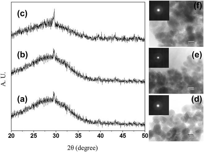
Bioactive materials can elicit a specific kind of biological response at the tissue–material interface, which results in the formation of a bond between the tissues and the materials. Using the method described in this paper, the synthesized bioglass material appears to be amorphous in the as-received condition at all the three compositions as depicted in the XRD profiles in Fig. 1. From the XRD pattern, it was not possible to identify any single phase in its entirety. Moreover, the peak broadening from 20° to 37° in 2θ was evidently due to the amorphous phase of the synthesized powders. Three compositions of the bioactive glass were selected based on the conventional bioglass composition. The composition shown in Table I is based on the precursor materials. After synthesis, the bioactive glass materials' compositions are shown in the EDS data in Table II. The Si content increased marginally from Bioglass A to Bioglass C in accordance with the proportion of the precursor composition and Ca and P. The precursor composition was maintained with constant SiO2/CaO and only the phosphorous content was in a decreasing order. The Na content was not significantly different among all the three compositions. The compositional variation is governed by the equilibrium condition of reaction in the aqueous solution. The anionic SiO3−2 and PO4−3 groups from sodium silicate and diammonium hydrogen phosphate solutions mostly precipitated with the available cationic Ca+2. The higher solubility of the Na+ meant that it did not precipitate well and a nominal amount was found in the precipitated powder. The TEM images and the electron diffraction patterns (EDPs) of the synthesized bioglass powders in the as-received condition in Figs. 1(d)–1(f) are shown for the Bioglass A, Bioglass B and Bioglass C compositions with the EDP profiles, respectively. The EDP profiles included ascertain that the powder materials were all in the glassy phase without any observable development of crystallinity. The absence of a definitive ring pattern in the EDP means that there was not any defined short-range order. The grain sizes from the TEM images were found to be around 20–50 nm for all the three bioactive glasses. This is particularly noteworthy because the ultrafine nanopowders have many unique properties that are not observed in much larger particles. Especially in the case of the melt-derived conventional bioglass, it is extremely difficult to achieve nanopowders of this scale. The feebly observed crystallites were stable in terms of their sizes during the heat treatment process. It has been reported that the development of crystallinity drastically alters the bioactivity of the silica-based bioglass and when the crystallinity was 89%, the bioglass did not show any biological apatite formation behavior in SBF even after soaking for 140 h.Reference Angelopoulou, Montouillout, Massiot and Kordas17 The stability of the amorphous state of the bioglass is a key criterion for further processes such as densification in making solid preforms for using in implant devices.
XRD profiles of as-received bioglass powders. (a) Bioglass A, (b) Bioglass B, and (c) Bioglass C powders. TEM image of the (d) Bioglass A, (e) Bioglass B, and (f) Bioglass C powders after synthesis. Insertions are corresponding electron diffraction patterns.
To achieve densification, the bioglass materials were sintered by microwave sintering at different temperatures, i.e., 1000 and 1100 °C. Higher temperatures were not taken into consideration due to the possibility of developing extensive crystallinity. The chemically etched surfaces of the sintered Bioglass A, Bioglass B, and Bioglass C samples at 1100 °C were imaged by SEM as shown in Figs. 2(a)–2(c). In all the images, a glassy phase is evident in all the samples. Usually for bioglass, a higher sintering temperature is prohibitive because of the possibility of losing the glass characteristics. As the silica content was increased, so was the prevalence of the glassy characteristics. The EDS profiles associated with the corresponding phases are shown in Fig. 2 for (f) Bioglass A, (d) Bioglass B, and for (e) Bioglass C for compositional profiles.
SEM images of the microwave-sintered bioglasses after polishing and chemical etching (a) Bioglass A, (b) Bioglass B, and (c) Bioglass C compact. Corresponding EDS profiles of the bioglasses are shown below in (d), (e), and (f), respectively.
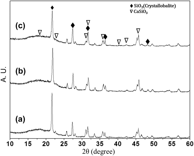
The XRD profile of the sintered bioactive glass compact is shown in Fig. 3. These profiles showed that after sintering some crystallinity developed. The major peak was due to crystallobalite (SiO2). The peaks of CaSiO3 phase were also present although the intensities were lower than that related to SiO2. The development of crystallinity was attributed to the presence of low levels of alkaline materials in the composition. The Na2Ca2Si3O9 phase was recently reported to be the crystalline phase in the hexagonal system in previous studies that examined sintered 45S5 bioactive glass.Reference Filho, LaTorre and Hench26,Reference Li, Yang, Zhang and Kokubo27 Some researchers also suggested that the major crystalline phase from 45S5 bioglass was nonstoichiometric (Na0.951Ca0.049)2Ca(Si0.899P0.101)2O6 of Na2CaSi2O6-type crystal structure.Reference Huang, Lin and Shen28 Even NaCaPO4 crystalline phase was reported with excess Na2O.Reference Mirhadia and Mehdikhanib29 However, our system contained a very low Na content, which was defined by the solubility of Na+ in the aqueous reaction environment. Thus, the eventual development of crystallinity led to the formation of the SiO2 and CaSiO3 phases. Nevertheless, the variation was negligible. Usually the SiO2 phase shows a high intensity peak in XRD, which may dwarf the visibility of peaks with lower intensity. However, the amorphous background with a bulge is clearly visible in Fig. 3 at 13°–25° of 2θ value. The XRD implies that there are significant glassy phases present as observed in the SEM image of the chemically etched sample in Fig. 2. The strong peak of SiO2 dominates the profile disproportionately and the glassy phases of the compositions are evident. In accordance with the SEM image of Fig. 2 the Bioglass C composition showed a better glassy composition. It seems that with a lower content of phosphorous the sodium lean bioglass shows a tendency to be glassier than with a marginally higher content of phosphorous.
XRD profiles of bioactive glass compacts for (a) Bioglass A, (b) Bioglass B, and (c) Bioglass C bioactive glasses sintered at 1100 °C in microwave furnace.
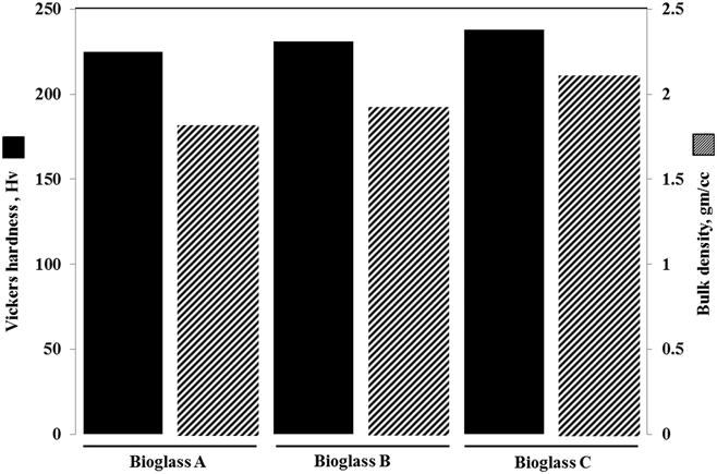
The material properties of the bioactive glass materials were investigated and the density and hardness values are summarized in Fig. 4. It is evident that the hardness increased marginally from Bioglass A to Bioglass C. In addition, these values were comparable with that of the conventional commercial 45S5 bioglass.Reference Kokubo, Kim and Kawashita30 The density values also improve marginally showing better densification behavior as evidenced in the SEM images of the chemically etched samples. The surface characteristics of an implant material are of crucial importance as it dictates the tissue–material interaction. The unique nanophase form of the synthesized powders enables this material to be used in coating applications for many less biocompatible but mechanically strong bioimplant materials.
Vicker's hardness and bulk density values of the sintered bioglass depending on composition variation.
To investigate the biological response on the bioglass after implantation and formation of the apatite, the sintered bioglass compacts were immersed in SBF for 1, 7, 14, and 28 days and investigated by SEM. We have reported a representative one of that of 7 days only in Fig. 5, and apatite formation was found prolific as seen on the surface of dense glass materials in Figs. 5(a)–5(c). After 7 days of SBF immersion, the compact surfaces were fully covered with the biological apatite. The surface displayed intense apatite formation with dome-shaped growth at some places. Prior to immersion of the sample in SBF, the surface was polished using a diamond plate polisher for leveling and to produce a similar nucleation environment across all samples. The prolific growth of an apatite-like phase confirmed the bioactivity of the synthesized bioglass powders. In all the three bioglass compacts, apatite formation started from a nucleation zone, which spread to the surrounding area and initially produced columnar growth. The intense apatite formation eventually leveled out this columnar growth and a textured surface of biological apatite was produced. However, in some places, nucleation continued to form in the upward direction, resulting in the formation of dome-shaped sites seen in the low magnification images. In the enlarged images, an ultrafine morphology was revealed where nanometric crystallites were observed. This extremely porous surface morphology indicates that there was a strong interaction between the material surface and the ions present in the SBF. In reality, natural bone is also composed of nanocrystalline biological apatite dispersed in a matrix of collagen protein. These nanocrystalline apatite materials are thus advantageous to bone formation and the bone bonding ability of the fabricated bioglass. It is obvious that a real biological environment would be much more complex around the implant site with several different biochemical agents and living cells acting directly on the implant material along with the ionic constituents of the body fluid, which the SBF mimicked in our experiment in terms of ionic species and their concentrations. Figures 5(d)–5(f) shows the XRD profiles of the apatite layers. It is evident that some HAp peaks were present but were very low compared to the background reflection of the dense compact bioglass. The relative obscurity of the HAp peaks in the XRD profiles in Figs. 5(d)–5(f) is due to the presence of the very high intensity peak of SiO2 at 2θ = 22°. SiO2 is observed to show a very high level of crystallinity as well as a very high intensity XRD peak at this 2θ value. This high intensity produced by the SiO2 dwarfed the relatively weaker peaks of HAp phase of the formed apatite coating. Even the apatite-coating mask could not affect the attenuation of the diffracted x-ray beam. Although the SEM images of the samples after SBF immersion clearly showed the presence of apatite formation we investigated further to confirm the presence of apatite by chemical composition analysis. To address this we performed the EDS analysis to determine the elemental composition of the apatite coating. As shown in Figs. 5(d)–5(f) with the corresponding XRD profiles, the EDS spectra showed the presence of Ca, P, and O as the primary elemental constituent, which is the definitive proof of the presence of the HAp in the apatite coating. The sintered surface of the Bioglass compacts prior to the SBF immersion showed Si, Ca, and O as the prominent elemental constituents, whereas, understandably, Na and P was very low as expected from the Bioglass composition. However, even though the XRD profile showed a very strong peak intensity of SiO2, the EDS data did not show the presence of Si in significant quantity. This was because the x-ray penetration depth was much higher compared to the electron beam and the x-ray interacted with both the apatite and the bioglass compact underneath, whereas the EDS could only interact with the apatite coating. The small amount of Si is thought to appear from the bioglass compact underneath or from the Si inclusion within the apatite deposition which appears in the SBF solution from the bioglass surface dissolution.
SEM of bioactive glass surface after 7 day of SBF immersion for (a) Bioglass A, (b) Bioglass B, and (c) Bioglass C. XRD profiles of the apatite coating on the bioglass compacts and the associated EDS.
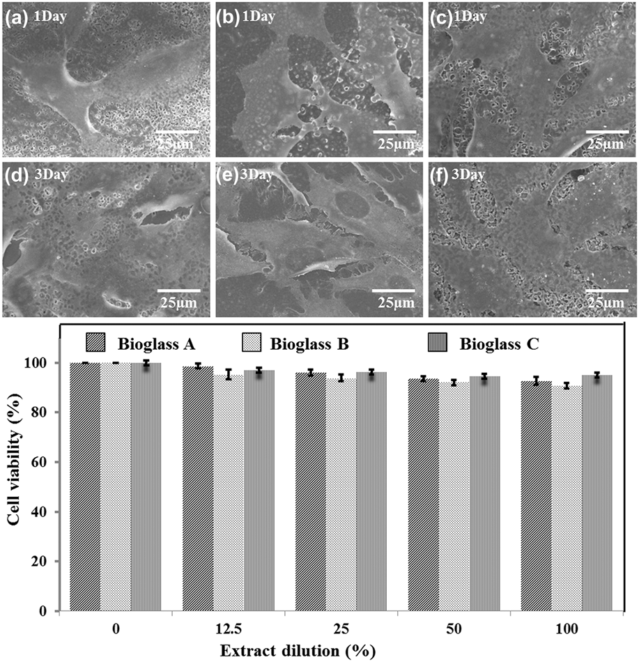
The cell adhesion and spreading response on the surface of the biomaterials is a very important indication of the final behavior of the implant materials regarding the osteocompatibility and host–material interaction for interfacial integration. Growth and differentiation of anchorage-dependent cells are strongly influenced by the substrate materials. A number of factors are responsible for allowing tight adhesion of the cells and the chemical composition of the materials is the most important. In our experiment, the osteoblast-like MG63 cells, which were cultured on all three kinds of bioglass materials, showed superior cell adhesion and spreading behavior. The cell viability and cell proliferation were assessed after 1 and 3 days of culture and the results are shown in Figs. 6(a)–6(f). In all the images, it was evident that cell proliferation was as good on these bioglass materials as on the control surface of a commercial cell culture plate. The interaction between the cell and the material was found to be superior. The cell viability was assessed by MTT assay for 3 days with the three bioglass compositions. Using this assay, all bioactive glass compositions were shown to produce good cell viability. The minimum limit of good cell viability using this protocol was 70% and the cell viability on all the compositions and all extract dilutions were greater than 90%. This demonstrates that the synthesized bioglass compacts were highly biocompatible and the proposed synthesis method was able to produce glass materials with sufficient biocompatibility. All the glass compositions showed superior biocompatibility and Bioglass C showed a marginally improved biocompatibility. This clearly indicates the excellent biocompatibility of the synthesized materials. This trend was particularly interesting because the bioactivity increased at a lower phosphorous content. In previous studies, a decrease in biocompatibility was reported at higher Si concentrations for the conventional SiO2–CaO–P2O5–Na2O bioactive glass system.Reference Nicholson31 In this study, a marginally higher Si content and a decrease in phosphorous content showed a marginally improved biocompatibility in this sodium-less bioglass systems. This result leads us to conclude that the amorphous powder phase at the beginning of the fabrication process and eventual dense body fabrication by the microwave sintering have influenced the structural features of the formed bioglass to elicit a better biological response.
Cell proliferation behavior of MG63 cells on the bioglass compacts for 1 day (a–c) for Bioglass A, Bioglass B, and Bioglass C and for 3 days (d–f), respectively. Biocompatibility of the synthesized powders by MTT assay with L-929 fibroblast cell line.
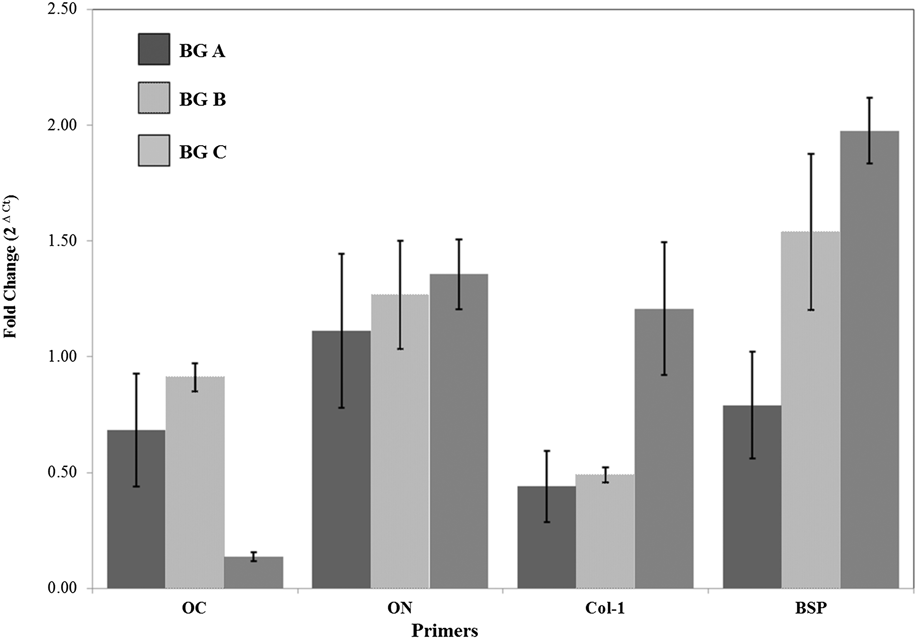
In terms of molecular-level microbiology, for the biocompatibility of a material the specific gene expression is vital. The total RNA of the MG63 cell line was cultivated on the surface of the fabricated materials and was subjected to RT-PCR. A positive control was established with MG63 that grew in culture media only. In this study, the GAPDH gene primer was chosen as control for intact extraction of the total RNA. GAPDH is usually stable across treatments and was used to normalize the data attained from the target genes which, in this case, are osteocalcin (OC), osteonectin (ON), collagen 1 (Col-1), and bone sialoprotein (BSP). These primers represent the genes that are found in the bone-specific cells and may account for the ability of the fabricated material to harbor the said cells and aid in assisted bone regeneration. The gene expression is reported as fold change (2∆Ct) in relation to the positive control or the calibrator. From the results shown in Fig. 7, it can be observed that different genes used were expressed during the experiment and justify the proliferation of the MG63 cells in the surfaces of the fabricated materials. This will show the ability of the material to accommodate cell growth and proliferation on its surface, thus qualifying it as a potent biomaterial. It can be also observed that no significant difference can be observed in the gene expression of cells seeded in the different materials though it can be noted that there is mostly better expression of genes in the cells grown on the surface of the Bioglass C sample. Especially the BSP and collagen expression were higher in the Bioglass C sample. Together with the cell proliferation and cell viability data it can be said that with a lower content of phosphorous in the aforesaid bioglass compositions with limiting sodium content the biocompatibility is slightly improved.
Relative gene expression MG63 cell line grown on the surface of Bioglass A, Bioglass B, and Bioglass C after 7 days of incubation as detected by RT-PCR.
Based on the results described above, a decrease in the phosphorous content in the composition was shown to marginally improve the bioactivity of the bioactive glass materials synthesized by the ultrasonic-assisted hydrothermal method. The molecular organization of the fabricated materials must play a role in the favored biological response of the materials. Thus, further investigations are needed to better understand the structural organization of the bioglass at the atomic level.
IV. CONCLUSION
Bioactive glass materials were synthesized using the ultrasonic energy-assisted hydrothermal method with a low sodium content. Three different compositions were used to investigate the effect of phosphorous content on the final biocompatibility of the materials. Superior biocompatibility was observed for all the bioglass materials and vigorous biological apatite formation was observed in the presence of a physiological fluid. However, marginal improvement in biocompatibility was observed with a decrease in the phosphorous content. The biocompatibility from the cell material interaction showed that the initial cell response on the surface was excellent and the cell spreading and proliferation behavior were also excellent, demonstrating the superior biocompatibility of the synthesized bioactive glasses.
ACKNOWLEDGMENT
This work was supported by the Mid-career Research Program through an NRF grant funded by the MEST (NO 2009–0092808), Republic of Korea.