I. INTRODUCTION
Since the first discovery of carbon nanotubes,Reference Iijima1 there have been significant research efforts to controllable fabrication of nanostructured materials with well-defined shapes and dimensions due to their novel properties and promising applications in electronics, mechanics, physics, and chemistry arising from shape-specific and quantum size effects.Reference Whitesides, Mathias and Seto2–Reference Hao, Meng, Wang, Ye and Zhang4 In the past, these researches have concentrated on the compounds with simple composition. Comparatively speaking, little work has been performed on the fabrication of technologically and practically important polynary complex nanostructures because it is generally difficult to match the reactivity of the different metal precursors during synthesis.Reference Pinna and Niederberger5–Reference Doeff, Chen, Conry, Wang, Wilcox and Aumentado7
SrAl2O4:Eu2+,Dy3+, served as a completely new generation of persistent luminescent phosphor, has attracted much interest due to its high luminescent intensity, long-lasting time, chemical stability, and environmental capability. The afterglow (or phosphorescence) phosphor has been widely used in traffic signs, emergency signs, watches, clocks, paintings, and textile printing as well as in “glow in the dark” toys.Reference Matsuzawa, Aoki, Takeuchi and Murayama8, Reference Yamamoto and Matsuzawa9 However, it is more significant that the nanostructured phosphors with long afterglow will have many potential tremendous applications in biological and medical science,Reference Chen and Zhang10–Reference Ma and Chen12 e.g., they can be used for in vitro and in vivo imaging while no excitation light is needed because some afterglow phosphors can last long time after activation. In addition, they are also used as a light source for photodynamic therapy (PDT) activation to enable PDT for deep cancer treatment, in which a photosensitizer is activated by afterglow light to produce singlet oxygen for cancer cell destruction. At present, many effective techniques, including solid state reaction,Reference Matsuzawa, Aoki, Takeuchi and Murayama8, Reference Zurba, Bdikin, Kholkin, Golberg and Ferreira13 sol–gel,Reference Kurihara and Suib14, Reference Chen and Chen15 coprecipitation,Reference Lin, Zhang, Zhang, Tang and Chen16–Reference Chen, Ma, Li, Shi, Li and Lu18 combustion,Reference Zhao, Chen, Yuan and Wu19–Reference McDonald, Rodriguez, Jones and Adams22 reverse microemulsion method,Reference Lu, Chen and Hsu23 and so forth, have been developed to synthesize the phosphors. However, the phosphor synthesized by solid state reaction route usually possesses larger size and higher hardness. For practical applications, smaller particles must be obtained by grinding the larger phosphor particles, which can easily introduce additional defects and greatly decrease the luminescence efficiency.Reference Rao24 For a traditional combustion method, it generally uses urea as a fuel, and moreover, it is suitable to synthesize fast the phosphor but often difficult to achieve well-defined morphologies and smaller sizes. Additionally, the optical properties of luminescent materials are not only closely related to their native lattice structure and size but are also strongly dependent on their morphology. Therefore, it is of great significance to effectively control the size and morphology of luminescent materials by some facile but efficient methods.
Nanobelts can be regarded as a new class of materials possessing features such as single crystalline quality, well-defined chemical composition, and extremely high anisotropy with a nanoscale dimension. In particular, the morphology of nanobelts is suitable for fabricating optoelectronic devices such as electroluminescence panels that consist of a stack of functional layers or sheets.Reference Frey, Reynolds and Friend25 Until now, however, to the best of our knowledge, there are no available reports on SrAl2O4:Eu2+,Dy3+ phosphor nanobelts with long-lasting phosphorescence. In this study, we develop a new method for the synthesis of the polynary complex nanobelts with long-lasting phosphorescence via a facile but efficient combustion synthesis approach followed by a postannealing reaction. The effects of the postannealing on the relevant photoluminescence and afterglow phenomenon were investigated in detail.
II. EXPERIMENTAL DETAILS
SrAl2O4:Eu2+,Dy3+ nanobelts were synthesized through a facile but efficient combustion method via a highly exothermic redox reaction between metal nitrates and organic fuels and then followed by postannealing at different temperatures. The concentration of both dopant Eu and Dy was selected as 3%. The procedure for the synthesis is briefly given as follows. First, Sr(NO3)2, Al(NO3)3, Eu(NO3)3, and Dy(NO3)3 were dissolved in a minimum amount of deionized water at a molar ratio of 1:2:0.03:0.03. Second, a proper amount of organic fuel triethanolamine (TEA) was added in the above solution. After mixing, the starting materials placed in a beaker were introduced into a muffle furnace and then heated to about 300 °C, where the organic fuels and metal nitrates were automatically ignited and reacted to burn, yielding a black, voluminous, and foamy precursor. Final, the resultant precursor was precalcined at 700 °C in air atmosphere for 3 h to remove residual carbon and subsequently calcined under a weak reducing atmosphere of flowing 5% H2 and 95% Ar gas at various temperatures for 3 h to get the effective conversion of the precursor to SrAl2O4:Eu2+,Dy3+ phosphors and to allow the variation of nanocrystal size.
The phase compositions of the as-synthesized phosphors were analyzed by an x-ray diffractometer (XRD; Rigaku D/max-3b diffractometer with Cu Kα radiation, Rigaku, Tokyo, Japan). The morphology and microstructure of the as-synthesized products were studied by means of a field emission environmental scanning electron microscope (FE-ESEM; FEI Quanta 200F, Oregon, USA) and a transmission electron microscopy (TEM; Hitachi H-800200 kV, Hitachi, Tokyo, Japan). The excitation, emission, and afterglow spectra were recorded using an Edinburgh FLS920 fluorescence spectrophotometer (Edinburgh, UK) equipped with a 450 W Xe lamp as excitation source. For the measurement of afterglow decay curves, the sample was first irradiated for 10 min using an Hg lamp with a wavelength of 365 nm and a power density of 6 mW/cm2, and then the excitation light was automatically switch off and the measurement was carried out by a photometer.
III. RESULTS AND DISCUSSION
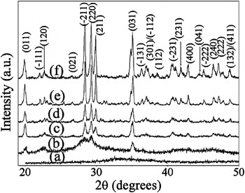
The structural analysis by XRD for the samples, postannealed at different temperatures, is presented in Fig. 1. As can be seen, there is no obvious diffraction peaks in the curve as the calcination temperature is less than 850 °C, suggesting that the sample does not crystallize and exists in the amorphous form. However, as the annealing temperature achieves 900 °C, the sample has crystallized completely and all the diffraction peaks can be neatly indexed to the pure monoclinic symmetry β-tridymite SrAl2O4 (JCPDS No. 74-0794). With further increasing heat treatment temperature, the width of the Bragg reflections reduces but its corresponding intensity increases, indicating the increase of crystallite size with the calcination temperature. This also demonstrates that nanostructures are beneficial to the formation of crystalline phase at relatively low temperature in contrast with the traditional solid state reaction which requires quite long reaction time (4–10 h) in a high temperature (i.e., 1400–1600 °C).Reference Takasaki, Tanabe and Hanada26 In addition, the lattice constants increase with the increase in calcination temperature, and after annealing at 1300 °C, they are close to the values in the JCPDS No. 74-0794. In the sample annealed at low temperature, the lattice contraction and deformation mainly originate from poor crystallinity, which results in some vacancy defects and the additional pressure imposed by the surface tension on the crystal.27,289 In nanostructures with lower crystallinity, therefore, there exists a larger lattice contraction and deformation due to higher densities of defects and larger specific surface area.
X-ray diffractometer patterns of the samples annealed at different temperatures: (a) 800 °C; (b) 850 °C; (c) 900 °C; (d) 1000 °C; (e) 1100 °C; and (f) 1300 °C.

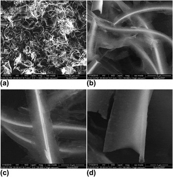
Figure 2 shows the various magnification FE-ESEM images of the sample postannealed at 1000 °C. As can be seen, the sample is mainly composed of semicurly nanobelts. The lengths of belts range from several tens to hundreds of micrometers, and the widths change from hundreds of nanometers to several micrometers. The high magnification FE-ESEM image [Fig. 2(d)] reveals that their thickness is less than 100 nm.
Various magnification scanning electron microscopy images of the sample annealed at 1000 °C.

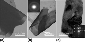
TEM analysis has provided further insight into the microstructural details of these nanostructures. Figure 3 shows the bright field TEM images of the samples prepared by a combustion method followed by a heat treatment at 800 and 1200 °C, respectively. The typical large belt-like structures reveal that the thickness is less than 100 nm. The selected area electron diffraction patterns recorded from these nanobelts, as inset in Figs. 3(b) and 3(c), correspond to the samples annealed at 800 and 1200 °C, respectively. As can be seen, it is mainly composed of one concentric diffuse circle in the sample annealed at 800 °C, implying amorphous nature. After annealing at 1200 °C, however, the obvious electron diffraction patterns, inset in Fig. 3(c), can be obtained, which can be indexed to the reflection of monoclinic SrAl2O4 crystals, indicating that the nanobelts can crystallize well after calcining at higher temperature.
Transmission electron microscopy images of the samples annealed at various temperatures: (a, b) 800 °C; (c) 1200 °C. The insets in (b) and (c) correspond to their selected area electron diffraction, respectively.

TEA is a surfactant and strong chelating agent.Reference Evans, Kozhevnikov, Kozhevnikova, Claridge, Vaidhyanathan, Dickinson, Wood, Cooper and Rosseinsky29, Reference Lin and Yen30 First, it can form a metal–TEA complex, which will be helpful for the homogeneous distribution of metal ions in the solution. Subsequently, in the course of the fast combustion reaction, TEA not only can serve as a fuel but also can serve as a shape-controlled agent to prepare the belt-like precursors.Reference Cheng, Zhang, Liu, Han, Xiao and Lei31 In addition, the products can still remain in their initial morphology during the conversion process of these precursors into the phosphors.
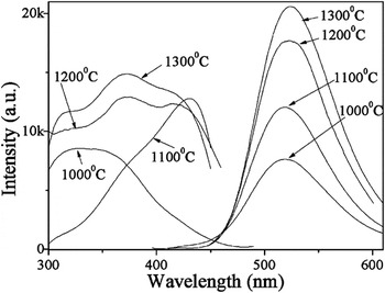
The room temperature photoluminescence excitation, photoluminescence emission, and afterglow spectra of the phosphors, prepared via a combustion method followed by a postannealing reaction at various temperatures, are given in Fig. 4. All the phosphors show strong absorption in the ultraviolet (UV) region from about 300 to 450 nm, and moreover, three distinguished absorption peaks emerge at around 310, 370, and 410 nm in the broad bands, corresponding to the crystal field splitting of the Eu2+ d-orbital. However, their relative intensity ratio is different by comparison between the phosphors annealed at various temperatures, indicating the different natures of Eu2+ surrounding. All the samples emit greenish-yellow light with a very broad band when they were illuminated by the UV light of 365 nm, which can be identified as the 4f 65d 1-4f 7(8S 7/2) electric dipole-allowed transition of Eu2+. However, a slight redshift in wavelength can be observed as the calcination temperature rises, which changes from about 517 to 524 nm with the variation of calcination temperature from 1000 to 1300 °C. With increasing calcination temperature, the crystallinity increases, resulting in increase in crystal symmetry and size. As a consequence, their emission spectra may redshift due to the enhancement of crystal field strength and average optical path around Eu2+ ions.
(a) Photoluminescence excitation spectra (left) and photoluminescence emission spectra (right) of SrAl2O4:Eu2+,Dy3+ nanobelts calcined at various temperatures.

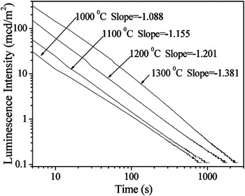
The measurement of afterglow was performed at room temperature, and the decay curves are shown in Fig. 5 on a logarithmic scale. As seen from the decay curves, all the annealed samples show relatively long decay time (i.e., lifetime) when they were efficiently activated by using an Hg lamp with a wavelength of 360 nm for 10 min. With the increase in the calcination temperature, the initial lightness and afterglow time of the phosphors increase, e.g., the afterglow of nanostructured phosphor annealed at 1300 °C lasts for over 1500 s after the source lamp is switched off. In addition, the luminous intensity decays exponentially and it can be expressed by the following equation at the whole stage

Afterglow decay curves of SrAl2O4:Eu2+,Dy3+ phosphor nanobelts annealed at different temperatures, displayed on a logarithmic scale.

where I is the luminous intensity at time t after the removal of activating light, A is the luminous intensity at t = 1, and n is the decay constant. The longer the luminous time is, the smaller the n value is, usually 1 < n < 2. On the basis of Eq. (1), the decay constant gradually increases from 1.088 to 1.381 as the annealing temperature varies from 1000 to 1300 °C, suggesting that the afterglow decay rate of the nanophosphors becomes faster and faster with an increase in the annealing temperature.
In general, the long afterglow characteristics of SrAl2O4:Eu2+,Dy3+ can be attributed to the electron and hole trapped-transported-detrapped process.Reference Matsuzawa, Aoki, Takeuchi and Murayama8, Reference Clabau, Rocquefelte, Jobic, Deniard, Whangbo, Garcia and Le Mercier32 Once the irradiation source was removed, the trapped holes would be released thermally to the valence band and migrate to recombine with the excess electrons in the metastable state (Eu+)* sites and lead to the long afterglow. Therefore, a deeper trap level and a higher trap density will normally lead to a higher initial brightness and longer persistence. Owing to the similar valence and ion radius between Sr2+ and Eu2+, Eu2+ substitutes readily for Sr2+ sites in SrAl2O4 lattice. With respect to obtained SrAl2O4:Eu2+,Dy3+ nanostructures in this case, not only the incorporation of Dy3+ can generate electron traps but also the lattice defects can increase the number and the depth of electron traps in nanostructure. Under UV irradiation, some electrons and/or holes generated by the excitation are trapped in such localized levels. Moreover, defect density will reduce with the calcination temperature. Thus, compared to the sample annealed at high temperature, the decay constant of the one annealed at low temperature can show a small value owing to the presence of high densities and depths of effective electron traps donated by lattice defects. However, for the sample annealed at low temperature, the volume fraction is small due to relatively low crystallinity, and therefore, its initial brightness is lower than that of the sample annealed at high temperatures.
IV. CONCLUSIONS
In summary, SrAl2O4:Eu2+,Dy3+ nanobelts with long-persistent phosphorescence were synthesized via a facile but efficient combustion approach followed by a postannealing reaction at the temperature higher than 900 °C. This method is very suitable for the synthesis of complex multimetal oxides with belt-like nanostructures. All the postannealed phosphors emit greenish-yellow light associated with the 4f 65d 1-4f 7 transition of Eu2+; with increase in calcination temperature, the emission peaks emerge slight redshift since the increase of crystal size and the improvement of crystalline quality cause large average optical path and high crystal symmetry, respectively. Additionally, in the sample annealed at relatively low temperature, quantities of host defects generate higher densities and depths of electron traps, resulting in a relatively slow decay rate compared to the one annealed at high temperature. On the contrary, for the sample annealed at low temperature, its initial brightness is relatively low since the volume fraction is small owing to relatively low crystalline quality. These experimental results demonstrate that SrAl2O4:Eu2+,Dy3+ nanobelts exhibit relatively strong emission and initial lightness, suggesting potential applications in future optoelectronic nanodevices and in biological and medical science.
ACKNOWLEDGMENTS
This work was supported by the Foundation of Jiangxi Educational Committee (GJJ09024), the Natural Science Foundation of Jiangxi Province (00008559), and the Start-up Funds of Nanchang University.