Introduction
Transcanal endoscopic ear surgery has gained widespread acceptance as an alternative method to traditional microscopic ear surgery. The otoendoscope provides a wider field of view and higher magnification, and has fewer anatomical limitations than the traditional microscope. The wider field of view provided by the endoscope allows accessibility to the so-called hidden areas of the tympanic cavity like the sinus tympani, facial recess and epitympanum. As a result, otoendoscopy has been shown to decrease the rate of canal wall down mastoidectomy. Otoendoscopy has also been used to detect residual cholesteatoma following microscopic dissection, leading to a decreased rate of residual cholesteatoma. Endoscopes have been used in the ear as an adjunct to microscopic dissection, and for exclusive endoscopic middle-ear surgery including tympanoplasty, atticotomy and ossiculoplasty, allowing incisionless surgery.
The wide view that transcanal endoscopic ear surgery provides enables the surgeon to access the attic, facial recess, hypotympanum and sinus tympani without the need for a post-auricular incision. Transcanal endoscopic ear surgery can also decrease operating time as less time is needed to access the middle ear. One of the limitations of transcanal endoscopic ear surgery is the inability to use standard drills because of the risk of damage to the endoscope and the limited workspace in the external auditory canal.Reference Kanona, Virk and Owa 1
Most transcanal procedures have been performed with curettes, which may not provide precise control during bone removal and can be quite time consuming. The Sonopet® ultrasonic bone aspirator is a novel device that enables minimal bone removal and maximal mucosal preservation. It has been suggested that mucosal preservation can improve mastoid cavity re-aeration. However, data on the use and safety of the Sonopet in middle-ear procedures are limited.Reference Kakehata, Watanabe, Ito, Kubota and Furukawa 2
The Sonopet's mechanism of action is a torsional oscillation of a metal bone rasp at 25 kHz. The frequency is ideal in that it only cuts mineralised tissue. Soft tissues are cut at frequencies of more than 34 kHz. The handpiece, which is 140 mm in length and 20 mm in diameter, weighing 110 g, is very easy to use. Irrigation fluid emerges from a sheath near the tip of the handpiece, and is adjustable, with a rate of 3–40 ml/minute. Aspiration occurs at the tip of the handpiece, with a maximum aspiration pressure of 500 mmHg.
This study aimed to determine the feasibility and safety of the Sonopet ultrasonic bone aspirator as an alternative to standard curettes in transcanal endoscopic ear surgery.Reference Vrcek, Starks, Mancini and Gilliland 3
Materials and methods
This cadaveric study was exempt from institutional review board approval.
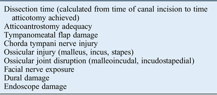
The study was conducted at the Louisiana State University Health Sciences Center Temporal Bone Laboratory. The set up included a Stryker endoscopy video tower with a light source and a camera (Kalamazoo, Michigan, USA). A 0-degree 16 mm endoscope was used for the transcanal endoscopic dissections. A complete set of endoscopic ear instruments was available. The Sonopet was set up along with all available attachments. All dissections were performed by a single surgeon (SPK).The dissections were recorded and analysed in terms of the parameters shown in Table I.
Parameters studied

A total of 10 adult cadaveric temporal bones were dissected by an otolaryngologist. One of the bones was not included in the analysis because of chorda tympani nerve damage that occurred during tympanomeatal flap elevation. Five bones were dissected using the Sonopet ultrasonic bone aspirator. Four additional bones were dissected using standard curettes.
Dissection technique
All the temporal bones were mounted on a temporal bone holder and the ear canal was meticulously cleaned of any debris. A modified Rosen incision was made using a round knife, extending from 12 to 6 o’ clock for left temporal bones and from 12 to 6 o'clock for right temporal bones. The flap was atraumatically elevated to the middle ear. The annulus was elevated using an annulus elevator. The chorda tympani nerve was preserved in all specimens. Ossicular mobility was then tested.
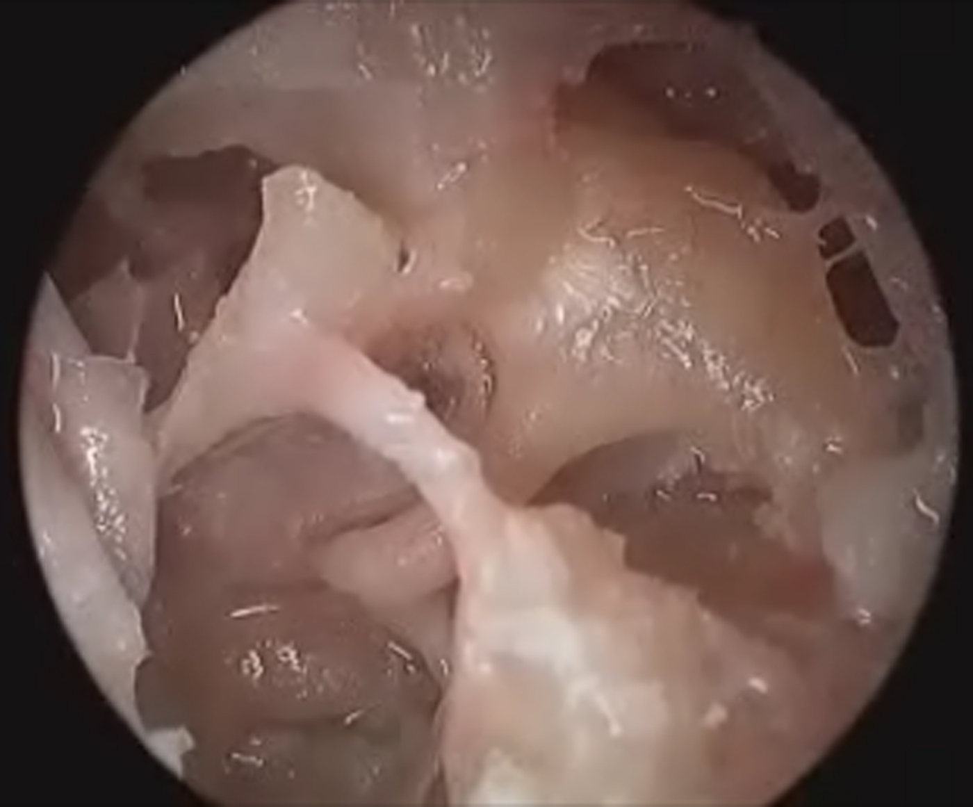
In the Sonopet group, the bone dissection was started in the attic region, with careful and meticulous dissection of the scutum. The dissection was continued until the antrum was reached. The facial nerve and tegmen were both identified. In the traditional curette group, curettage was started in the attic region and similarly continued until the antrum was reached. A video of the atticoantrostomy procedure performed in a left temporal bone, using the Sonopet, is available on The Journal of Laryngology & Otology website (Appendix 1). A still image taken from the supplementary video is shown in Figure 1.
Still image of atticoantrostomy using the Sonopet, taken from the supplementary video (Appendix 1).

An otologist who was blinded to the groups rated the temporal bones according to the study parameters described in Table I.
Statistical analysis
The Minitab® statistics package was used. Non-parametric variables were compared using the Mann–Whitney U test, with a significance level of p < 0.05.
Results
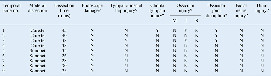
The atticoantrostomy was adequate in all nine temporal bones. Five temporal bones were left-sided and four were right-sided. The ossicular chain disruption rate was higher in the standard curette dissection group, occurring in three of the four bones (75 per cent). Use of a standard curette was associated with: tympanomeatal flap injury in one bone (25 per cent), chorda tympani nerve injury in one bone (25 per cent) and incus injury in two bones (50 per cent). There was no damage to the facial nerve or dura. There was also no damage to the endoscope during the dissections. The same endoscope was used for all dissections. The dissection results are presented in Table II.
Temporal bone dissection results

No. = number; mins = minutes; M = malleus; I = incus; S = stapes; N = no; Y = yes
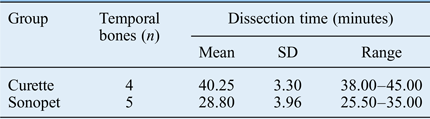
The mean dissection time for the curette group was significantly higher than that for the Sonopet group, as shown in Table III. The mean dissection time using the standard curettes was 40.25 minutes. The mean dissection time using the Sonopet was 28.80 minutes. Dissection duration did seem to decrease over time as we became more accustomed to using the Sonopet.
Dissection times for both groups*

* Difference between groups was statistically significant (p = 0.02). SD = standard deviation
Discussion
Transcanal endoscopic ear surgery allows for a minimally invasive approach to the middle ear compared with traditional post-auricular mastoidectomy, with potentially less post-operative pain, no auricular numbness, no auricular displacement, no visible surgical scar, potentially shorter intra-operative time and reduced hospitalisation time. The use of a microscope in traditional microscopic ear surgery offers a straight-line view, which necessitates a wide transcortical mastoidectomy.Reference Sarcu and Isaacson 4 Despite a more invasive approach, there are still anatomical dead corners such as the sinus tympani, facial recess and the epitympanum.Reference Kakehata, Watanabe, Ito, Kubota and Furukawa 2 Endoscopes limit these anatomical dead corners. Many surgeons have reported using endoscopes as an adjunct to conventional microscopic ear surgery, but it can be time consuming and cumbersome to switch back and forth from microscope to endoscope.Reference Kanona, Virk and Owa 1
Transcanal endoscopic ear surgery is still limited in middle-ear procedures because of the inability to drill. The Sonopet may offer a safe and effective alternative to a drill; its use is associated with less damage to the tympanomeatal flap and other soft tissues.Reference Kakehata, Watanabe, Ito, Kubota and Furukawa 2 , Reference Pagella, Giourgos, Matti, Colombo and Carena 5 – Reference Hadeishi, Suzuki, Yasui and Satou 7 One of the drawbacks of the drill is the damage caused to tympanomeatal flaps and/or to the endoscope itself. Sonopet use may prevent this damage, partly because the claw on the device faces the endoscope and partly because of the more precise control. There were no injuries to either the chorda tympani nerve or tympanomeatal flap in the Sonopet group.
The use of an ultrasonic bone aspirator may expand the indications for transcanal endoscopic ear surgery. The ultrasonic bone aspirator has been used in several other types of surgery, including neurosurgery, sinus surgery and spinal surgery, with good results.Reference Pagella, Giourgos, Matti, Colombo and Carena 5 – Reference Hadeishi, Suzuki, Yasui and Satou 7 It has been shown to permit safer and faster procedures for the surgeon as opposed to drill use in other anatomical areas.Reference Pagella, Giourgos, Matti, Colombo and Carena 5 Data on the safety and use of an ultrasonic bone aspirator in middle-ear surgery are limited. Ito et al. investigated skull vibration levels generated by the ultrasonic bone aspirator and determined that they were comparable or even lower than those generated by conventional drills, and thus they were deemed safe for use.Reference Ito, Mochizuki, Watanabe, Kubota, Furukawa and Koike 8 These authors did not explicitly record all parameters measured, but did not find any clinical evidence of sensorineural hearing loss, facial palsy or dural injury.Reference Ito, Mochizuki, Watanabe, Kubota, Furukawa and Koike 8
The non-rotational design of the Sonopet makes it a feasible alternative for small surgical fields such as the ear canal. Kakehata et al. reported on a series of cases in which the ultrasonic bone curette was used in middle-ear surgery.Reference Kakehata, Watanabe, Ito, Kubota and Furukawa 2 Previous literature had indicated that transcanal endoscopic ear surgery could be used to successfully remove early cholesteatomas.Reference Tarabichi 9 Kakehata et al. noted that transcanal endoscopic ear surgery was limited to these early cholesteatomas because transcanal atticotomy procedures were performed with curettes and/or drills.Reference Kakehata, Watanabe, Ito, Kubota and Furukawa 2 The authors proposed that Sonopet use could offer a feasible alternative to standard curettes or drills. Retrograde mastoidectomies were performed using the ultrasonic bone aspirator with no significant adverse effects. Kakehata et al. found that transcanal endoscopic ear surgery could be used for cholesteatomas extending up to the antrum with the use of the ultrasonic bone aspirator.Reference Kakehata, Watanabe, Ito, Kubota and Furukawa 2 If the Sonopet is indeed a safe and feasible alternative to traditional curettes and drills, indications for transcanal endoscopic ear surgery could be expanded.
In the current study, the mean dissection time was significantly lower using the Sonopet as compared to traditional curettes. Time decreased with each dissection as we became more accustomed to using the device. One of the most important benefits of using the Sonopet is the time that it saves during dissection, which could result in overall cost savings. This study showed no injury to vital structures using the Sonopet. In fact, there was a significantly higher percentage of ossicular chain disruption in the standard curette group. This may in part be explained by the lower precision and control associated with using standard curettes.
The main drawback of the study is the limited number of temporal bones dissected, which precludes any statistical analysis between the two groups. In addition, it is a cadaveric temporal bone study, and clinical correlation is required. Nevertheless, the study indicates advantages of using the Sonopet, including shorter operative time and greater control in bone removal in endoscopic ear surgery, with a decreased complication rate.
-
• Ultrasonic bone aspirator use is a feasible option in endoscopic ear surgery
-
• It enables easier bone removal, with no additional complications, as compared to traditional curettes in a temporal bone model
-
• The time taken to perform atticoantrostomy was significantly lower using a Sonopet than curettes in a temporal bone model
-
• An ultrasonic bone aspirator can be part of the armamentarium for transcanal endoscopic ear surgery
Disadvantages of the Sonopet include the cost and the learning curve, as seen with any new instrument and procedure. The size of the device might limit its use, as space for the endoscope and Sonopet can be constrained. As mentioned earlier, with the faster dissection times, the overall cost of the device may result in overall cost savings associated with the decreased operative time. Clinical experience is necessary to determine the clinical value of the device indicated by the findings of this anatomical study.
Conclusion
Ultrasonic bone aspirator use is a feasible option in endoscopic ear surgery. It may enable easier bone removal, with no additional complications and greater efficacy than traditional bone curettes. The device can be a part of the armamentarium for transcanal endoscopic ear surgery.
Appendix 1. Supplementary video material
A video demonstrating atticoantrostomy using the Sonopet in a left temporal bone is available online at The Journal of Laryngology & Otology website, at https://doi.org/10.1017/S0022215117001955.