CHD is the commonest birth defect worldwide, and atrial septal defect makes 15.37% of all CHD.Reference Liu, Chen and Zühlke1 When the normal closure process and tissue integrity of the interatrial septum are disturbed in the embryo, an ASD, which is classified into five types according to the localisation, develops. Secundum type ASD (ASD2), which is relatively in the middle of IAS is the most common form of childhood ASDs.Reference Celermajer2 The symptoms of secundum ASD depend on the shunt-related consequences on the cardiac chambers and great vessels.Reference Torres3 Most paediatric secundum ASD cases with smaller size defects are asymptomatic and expected to close spontaneously. However, the persistence of secundum ASD beyond 3–5 years of age, marked left-to-right shunting leading to an increased right cardiac load, pulmonary hypertension, and heart failure necessitate secundum ASD closure.Reference Knop, Szkutnik, Fiszer, Białkowska, Głowacki and Białkowski4
Although the surgical approach for secundum ASD closure proved to be effective and feasible, it inherits procedural disadvantages, such as a long recovery time and a relatively high potential of complications due to iatrogenic trauma, i.e., emotional burden in children caused by an apparent incision scar.Reference Ooi, Kelleman and Ehrlich5 Alternatively, percutaneous transcatheter closure, which is used in more than 80% of isolated secundum ASD cases and overcomes some disadvantages of surgery, has been established as a standard intervention in selected patients.Reference Prieto6 The FDA-approved Amplatzer Septal Occluder ranks amongst top available devices for secundum ASD PTC, and there is an extensive literature on its technical aspects, i.e., optimal ageReference Han, Zhang and Zhang7 and defect size for intervention,Reference Houeijeh, Hascoet and Bouvaist8 intraprocedural guidance by echocardiography or fluoroscopy,Reference Baykan, Pamukcu and Ozyurt9 as well as comparative studies amongst various types of ASD occluder devicesReference O’Bryne and Levi10 and between PTC intervention versus surgical approach.Reference Castaldi, Vida and Argiolas11 The PTC procedure of secundum ASD via ASO requires an assessment of the defect prior to and guidance of the device during the intervention. For this purpose, fluoroscopy and echocardiography, either transthoracic (TTE) or transesophageal (TEE) can be used. Studies in different age groups of patients demonstrated the benefits and limitations of each technique.Reference Baykan, Pamukcu and Ozyurt9,Reference Narin, Baspinar and Pamukcu12 The post-interventional effects of secundum ASD PTC on cardiac remodelling in geometry and function have been researched in some studies; however, most research focus on not only adult,Reference Jategaonkar, Scholtz, Butz, Bogunovic, Faber and Horstkotte13–Reference Stephensen, Ostenfeld and Kutty16 or heterogeneous age group of patients,Reference Mainzer, Braver and Khoury17–Reference Yoo, Kim, Eun, Choi and Dong19 but also on only right or left cardiac remodelling.
To the best of our knowledge, there is no comprehensive study on haemodynamic response and both right and left cardiac remodelling after secundum ASD PTC with ASO in children. In this study, we aimed to assess the immediate haemodynamic response and the timing of right and left cardiac remodelling in paediatric secundum ASD patients who underwent PTC with ASO.
Patients and methods
Study groups
This longitudinal cohort study was conducted on 41 paediatric secundum ASD patients (patient group) and 39 age- and gender-matched healthy children without any cardiovascular diseases (control group). The control group included asymptomatic patients who were referred for murmur, and ECG and ECHO findings were normal. The patients included in the study had PTC intervention for an isolated secundum ASD using an ASO device. The exclusion criteria consisted of the presence of concomitant CHD (i.e., VSD, PDA) and complex CHD, infections, or bleeding disorders. Patients with large defects, cribriform, or multiple defects were not excluded from the study. The signed informed consent forms were obtained from parents of participants, and the study protocol, which adhered to the tenets of the Declaration of Helsinki, was approved by the institutional ethics committee.
Procedure
Demographic data consisting of the age, the gender, the weight, and the height of the participants were recorded initially, and body mass index was calculated using the standard formula. Pre-interventional TTE was performed to obtain basal values of the control group subjects and the patients who were scheduled for PTC 24 hours later.
The procedure was performed under general anaesthesia in room air without intubation. On the day of PTC, invasive haemodynamics of patients were assessed by cardiac catheterisation 15 minutes before and after the intervention. The haemodynamic workup included the right atrium, the right ventricle, and the pulmonary artery pressures (RAP, RVP, PAP), as well as oxygen saturation levels from RA, RV, vena cava superior, vena cava inferior, and PA. The data obtained in invasive haemodynamic studies were used for estimating the pulmonary (Qp) and systemic (Qs) flow, pulmonary vascular resistance, and for calculating the ratio of PVR to systemic vascular resistance.
The PTC intervention was performed by using Amplatzer Septal Occluder device (St Jude Medical, Inc, Minnesota, United States of America) following the standard procedure according to the manufacturer’s manual.
All echocardiographic studies were performed by the TTE approach based on previously published guidelinesReference Lopez, Colan and Frommelt20 and using the VIVID 7 ultrasound (GE, Horten, Norway) machine with multi-frequency transducers and GE EchoPAC (BT 09, Horten, Norway) system. Multi-modal TTE studies for evaluating the cardiac geometry and functions in patients and controls were performed 1 day before the PTC intervention, and at the post-interventional 1st, 3rd, 6th, and 12th months for follow-up purposes.
TTE M-mode using apical four-chamber views was used to evaluate the size, location, and rims of the ASD and the size, volume, and area of the left and right cardiac chambers as described previously.Reference Castaldi, Vida and Argiolas11 The tricuspid valve and pulmonary annulus diameters were measured in the parasternal short-axis view. Pulsed Wave Doppler was used to measure the right ventricle myocardial index (Tei index), and tricuspid valve’s E peak (early diastolic flow), A peak (late diastolic flow), and S (peak systolic flow) waves. The LV ejection fraction was calculated. Right ventricular fractional area change was estimated via the formula (end-diastolic area – end-systolic area)/end-diastolic area × 100%.Reference Anavekar, Skali and Bourgoun21 Apical four-chamber view was used to record the tricuspid annular plane systolic excursion by placing the M-mode cursor at the junction of the RV free wall and the tricuspid valve to provide a recording of the maximal longitudinal shortening and relaxation of the RV free wall. TAPSE was defined as the systolic amplitude of the tricuspid annulus excursion from the base towards the apex. RV Tei index was measured as described previously.Reference Ammar, Arab, Hammady and Sayed22 Similar measurements for the left cardiac side were performed to record the mitral annular plane systolic excursion. During the follow-up examinations, colour Doppler was used to assess the presence of residual shunting, and it was classified as trivial when the width of the residual jet was below 1 mm.Reference Cao, Du and Joseph23
Statistical analysis
Histogram, Q–Q plots, and Shapiro–Wilk’s test were used to assess data normality. Levene test was used to test variance homogeneity. Mauchly’s test was used to test sphericity assumption. To compare the differences of demographics and cardiac echocardiographic (TTE) data between patient and control groups, a two-sided independent samples t-test was conducted. To compare pre- and post-procedural invasive haemodynamics of the patients, paired t-test was applied. To compare pre- and post-interventional cardiac echocardiographic (TTE) data, either one-way repeated measures of ANOVA or Friedman tests were performed. Bonferroni and Nemenyi tests were applied for post hoc comparisons. All analyses were conducted using R 3.4.0 (www.r-project.org) statistical software. A p-value less than 5% was considered as statistically significant.
Results
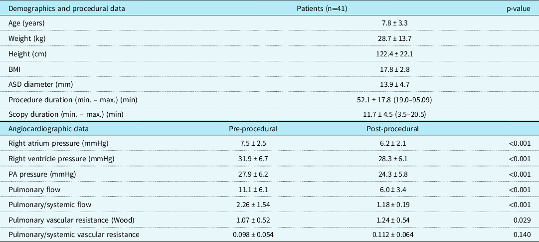
The study was completed with 41 patients (21 girls, 20 boys) and 39 healthy children. No significant difference in mean values of weight, height, and BMI were found between the patient group and the age- and gender-matched control group. The mean value of secundum ASD size was calculated by using data of each patient evaluated with TTE before the intervention and found as 13.9 ± 4.7 mm. The PTC of secundum ASD in all patients was completed without any peri-procedural complications. Demographic and intervention-related data of all participants were presented in Table 1.
Demographics and procedural data and comparison of pre- and post-procedural invasive haemodynamics of the patients.

Values are expressed as mean ± SD.
ASD = atrial septal defect; BMI = body mass index; PA = pulmonary artery.
Invasive haemodynamic evaluation results
The invasive haemodynamic parameters of each patient was measured 15 minutes before and after the intervention, and were recorded and calculated to obtain an average value for all variables. The average pre-interventional Qp/Qs ratio of 2.26 ± 1.54 was significantly reduced to a post-interventional average of 1.18 ± 0.19 (p < 0.001). The reductions in mean RAP, RVP, and PAP were statistically significant compared to pre-interventional values (p < 0.001). We observed a significant increase in pulmonary vascular resistance after the intervention (p = 0.029), while the increase in the ratio of pulmonary to systemic resistance was not statistically significant (p = 0.140). The post-interventional 24th-hour assessment with PW Doppler did not reveal any signs of residual shunting. The data from invasive haemodynamic studies were summarised in Table 1.
Right cardiac remodelling
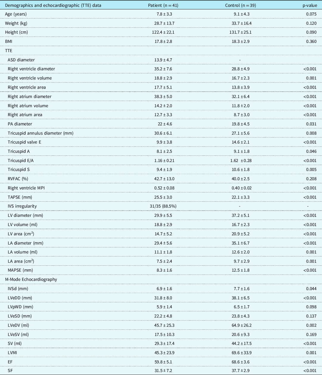
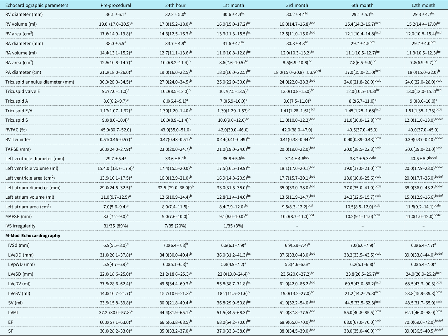
Before the intervention, TTE revealed that the mean values of RA and RV diameters, volumes, and areas of the patient group were significantly more than those of the control group (p≤0.001). Pre-interventional tricuspid velocities of E, A, and S waves, and E/A values were significantly lower, while the diameters of the tricuspid annulus and PA, right ventricular MPI, and TAPSE were significantly higher than those of the control group (p < 0.05). The echocardiographic follow-up evaluations of the patients in the post-interventional 24th hour, 1st, 3rd, 6th, and 12th months demonstrated that the right cardiac geometrical and functional markers, except for the RV Tei index, tricuspid E/A ratio, and tricuspid S wave, started to improve in the 24th post-interventional hour and remodelling with normalised values comparable to those of the control group was completed in the 6th post-interventional month. The IVS irregularity in all patients disappeared in the post-procedural 1st month. The RV Tei index, tricuspid E/A ratio, and S wave values normalised in the post-interventional 12th month. Data on right cardiac functional improvement and geometric remodelling were presented in Tables 2 and 3.
Demographics and cardiac echocardiographic (TTE) data comparison between patients and controls.

Values are expressed as mean ± SD.
ASD = atrial septal defect; BMI = body mass index; EF = ejection fraction; IVS = Interventricular septum; IVSd = Interventricular septum diastolic; LA = left atrium; LV = left ventricle; LVMI = left ventricular mass index ; LVeDD = left ventricular end-diastolic diameters; LVeDV = left ventricular end-diastolic volume; LVeSD = left ventricular end-systolic diameters; LVeSV = left ventricular end-systolic diameters; LVpWD = posterior wall thickness in end-diastole; MAPSE = Mitral Annular Plane Systolic Excursion ; MPI = myocard performance index ; PA = pulmonary Artery; RVFAC = right ventricle area change; SV = stroke volume; SF = shortening fraction (LVSF); TAPSE = Tricuspid Annular Plane Systolic Excursion.
Pre- and post-interventional cardiac echocardiographic (TTE) data comparison in patients.

Different superscripts in the same row indicate a statistically significant difference between groups.
Values are expressed as mean ± SD.
Echocardiographic parameters of pre-procedural, 24th hour, 1st month period are compared and shown in the Table. Numeric variables are expressed as mean ± SD.
aP<0.05, when compared with the pre-procedural period; bp < 0.05, when compared with the 24th hour after procedure; cp < 0.05, when compared with the 1st month after procedure; dp < 0.05, when compared with the 3rd month after procedure; ep < 0.05, when compared with the 6th month after procedure; fp < 0.05, when compared with the 12th month after procedure.
Left cardiac remodelling
Pre-interventional evaluation of left cardiac echocardiographic parameters showed that the mean values of LA and LV diameters, volumes and areas, and MAPSE value of the patient group were significantly less than those of the control group (p≤0.001). M-mode echocardiography revealed that LV end-diastolic diameter and volume, LV mass index and SV, diastolic IVS diameter, EF, SF were significantly lower than the mean values of the control group (p < 0.05). Post-interventional evaluation and follow-up examinations showed that the left cardiac markers of remodelling started in the 24th hour and significantly and periodically improved afterwards. While the irregularity of the IVS disappeared and normalised in the 1st post-interventional month, the rest of the left cardiac remodelling was completed, and all parameters normalised compared to those of the control group at the end of the post-interventional 12th month. Data on left cardiac functional improvement and geometric remodelling were presented in Tables 2 and 3.
Discussion
In this study, we assessed the immediate haemodynamic response and the timing of right and left cardiac remodelling in children with secundum ASD who underwent PTC via ASO. As per the standard of our practice, the shunt elimination and normalisation of cardiac geometry and functions due to secundum ASD-caused cardiac overload were set as the main management targets in children. We defined interventional success as occluder implantation with no signs of embolisation, malposition, and peri-procedural complications, as described previously.Reference Jalal, Hascoet and Gronier24 The management of secundum ASD in adults and children has been extensively studied with an abundance of literature comparing surgical versus percutaneous transcatheter closure using various occluder devices. PTC of secundum ASD is an established technique with a low incidence of mortality and morbidity, and TTE guidance is satisfactory in case selection eligible for PTC. ASO use in paediatric secundum ASD closure was found effective and suggested to be the first option for interventional purposes.Reference Behjati, Mirhosseini, Hosseini and Rajaei25 We used the ASO device in this study, and no peri-procedural complications or signs of residual shunting were observed in patients. The 100% success and 0% complication rates in our study were highly superior to a similar study that reported a success rate of 95.3% and a complication rate of 1.8% as excellent.Reference Jalal, Hascoet and Gronier24
ASD haemodynamics in children greatly depends on the shunting, right heart geometry, and PAP. Cardiac catheterisation for diagnostic purposes is indicated on suggestive signs of pulmonary hypertension on echocardiography. RV dilatation and diastolic IVS shift towards the left side impair the diastolic filling of the LV, so the size of the ASD and interventricular diastolic compliance differences determine the degree of shunting. Chronic volume overload remodels the pulmonary vasculature bed, ultimately leading to PH due to narrowing and increased resistance.Reference Torres3 The pre-interventional cardiac catheterisation demonstrated that the average Qp/Qs (2.26 ± 1.54) in the patient group was higher than the threshold value of 1.5 and indicated PH in patients. Additionally, pre-interventional mean PAP of 27.9 ± 6.2 mmHg and major signs of right cardiac overload further supported the presence of PH. In some studies that evaluated the changes in cardiac haemodynamics 1 day after the PTC of secundum ASD, RV dimensions started to decline in 24 hours after the intervention and continued to normalise during the 6 months following PTC.Reference Yilmazer, Guven and Vupa-Cilengiroglu26–Reference Xu, Sun and Zhou29 Using cardiac catheterisation, we assessed the immediate response of cardiac haemodynamics after the PTC intervention, and observed signs of improvement starting in seconds due to a marked reduction in Qp. As the first control was made 15 minutes after the intervention, the Qp/Qs ratio had not reverse to the normal at that time. We suspect that it might be due to negligible intradevice microshunts, as reported in previous studies.Reference Wyss, Quandt and Weber27 The increase in post-interventional PVR and PVR/SVR values in our study could be either due to unresponsiveness of pulmonary bed to increased flow as early as 15 minutes after closure or to secondary vascular adaptations against the dramatic reduction in blood flow caused by the sudden disappearance of shunting.
The morphologic, conductive, and haemodynamic changes in various time intervals, which occur in few years after PTC of secundum ASD, cause cardiac remodelling and result in the structural and functional normalisation of the heart.Reference Mainzer, Braver and Khoury17 The time course of cardiac remodelling in the literature has a broad range from 3 monthsReference Xu, Sun and Zhou29 to 2 years.Reference Agha, El-Saiedi and Shatout30 In one study on children weighing less than 10 kg, the RV dimensions and the RV/LV proportion gradually decreased and normalised in the first year after the PTC.Reference Knop, Bialkowski and Szkutnik28 On the other hand, in a recent study on the evaluation of RV myocardial strains after PTC of secundum ASD in children, the researchers showed that the intervention improved the RV strain indices and RV function normalised in 3 months. Those researchers also showed that the RA and RV size decreased in post-interventional 24 hours. and the RV volume overload disappeared immediately after the intervention and before the further improvement in RV function in the following 3 months.Reference Xu, Sun and Zhou29 In another study, a 3-month follow-up period was suggested to be sufficient to determine the RV remodelling as it appeared to be an early post-interventional effect that occurred in the post-interventional first few months.Reference Jategaonkar, Scholtz, Butz, Bogunovic, Faber and Horstkotte13 In a comparative study of PTC versus intraoperative device closure of secundum ASD in children, RV remodelling was successfully demonstrated by structural and haemodynamical parameters, i.e., RV dimensions and RV/LV decreased in all patients at the 2-year follow-up evaluation.Reference Han, Zhang and Zhang7 Our results demonstrated that right cardiac remodelling was completed in the post-interventional 6th month, and no statistically significant change in the echocardiographic parameters appeared afterwards. As we demonstrated in our study, echocardiographic improvement at right atrium and right ventricle that was relieved from the volume load beginning from the first minute after closure was completed at 6th month (there was no difference between 6th and 12th-month values) except for a few parameters (RV Tei index and tricuspid valve Doppler S value). This may be explained by the fact that cardiac compliance is good and cellular regeneration is fast in children.
Ammar et al underlined the importance of measuring post-interventional RV systolic and diastolic functions by using the Tei index, TAPSE, and RV FAC.Reference Ammar, Arab, Hammady and Sayed22 Agha et al showed that TDI velocities and isovolumetric indices started reducing 1 month after PTC of secundum ASD; however, RV and LV Tei indices although decreased during the 1-year follow-up, did not reverse to normal values.Reference Agha, El-Saiedi and Shatout30 Akula et al reported that the deterioration in RV Tei index after PTC of secundum ASD in adult patients might not reflect a true functional loss, instead might be due to cardiac geometry normalisation after shunt disappearance, and the authors reported that right cardiac remodelling in adult patients of that study was completed in the post-interventional 6th month.Reference Akula, Durgaprasad, Velam, Kasala, Rodda and Erathi15 The RV Tei index was one of the parameters in which the normalisation process lasted longer than the others and was completed in the 12th post-interventional month. Hence, we might conclude that global RV functions in children normalise later in the remodelling period after the PTC intervention. A period of 6 months as the average duration for right heart remodelling and geometric normalisation observed in our patients can be considered as shorter compared with previous studies,Reference Han, Zhang and Zhang7,Reference Knop, Bialkowski and Szkutnik28,Reference Agha, El-Saiedi and Shatout30 and support the results of other studies.Reference Akula, Durgaprasad, Velam, Kasala, Rodda and Erathi15,Reference Yilmazer, Guven and Vupa-Cilengiroglu26,Reference Xu, Sun and Zhou29
ASD in children causes a reduction in LV systolic functions and stroke volume due to the decrease of diastolic LA and LV volumes. We found that the improvement in the dimensions and end-diastolic volumes of the LA and LV and their systolic functions started in a few hours after PTC intervention and normalised in the 12th month. In a study comparing the timing differences in the outcome of PTC of secundum ASD on cardiac geometry and function amongst various age groups, the researchers found that systolic and diastolic LV volume increased while EF did not change in any of the age groups.Reference Tashiro, Suda, Iemura and Teramachi18 In a study on left heart function in children after PTC of secundum ASD, the researchers found a significant increase in the size of the LA from the baseline value at the 3rd-month follow-up visit. The research suggests that the decline in LA volume after closure might be due to an improvement in the LV diastolic filling.Reference OBryne, Kennedy, Rome and Glatz31 It was shown that PTC of secundum ASD in adults resulted in differences in adaptation between right and left ventricles. A very recent longitudinal study with serial cardiac MRI examinations revealed that the LV adaptation in adult secundum ASD patients who underwent PTC was earlier (1st-day post-interventional) than RV adaptation (3rd-month post-interventional).Reference Stephensen, Ostenfeld and Kutty16 We have demonstrated that the increase in LV and LA geometry, MAPSE, EF, and SF started in the post-interventional 24th hour, and the statistically significant increase checked periodically in 3-month intervals continued till the end of the follow-up period of 12th month. The IVS irregularity gradually decreased and improved in the 1st month. The left cardiac remodelling was completed and normalised at the end of the post-interventional 12th month. Our study has the opposite outcomes with an earlier right cardiac improvement than the left when compared to the results of studies with adult patients.Reference Stephensen, Ostenfeld and Kutty16 The longer exposure to an increased Qp might be the basis of this discrepancy; alternatively, the difference in secundum ASD sizes amongst study populations might be responsible for the contradictory results.
Despite the vast amount of literature on PTC procedure-related studies, the research on cardiac remodelling effects of secundum ASD occlusion is scarce. Even further complicating the issue, the available data usually belong to studies on adultsReference Balci, Balci and Aksoy14–Reference Stephensen, Ostenfeld and Kutty16 or mixed-aged groups.Reference Yoo, Kim, Eun, Choi and Dong19,Reference Ammar, Arab, Hammady and Sayed22 Additionally, as determined in a retrospective multicentre observational study of data from the IMPACT registry, there are measurable and significant practice variations in PTC of ASD amongst institutions.Reference OBryne, Kennedy, Rome and Glatz31 Therefore, it is difficult to compare the results of studies with mixed-age populations from different institutions to our study due to great variation in definitions and thresholds for secundum ASD dynamics between adult and children.
There are a few limitations and more strengths of our study that needed to be acknowledged. This is a single-centre study with a small number of patients. Nevertheless, the longitudinal design, the focused selection of only children, and the comprehensive workup procedures to obtain data on haemodynamics and right and left cardiac remodelling can be counted as the particular strengths of the current study.
Conclusion
This study with very high interventional success rates can be counted as the first example of research on the haemodynamic response and timing of cardiac remodelling after PTC of secundum ASD in children. We suggest that future multicentric studies with larger cohorts and a comprehensive methodology like ours with longer follow-up periods would better serve to further assess the cardiac remodelling in children after PTC of secundum ASD.
Acknowledgements
None.
Financial support
This research received no specific grant from any funding agency, commercial, or not-for-profit sectors.
Conflicts of interest
None.
Ethical standards
The authors assert that all procedures contributing to this work comply with the ethical standards of the national guidelines of Turkey on human experimentation (please name) and with the Helsinki Declaration of 1975, as revised in 2008, and has been approved by the local institutional committee.