Introduction
Regenerative medicine describes research and clinical applications that aim to repair, replace or regenerate cells, tissue and organs via cell therapies, gene editing or tissue engineering. Over the past two decades, research activity in regenerative medicine has grown exponentially across all medical disciplines.Reference Cossu, Birchall, Brown, Culme-Seymour, Gibbon and Hitchcock1 Despite well-publicised examples of functional restoration across a wide range of organs,Reference Atala, Bauer, Soker, Yoo and Retik2–Reference Nyame, Chiang, Leavitt, Ozambela and Orgill5 few regenerative treatments have gained licence for routine clinical use in the USA and Europe.Reference Pellegrini, Ardigò, Milazzo, Iotti, Guatelli and Pelosi6–Reference Yu, Gupta, Ronfard, Vertès and Bayon8 Many are withdrawn because of poor efficacy, safety concerns or financial insolvency.Reference Abou-El-Enein, Elsanhoury and Reinke9,Reference Boráň, Menezes-Ferreira, Reischl, Celis, Ferry and Gänsbacher10
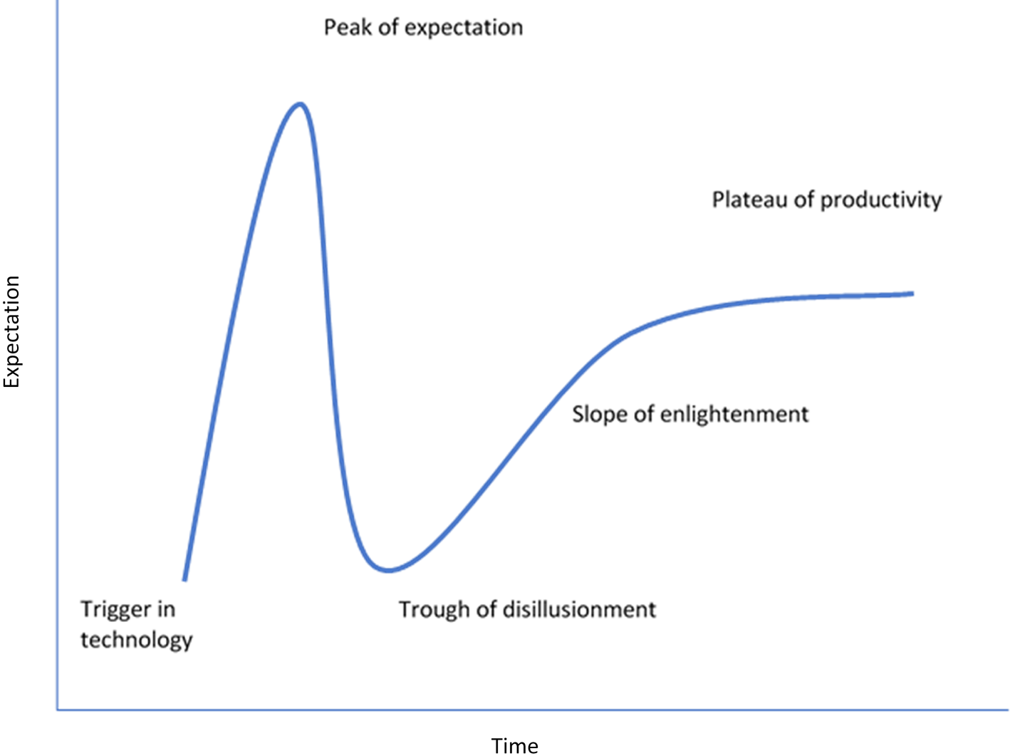
Regenerative medicine aims to harness intrinsic cellular mechanisms for tissue or organ regeneration. This variably involves tissue engineering, cell biology, gene therapy and stem cell therapy. Alongside other new technologies, regenerative medicine is claimed to follow the Gartner Hype Cycle (Figure 1).Reference Fenn and Raskino11,Reference Polykandriotis, Popescu and Horch12 Technological triggers in the 1980s and 1990s encouraged optimism, investment and expectations of swift mass-market success. However, limited existing technology and scalability issues prevented a breakthrough to routine clinical use, prompting disillusionment. Currently, regenerative medicine is reckoned to be in the third phase of the cycle,Reference Geris and Papantoniou13 a plateau of productivity before treatment will reach patients effectively and economically. Therefore, investments in tissue-engineered medicinal products are predicted to grow from $10.8 billion in 2016 to $22.3 billion in 2025.Reference Mikulic14
The Gartner Hype Cycle represents the maturity of emerging technologies. Regenerative medicine is believed to follow this pattern with initial high expectations giving way to disillusionment followed by a slower phase of progress and a plateau of productivity.

Regenerative medicine applications for regions of the upper aerodigestive tract (mouth, throat, larynx and upper airways) are growing. These regions permit rapid tissue retrieval for ex-vivo cell expansion. They allow for less invasive tissue delivery into and monitoring of treated areas than in organs within enclosed body cavities. There is clear demand: head and neck cancer is the sixth most common cancer globally, with approximately 630 000 new cases each year.Reference Torre, Bray, Siegel, Ferlay, Lortet-Tieulent and Jemal15,Reference Vigneswaran and Williams16 Despite relatively high cure rates and survival,Reference Torre, Bray, Siegel, Ferlay, Lortet-Tieulent and Jemal15 treatment with surgical excision, chemotherapy or radiotherapy results in tissue loss and fibrosis within the upper aerodigestive tract. Severe long-term disability may ensue, including loss of voice and swallow, and repositioning of the airway opening into the neck. The resultant health burden brings difficulties returning to fulltime employment and low quality of life.Reference Melo Filho, Rocha, Pires, Fonseca, Freitas and Martelli Junior17,Reference Verdonck-de Leeuw, van Bleek, Leemans and de Bree18 Benign disease may also result in end-stage fibrosis within the upper aerodigestive tract, with the same deleterious effect on function.Reference Kumai19,Reference Quinn, Gelbard, Sibley, Sirajuddin, Ferrada and Chen20 Regenerative treatments that address fibrosis or replace diseased tissue therefore present an important anatomical target for regenerative medicine over the next decade.
This review examines the past twenty years, attempting to identify current focuses in the field and progress toward clinical application and improved patient outcomes. It focuses on upper aerodigestive tract disease affected by end-stage fibrosis and tissue loss because of cancer, surgery, radiation, trauma, age or congenital defects, namely in the throat, salivary system, tongue, larynx and upper airway. Previous studies have demonstrated the complexity of the task involved.Reference Wormald, Fishman, Juniat, Tolley and Birchall21 Some areas of success for regenerative medicine, with established protocols and guidelines, are mentioned.
Materials and methods
A systematic literature review on the use of regenerative medicine targeted to the upper aerodigestive tract regions involved in airway, swallow and voice was conducted. PubMed, Embase, Google Scholar, Cochrane Library, Ovid Embase, Ovid Medline databases and clinicaltrials.org were searched between 1 January 2000 and 16 July 2019 for English-language articles.
The following keywords were used: bioengineering, regenerative medicine, tissue engineering, cell therapy, regenerative surgery and upper aerodigestive tract, pharynx, oropharynx, larynx, trachea, vocal cord, tongue, and salivary glands. Articles concerning nerve regeneration, periodontology, ear, nose and maxillofacial disorders were excluded. Publications on immunotherapy for cancer were excluded as an exclusive field beyond the scope of this article. Notes, letters and editorials with no additional contribution were excluded.
Overall publication data were gathered, and original human studies on regenerative medicine for fibrosis or tissue loss in the trachea, larynx, salivary glands and tongue or pharynx were analysed further. Reference lists were examined, and international experts were consulted for relevant published and unpublished work. Papers were subcategorised by anatomical region (trachea, larynx, salivary glands, and tongue or pharynx) and by usage of animal models for in vivo studies. Author, year, country, study type, sample size, mean age, condition, follow-up, intervention type, technique, materials used, main outcome, complication and results were extracted from original human studies.
Primary and secondary outcomes
Primary outcomes from original human studies were: mechanical and functional properties of the treated site, integration of grafts, absence of immune response and mortality at the end of follow up. Secondary outcomes were: morbidity, epithelialisation, normal airway function, quality of life, complications, safety and efficiency of preliminary results.
Results
Global distribution and categorisation
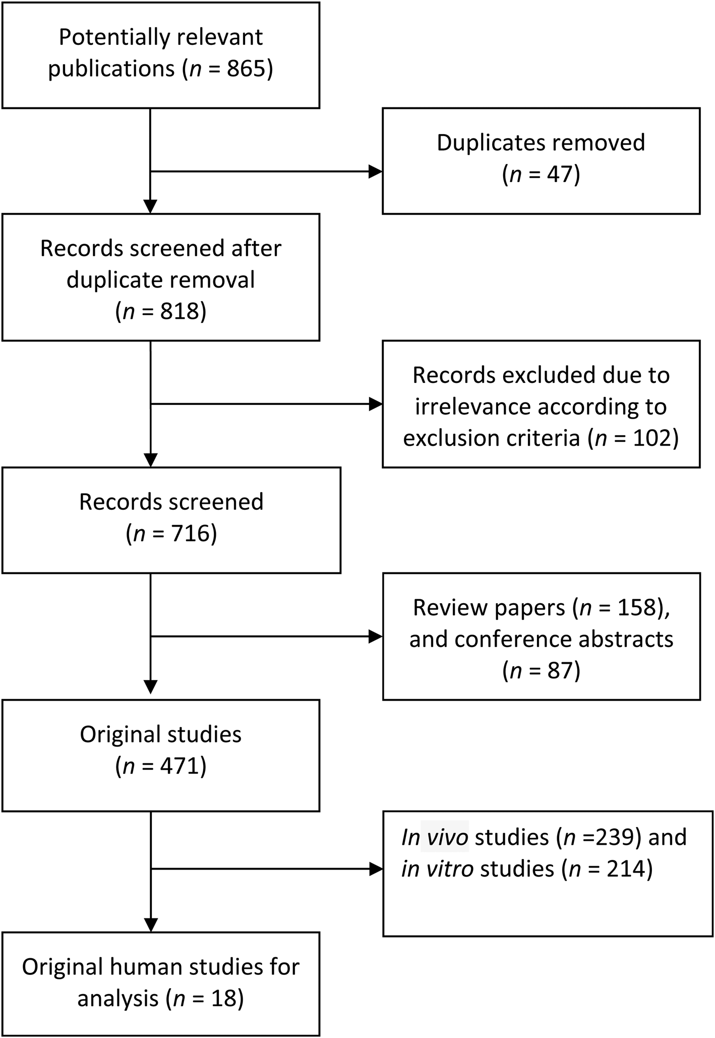
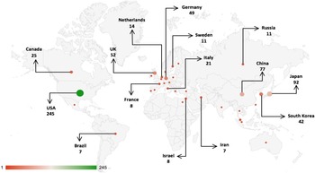
Of 865 articles found, 149 were excluded because of duplication, replication of a previous study or irrelevance, leaving 716 publications (Figure 2). Institutions in the USA contributed 245 publications; institutions from East Asia and Western Europe also contributed substantial numbers. Figure 3 shows an annotated map that highlights countries that contributed over five publications.
Selection of articles for further analysis.

Annotated map highlighting countries that contributed at least five publications.

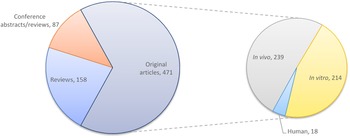
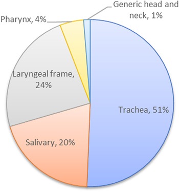
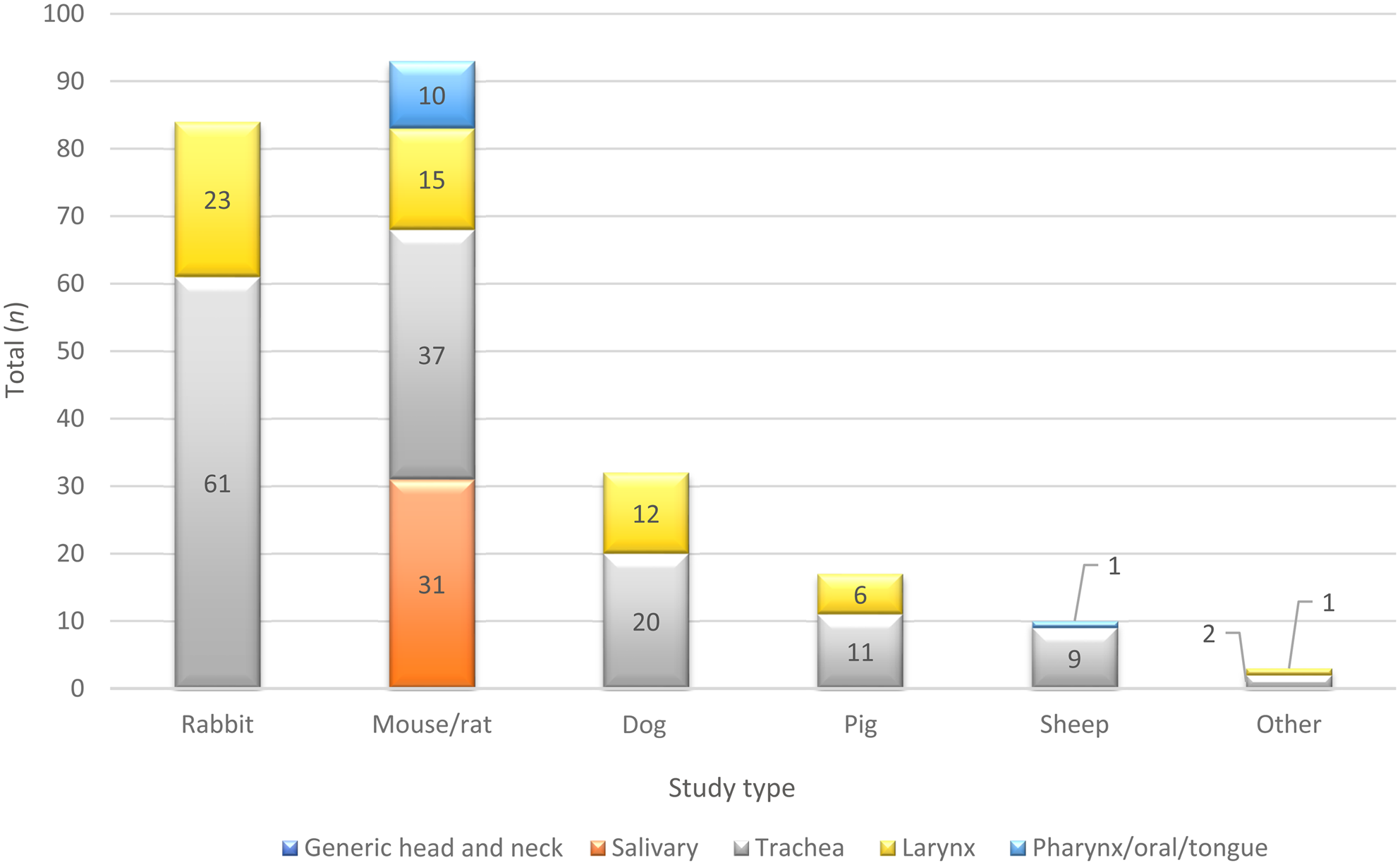
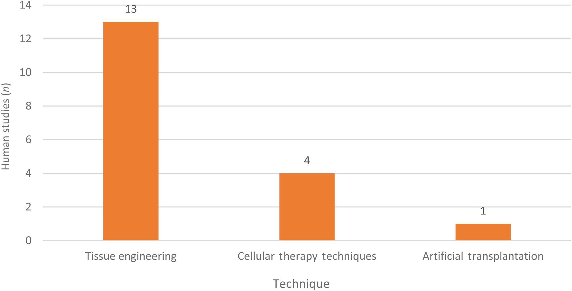
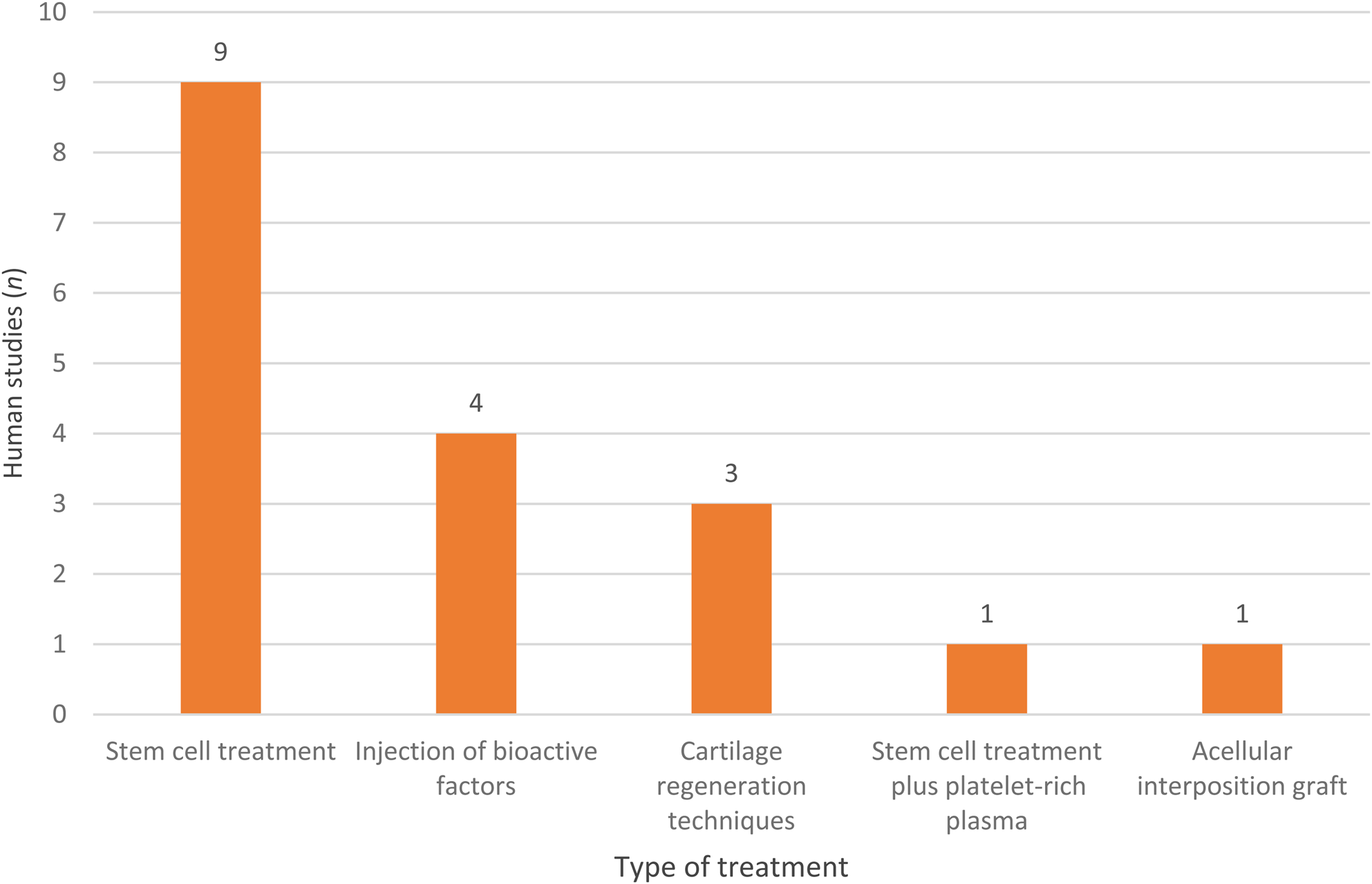
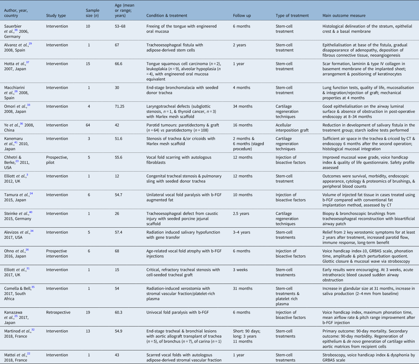
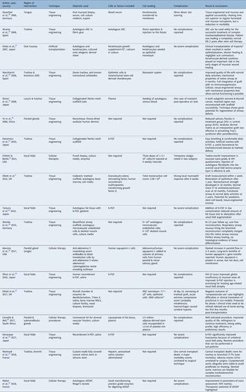
Figure 4 subdivides the 716 publications into 158 reviews, 87 conference abstracts and 471 original articles, of which 239 were in vivo, 214 in vitro and 18 were original human studies. Fifteen human studies were interventional, 1 was retrospective and 2 were prospective (Tables 1 and 2). Figures 5 and 6 represent the breakdown of original studies and animal models used for in vivo studies by anatomical region. Figures 7 and 8 represent the techniques and treatments used in the human studies included in this review.
Categorisation of type of publications reviewed.

Distribution of all 471 original studies by anatomical region.

Breakdown of in vivo studies by animal used and anatomical region.

Techniques used in included human studies.

Types of treatments in included human studies.

Basic characteristics of included human studies

CT = computed tomography; b-FGF = basic fibroblast growth factor; GRBAS = grade, roughness, breathiness, asthenia, strain
The impact of interventions on the clinical outcomes in included human studies

*Cascade Biologics, Portland, USA; †AlloDerm, LifeCell, Branchburg, USA. ASC = adipose derived stem cells; b-FGF = basic fibroblast growth factor; VHI = voice handicap index; CT = computed tomography; MSC = mesenchymal stem cell; ADSVF = adipose-derived stromal vascular fraction
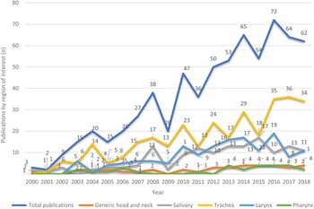
Within a general year-on-year increase in total publications, 2016 saw the largest number with 72 publications, of which 35 were trachea focused, a similar proportion seen overall (Figure 9). One noticeable dip in 2008 to 2009 coincided with a global economic recession.
Timeline of publications.

Upper aerodigestive tract disorders
For the purposes of this article, upper aerodigestive tract was defined as the trachea, larynx, salivary glands, and tongue and pharynx. Relevant disorders that were targets of human studies are outlined.
Vocal folds
The vocal folds are the functional unit of the larynx. Scar formation and fibrosis secondary to abuse, iatrogenic injury, irradiation or trauma may cause vocal loss or change. This review identified two main regenerative strategies to reduce scarring: the first is inhibition of fibrosis and repair of scarring, and the second is regeneration of the extracellular matrix. Biomaterials for correcting defects require similar viscoelastic mechanics to the natural lamina propria (the vibratory component of vocal folds) and must facilitate tissue regeneration. Through techniques alluded to later, these show potential for addressing vocal fold fibrosis and age-related vocal fold atrophy.Reference Mattei, Magalon, Bertrand, Grimaud, Revis and Velier22–Reference Ohno, Hirano, Yasumoto, Ikeda, Takebayashi and Miura26
Trachea
Tracheal defects, or congenital or acquired stenosis, may cause variable degrees of airway obstruction. Tracheal resection or aggressive radiotherapy may be necessary for benign or malignant disease, with resectable lengths under 30 per cent in children or 6 cm in adults.Reference Kirschbaum, Teymoortash, Suárez, Shah, Silver and Nixon27 Tissue engineering and stem cell implantation, the most prevalent techniques found, have had mixed success in treating tracheoesophageal fistula, lesions and stenosis.Reference Elliott, De Coppi, Speggiorin, Roebuck, Butler and Samuel3,Reference Macchiarini, Jungebluth, Go, Asnaghi, Rees and Cogan28–Reference Omori, Tada, Suzuki, Nomoto, Matsuzuka and Kobayashi33
Salivary glands
Radiation-induced salivary hypofunction has been the main target of studies attempting to restore salivary flow.Reference Alevizos, Zheng, Cotrim, Liu, McCullagh and Billings34,Reference Comella and Bell35 Studies have treated irradiated glands with gene editing,Reference Alevizos, Zheng, Cotrim, Liu, McCullagh and Billings34 adipose-derived stem-cells from stromal vascular fractionReference Comella and Bell35 and have used acellular dermal matrix as an interposition graft post-parotidectomy to target Frey's syndrome.Reference Ye, Zhu, Zheng, Wang, Zhao and Zhong36
Tongue
Tissue-engineered oral mucosa has been used to repair defects from tongue carcinoma and freeing of the tongue with promising mucosal integration of graft,Reference Hotta, Yokoo, Terashi and Komori37 tongue movability and quality of life indicators.Reference Sauerbier, Gutwald, Wiedmann-Al-Ahmad, Lauer and Schmelzeisen38
Regenerative techniques
The strategies found in human clinical studies over the last two decades are outlined and include: (1) cell transplantation, (2) injection of bioactive factors, (3) scaffolds and (4) bioengineering of laryngeal structures.
Cell transplantation
For vocal fold regeneration, both autologous fibroblasts and stem cells have been used (Tables 1 and 2).Reference Mattei, Magalon, Bertrand, Grimaud, Revis and Velier22–Reference Ohno, Hirano, Yasumoto, Ikeda, Takebayashi and Miura26 Stem cell-based tissue engineering and gene therapy is practised in the optimal cellular microenvironment under the influence of regulatory proteins. Culturing should be strictly monitored to prevent early genetic changes occurring within the stem cells, given that they may undergo carcinogenesis after implantation. They have been used for autologous tissue generation in grafts and transplantation for vocal fold regeneration, tracheoesophageal fistula, xerostomia, congenital tracheal stenosis and pulmonary sling.Reference Elliott, De Coppi, Speggiorin, Roebuck, Butler and Samuel3,Reference Alvarez, García-Arranz, Georgiev-Hristov and García-Olmo29,Reference Li, Stiadle, Lau, Zerdoum, Jia and Thibeault39 For example, in one case report, a bioartificial airway patch (BioVasc, Interfacial Engineering and Biotechnology, Stuttgart, Germany) seeded with autologous microvascular endothelial cells (9 × 10⁶ microvascular endothelial cells) and skeletal muscle cells (6 × 10⁶ skeletal muscle cells) was implanted into a defect extending from the trachea to the left main bronchus following caustic injury. The microvascular endothelial cells were grown under standard conditions (37°C, 5 per cent carbon dioxide) for 4 weeks in endothelial cell growth medium (Promocell, Heidelberg, Germany). The skeletal muscle cells were grown in 4°C Dulbecco's modified Eagle's medium supplemented with 15 per cent foetal calf serum. Bronchoscopic biopsies confirmed scar-free airway reconstruction, showed seeded cells merged into native airway mucosa and showed no histological evidence of tissue dedifferentiation.Reference Steinke, Dally, Friedel, Walles and Walles40
Injection of bioactive factors
Bioactive factors are mainly growth factors that regulate cell proliferation and differentiation. The most studied and clinically successful growth factor is basic fibroblast growth factor (Tables 1 and 2). In one report, 19 subjects were injected with 50 μg basic fibroblast growth factor into the vocal muscle layer. After six months, voice handicap index, maximum phonation time, mean airflow rate and pitch range significantly improved.Reference Kanazawa, Kurakami, Kashima, Konomi, Komazawa and Nakamura25 It is furthermore a relatively routine procedure. In another study for age-related vocal fold atrophy, 10 mcg basic fibroblast growth factor in 0.5 ml of saline was injected trans-orally into the superficial lamina propria on both sides of the vocal folds. Voice handicap index-10 scores, glottal insufficiency and mucosal wave scores all improved.Reference Ohno, Hirano, Yasumoto, Ikeda, Takebayashi and Miura26
Scaffolds
Scaffolds involve decellularised organ matrix, biological polymers, synthetic biomimetic hydrogels and synthetic polymers (Tables 1 and 2). Injectable scaffolds have been used for three-dimensional vocal fold lamina propria replacement,Reference Li, Stiadle, Lau, Zerdoum, Jia and Thibeault39 providing ease of application, requisite biomechanical properties, and delivery of cells and growth factors. They have also modulated the autoinflammatory response and facilitated remodelling of the extracellular matrix elsewhere. Rehydrated and shaped Renovtissue sheets (freeze-dried acellular human dermis; Beijing Qing Yuan Wei Ye Bio-tissue Engineering Company, Beijing, China) successfully reduced salivary fistula rates after superficial or partial parotidectomy (Tables 1 and 2).Reference Ye, Zhu, Zheng, Wang, Zhao and Zhong36
Marlex mesh (CR Bard, Billerica, USA) tube scaffolds with collagen sponge have been used for covering tracheal defects secondary to tracheal or subglottic stenosis, airway reconstruction, and thyroid cancer (Tables 1 and 2). Kanemaru et al. used polypropylene Marlex mesh covered with porcine dermal atelocollagen sponge of type I (70 per cent) and type III (30 per cent) collagen,Reference Kanemaru, Hirano, Umeda, Yamashita, Suehiro and Nakamura41 freeze-dried and cross-linked in a vacuum dry oven for several weeks. The scaffold is sutured to the resected or deficient tracheal segment along with venous blood and basic fibroblast growth factor.
Bioengineered laryngeal structures
Decellularised human larynges, infused with growth factors for angiogenesis and neovascularisation, have mechanical properties similar to original tissue.Reference Baiguera, Gonfiotti, Jaus, Comin, Paglierani and Del Gaudio42 Either cryopreserved allografts or cadaveric tracheal scaffolds have been used (Tables 1 and 2). If refined, these may provide functional partial laryngectomy reconstructive options or scaffold for total laryngeal regeneration.
Discussion
Over 90 per cent of the original studies originated from North America, Western Europe or East Asia, reflecting the global distribution of combined scientific output within the biotechnology sector.Reference Mikulic14 Alongside other industries, the centralisation of expertise and resources drives productivity, a positive sign for potential growth in regenerative therapies targeting upper aerodigestive tract disease.
Fifty-one per cent of original studies focused on airway regeneration and replacement. A steeper upward trajectory can be observed in airway research compared with other upper aerodigestive tract regions over the past 15 years (Figure 9). Eight of the 18 example human studies reported on airway replacement (trachea or a main bronchus).Reference Elliott, De Coppi, Speggiorin, Roebuck, Butler and Samuel3,Reference Macchiarini, Jungebluth, Go, Asnaghi, Rees and Cogan28,Reference Alvarez, García-Arranz, Georgiev-Hristov and García-Olmo29,Reference Elliott, Butler, Varanou-Jenkins, Partington, Carvalho and Samuel31–Reference Omori, Tada, Suzuki, Nomoto, Matsuzuka and Kobayashi33,Reference Steinke, Dally, Friedel, Walles and Walles40,Reference Kanemaru, Hirano, Umeda, Yamashita, Suehiro and Nakamura41 It is important to separate partial from total airway replacement therapy procedures. In partial tracheal replacement, the native trachea frame is left intact and provides mechanical support for the airway framework. The implant re-vascularises and re-epithelialises from native trachea vessels, countenancing loss of mechanical integrity and delayed epithelialisation, as reported by good functional outcomes and safety profiles.Reference Alvarez, García-Arranz, Georgiev-Hristov and García-Olmo29,Reference Omori, Tada, Suzuki, Nomoto, Matsuzuka and Kobayashi33,Reference Steinke, Dally, Friedel, Walles and Walles40,Reference Kanemaru, Hirano, Umeda, Yamashita, Suehiro and Nakamura41 However, total tracheal replacement requires that the replacement construct mechanically withstands the dynamic forces of the entire airway. The lack of distinct tracheal blood supply requires implant pre-vascularisation or a derivation of nutrients from in-growing vasculature from proximal and distal anastomoses.
This review found four human studies reporting total tissue-engineered tracheal or bronchial replacement.Reference Elliott, De Coppi, Speggiorin, Roebuck, Butler and Samuel3,Reference Macchiarini, Jungebluth, Go, Asnaghi, Rees and Cogan28,Reference Elliott, Butler, Varanou-Jenkins, Partington, Carvalho and Samuel31,Reference Martinod, Chouahnia, Radu, Joudiou, Uzunhan and Bensidhoum32 Three were single-patient case reports of decellularised tracheal segments repopulated with autologous epithelial cells. One patient died three weeks post-operatively; the other two required long-term stenting and periods of intensive hospital care.Reference Elliott, De Coppi, Speggiorin, Roebuck, Butler and Samuel3,Reference Macchiarini, Jungebluth, Go, Asnaghi, Rees and Cogan28,Reference Hamilton, Kanani, Roebuck, Hewitt, Cetto and Culme-Seymour43,Reference Gonfiotti, Jaus, Barale, Baiguera, Comin and Lavorini44 The other study used aortic homograft for tracheal replacement.Reference Martinod, Paquet, Dutau, Radu, Bensidhoum and Abad30,Reference Martinod, Chouahnia, Radu, Joudiou, Uzunhan and Bensidhoum32 Despite medical complications, no mortality causally related to the surgery was reported, and 80 per cent of recipients were free of airway prostheses after a mean of 18.2 months.Reference Martinod, Chouahnia, Radu, Joudiou, Uzunhan and Bensidhoum32 Although larger, multicentre trials are required for full effectiveness and safety profiling, the relative success and more mature development stage of aortic homograft as a scaffold for total tracheal or bronchi replacement warrants future targeted research.
Although refractory long-segment tracheal stenosis severely impacts sufferers’ life expectancy and quality of life, it is a relatively rare condition. Tracheal stenosis has been treated by tissue-engineered artificial trachea and stem cell based tracheal replacement for congenital tracheal stenosis and pulmonary sling.Reference Elliott, De Coppi, Speggiorin, Roebuck, Butler and Samuel3,Reference Elliott, Butler, Varanou-Jenkins, Partington, Carvalho and Samuel31,Reference Kanemaru, Hirano, Umeda, Yamashita, Suehiro and Nakamura41 Artificial tracheal implantation with collagenated Marlex mesh, performed via a staged operation in the presence of basic fibroblast growth factor, has shown promise in treating tracheal stenosis and defects. Patients reported easy breathing and pain-free daily activities.Reference Kanemaru, Hirano, Umeda, Yamashita, Suehiro and Nakamura41 In 2012, Elliott et al. reported a decellularised cadaveric tracheal scaffold for congenital tracheal stenosis and pulmonary sling.Reference Elliott, De Coppi, Speggiorin, Roebuck, Butler and Samuel3 Bone marrow mesenchymal stem cells were seeded (total mononuclear cell counts 2.56 × 10⁸) and grafted with patches of autologous epithelium. Topical human recombinant erythropoietin was applied to encourage angiogenesis and transforming growth factor β for chondrogenesis. During follow up, the patient developed a strong neutrophil response after eight weeks, generating luminal DNA neutrophil extracellular traps. At two years, the patient had a functional airway and normal daily activities.Reference Elliott, De Coppi, Speggiorin, Roebuck, Butler and Samuel3 As a paediatric study on a single patient, results are not fully applicable; validation is required with larger samples.
In this review of 111 laryngeal framework focused original studies, 80 concerned the vocal folds, and at least 45 primarily concerned the restoration or regeneration of scarred or fibrotic vocal tissue. Refractory vocal fold fibrosis occurs when scar tissue anchors the epithelial layer to the underlying vocal ligament, preventing effective propagation of the mucosal wave during phonation, inducing chronic hoarseness. This annually affects thousands worldwide and can disastrously impact quality of life. Cell therapies, small molecules and injected biomaterials all saw moderate success in overcoming fibrosis in human studies.
In one case, an autologous-derived stromal vascular fraction was injected locally into scarred vocal fold, resulting in improved voice measurements,Reference Mattei, Magalon, Bertrand, Grimaud, Revis and Velier22 an improvement on earlier studies using autologous fat or fascia alone.Reference Neuenschwander, Sataloff, Abaza, Hawkshaw, Reiter and Spiegel45–Reference Hsiung, Woo, Minasian and Schaefer Mojica47 Another group trialled multiple injections of autologous fibroblasts sourced from buccal mucosa into the lamina propria. Three doses of 1–2 × 10⁷ cells/ml were given at four-weekly intervals. Improvement was seen in voice handicap scores and mucosal wave grade in all five subjects, with minimal complications.Reference Chhetri and Berke23 Autologous fibroblast injection into scarred lamina propria was well tolerated, although two subjects reported transient otalgia post-treatment. Despite the potential for injectable regenerative therapies to address vocal fold fibrosis, the literature lacks consensus on the optimal cell type, material and delivery method for laryngeal regeneration. Any injectable requires durability and similar viscoelastic properties to the lamina propria.Reference Chhetri and Berke23 Future studies should target determining the most effective method, which would attract investment around a specific design, streamlining production towards scalable and efficacious novel therapies.
Xerostomia results from hyposalivation and commonly follows radiotherapy for upper aerodigestive tract cancer. Its relatively high incidence and paucity of effective treatments makes it a clear target for regenerative therapy to replace or repair damaged salivary tissue. Twenty per cent of studies in this review focused on the salivary system, although only one human case report and one series of five patients were found that aimed to restore salivary function.Reference Alevizos, Zheng, Cotrim, Liu, McCullagh and Billings34,Reference Comella and Bell35 Pre-clinical studies broadly divided into studies regenerating salivary tissue in vitro and re-implanting to restore function, and studies that implanted cells, such as adipose-derived stem cells,Reference Comella and Bell35 or used gene editingReference Alevizos, Zheng, Cotrim, Liu, McCullagh and Billings34 to stimulate regeneration of existing salivary tissue. The discovery of salivary stem cells that may repair or replace damaged salivary tissue provides another target for regenerative treatments. One example is work by Banh et al. in which activation of aldehyde dehydrogenase 3, a marker of submandibular stem cells, induced an increase in stem cell yield following extraction in a murine model.Reference Banh, Xiao, Cao, Chen, Kuo and Krakow48
Only eight original studies primarily concerned tongue regeneration.Reference Bunaprasert, Hadlock, Marler, Kobler, Cowan and Faquin49–Reference Zhang, Shi, Xu, Zhang, Shanti and Le56 This is surprising given fibrosis or loss of tongue volume commonly follows radiotherapy and surgery for upper aerodigestive tract cancer and significantly affects phonation and swallow. Cell therapy procedures and small molecules that can overcome fibrosis, as with the vocal folds, would benefit patients and would be relatively easy to deploy endoscopically as a graft or injectable. Furthermore, the use of biomaterials and tissue engineering to restore muscle bulk would be advantageous in upper aerodigestive tract cancer patients, alongside stroke patients with atrophy following denervation of musculature. Tongue tissue mainly consists of striated muscle that is highly vascular, rendering ex vivo fabrication and implantation more achievable than complex organs with different cellular layers and no distinct blood supply. In 10 cases of freeing of the tongue, oral mucosa was harvested, divided and resuspended in culture media. Cultured keratinocytes were transferred to the membrane (2 × 10 cells/cm2). The procedure was successful; however, healing time and the outcome did not appear to be superior to regular harvested oral mucosa transplants.Reference Sauerbier, Gutwald, Wiedmann-Al-Ahmad, Lauer and Schmelzeisen38 Regenerative therapies aimed at restoring tongue bulk and overcoming fibrosis are therefore clear additional targets for future research activity.
This review is not without limitations. Firstly, despite many studies pertaining to regenerative medicine or surgery in the upper aerodigestive tract, further analysis was restricted to human studies. Future review should compare the techniques and performance of different animal models for different anatomical sites. Secondly, assessing a risk of bias was not practical in the human studies because of the lack of retrospective designs. Finally, future review should focus on conducting a meta-analysis to observe the impact of techniques on outcome.
Conclusion
This review suggests that a future shift towards developing therapy procedures that address pathologies with a higher disease burden may be warranted, particularly with respect to the upper airways, the oropharyngeal mucosa and tongue. Reconstruction or repair of upper aerodigestive tract defects and restoring function will improve the lives of millions of ailing patients. Regenerative techniques may decrease operating time, eliminate the need of a donor site, improve healing and improve quality of life outcomes. Reconstructing laryngeal structures by tissue engineering allows patients to self-express, breathe unaided and in some instances, withstand the secondary effects of cancer treatment. This field of medicine is highly promising for the upper aerodigestive tract region, and its applications are only limited by our current knowledge. The demand for regenerative medicine will continue as upper aerodigestive tract cancer persists as the sixth most common cancer globally.
Competing interests
None declared