Introduction
Sensing environmental conditions allow organisms to detect different stimuli and adapt to changes accordingly (Chown & Terblanche, Reference Chown and Terblanche2006). Typically, various sensory modalities are involved in different behaviours. However, the main sensory modalities used depend on the specific organism and the particular task they perform. In the case of predators, vision and olfaction are involved in many different behaviours. Parasitoids can be considered a particular type of predators and, concerning host-location, they use similar strategies as insect predators for the location of a prey (Godfray, Reference Godfray1994). For parasitoids, mechanoreception and chemoreception are within the main sensory modalities used in the acquisition of information from the environment (Gullan & Cranston, Reference Gullan and Cranston2010; Chapman et al., Reference Chapman, Simpson and Douglas2013; Klowden, Reference Klowden2013).
Mechanoreception allows responses to different mechanical stimuli as air currents, touch, sound and gravity, but also to thermal, hygric and infrared light (Keil, Reference Keil1997; Ryan, Reference Ryan2002; Nation, Reference Nation2015). These stimuli are involved in different behaviours, such as intraspecific communication (e.g., the song of crickets), predator avoidance (e.g., moths avoiding bats), alarm behaviour (filiform hairs on the cerci of crickets and cockroaches), prey detection (e.g., antlion larvae in funnel-shaped pits in sand) or sensing environmental conditions. Bristles, campaniform sensilla and hair plates are within the main receptors involved in these behaviours (Ryan, Reference Ryan2002). Morphologically, a distinctive feature of mechanoreceptors is the absence of pores opening on the cuticular surface (Nation, Reference Nation2015).
Chemoreception involves responses to chemical stimuli that can vary in the distance at which they are detected and the kind of behaviours they elicit (i.e., attractants or repellents). Chemoreception is particularly important for orientation of light-deprived insects, such as nocturnal or soil-dwelling species (Gullan & Cranston, Reference Gullan and Cranston2010; Klowden, Reference Klowden2013). Chemoreceptors can be basically divided into olfactory and gustatory receptors. Olfactory receptors or basiconic sensilla tend to have multiple pores at the cuticular surface, while gustatory receptors or trichoid/chaetic sensilla tend to have a single pore, usually at the tip of a hair (Zacharuk, Reference Zacharuk1980; Ryan, Reference Ryan2002). Olfactory receptors are often concentrated on the antennae while gustatory receptors are often located on the palps (Nation, Reference Nation2015).
There are many studies that have shown the morphology of many different sensory structures of different insects, particularly adults, but there are few studies where the functionality of the sensory organs has been established (Eilers et al., Reference Eilers, Talarico, Hansson, Hilker and Reinecke2012). Furthermore, studies on larval insect stages are usually focused on agricultural pests such as scarab beetle larvae (Eilers et al., Reference Eilers, Talarico, Hansson, Hilker and Reinecke2012; Xu et al., Reference Xu, Zhang, Yang, Ren, Wang and Zong2015). In the case of parasitoids, the studies conducted deal only with hymenopteran parasitoids where the adult stage locates the host (Baaren et al., Reference Baaren, Boivin, Bourdais, Roux, Mendez-Vilas and Dias Alvarez2007). In this work, we studied the morphology and function of the sensory structures of the larva of a dipteran parasitoid.
Mallophora ruficauda (Diptera: Asilidae) is an ectoparasitoid that during its larval stage attacks scarab beetle larvae. Female M. ruficauda lays eggs on clutches on tall grasses and once larvae are born, they are wind dispersed and fall to the ground (Castelo et al., Reference Castelo, Ney-Nifle, Corley and Bernstein2006). Once in the ground, they bury themselves and, after 7 days in average, moult to the second instar (Crespo & Castelo, Reference Crespo and Castelo2010). It is noteworthy that these larvae are capable of moulting to the second instar even in absence of any host-related cue. Once larvae moult to the second instar, they start searching for its preferred host, third-instar larva of Cyclocephala signaticollis (Coleoptera: Scarabaeidae). Larvae detect their hosts with sensilla located in the maxillary palps (Crespo et al., Reference Crespo, Lazzari and Castelo2011). These sensilla detect a specific odour produced in the fermentation chamber of their hosts (Castelo & Lazzari, Reference Castelo and Lazzari2004; Groba & Castelo, Reference Groba and Castelo2012). After orienting towards a suitable host, the larva touches the host body and decides whether to accept it, hence attaching its mouthparts to the integument or not. If the host is accepted, both parasitoid larva and host remain through the whole winter in this state. When springtime comes, soil temperature increases and the parasitoid accelerates its development rate, moults until reaching the fifth instar in a very short period of time (ca. 3 weeks) and kills the host (Crespo & Castelo, Reference Crespo and Castelo2010). After, the parasitoid larva pupates and the adult emerges some time later. Regarding orientation to the host, it has been determined that only second-instar larvae are attracted to host odours (Crespo & Castelo, Reference Crespo and Castelo2008). In order to complete its development, the host must be found and parasitized, while the larva is a second instar since moulting to the third instar only occurs when a host is parasitized (Crespo & Castelo, Reference Crespo and Castelo2010). Different experiments show that larvae can orient to hosts odours with only one maxillary palp functional but not without them (Crespo et al., Reference Crespo, Lazzari and Castelo2011). Additionally, it has been seen that first-instar larvae (that cannot orient to hosts) have increased levels of exploratory movements when exposed to hosts odours indicating that other sensory structures may be involved in the detection of hosts (Crespo & Castelo, Reference Crespo and Castelo2008). In fact, optical microscope images show that first-instar larvae have their cephalic structures much less developed, particularly the maxillary palps (Crespo & Castelo, Reference Crespo and Castelo2010).
Given that almost no information is available on the morphology and function of active host-seeking Asilidae larvae sensory structures, we used larvae of M. ruficauda to study the importance and influence of both antennae and maxillary palps during host orientation.
Materials and methods
Insects
Larvae of M. ruficauda were obtained from egg-clutches collected in grasslands in Moreno (60°34′S, 58°79′W) a locality associated with apiculture and agricultural practices in a suburban zone in Buenos Aires province, Argentina. Immediately after hatching, neonatal larvae were separated in flasks at a density of 600 larvae per 100 ml of potting soil. This density ensured that larvae were non-selective and would orient to hosts odours even if recognized as low-quality hosts (Crespo et al., Reference Crespo, Martínez and Castelo2015). Since larvae live buried in the soil, flasks where kept in darkness, at room temperature (26 ± 1 °C, 65 ± 5% RH) until larvae moulted to the second instar for their use in the experiments. Only second-instar larvae were used because host orientation only occurs after moulting (Crespo & Castelo, Reference Crespo and Castelo2008).
As hosts, third-instar larvae of C. signaticollis were used. Hosts were collected in Navarro (34°97′S, 59°25′W), a locality in the Pampas region of Argentina, and obtained directly by digging the soil at a depth of 30 cm. Each host was manually collected and put in black tubes and taken to the laboratory. Once in the laboratory, hosts were numbered and identified using the taxonomic key of Alvarado (1980), kept individually at room temperature (26 ± 3.5 °C) in black tubes filled with potting soil and fed weekly with fresh pieces of carrot.
Morphology of sensory structures
In order to describe the sensory structures of second-instar larva, we fixed larvae in absolute ethyl alcohol for 2 days before sticking them in carbon tape in aluminium stubs. Larvae were then coated with gold and photographs were taken with a scanning electron microscope (SEM, Carl Zeiss NTS-Supra 40). The description and classification of the different sensory structures and sensilla were made by direct comparison with the bibliography (Zacharuk et al., Reference Zacharuk, Yin and Blue1971; Nicastro et al., Reference Nicastro, Melzer, Hruschka and Smola1998; Giglio et al., Reference Giglio, Ferrero, Perrotta, Tripepi and Brandmayr2003; Eilers et al., Reference Eilers, Talarico, Hansson, Hilker and Reinecke2012).
Function of sensory structures
In order to determine the role of the sensory structures and sensilla described in the previous section, we occluded the different structures in second-instar larvae of M. ruficauda and then experimentally tested for the orientation to the host in an air-stationary olfactometer.
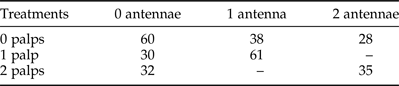
The occlusion of the putative sensory organs was achieved by placing a larva on a dry cooling device developed ad-hoc under a stereomicroscope (40×) to anaesthetize them and easily manipulate them. In brief, we controlled a Peltier element with an Arduino UNO device to expose larvae to low temperatures (4 ± 1 °C). After 30 s in the cold plate, a capillar with a blunt tip was used to gently occlude the sensory organs with a translucent, medium viscosity silicone adhesive (Kwik-Sil, World Precision Instruments). This silicone is suitable for working with living animals because it is non-toxic and has a very high curing speed. After occluding the specific structure with the Kwik-Sil, the larva was allowed for a few minutes to recover and then used in the trial. Either antennae, maxillary palps or both structures were occluded (table 1).
Combination of treatments tested in the present study. 0, 1 and 2 antennae or palps correspond to the amount of structures that were occluded with the silicone.

Note that the combinations 2 antennae–1 palp occluded and 1 antenna–2 palps occluded were not done because when a structure is totally occluded (either two antennae or two palps) no orientation is observed. Numbers indicate trials performed for each treatment.
The orientation behaviour was tested in an experimental arena (15.5 × 4.0 × 3.5 cm3) divided into three equal size zones (one central and two lateral) along the long axis. In every trial, a live host was placed in one of the lateral zones (stimulus zone), while in the opposite zone no host was placed (control zone). A plastic mesh was placed in the lateral zones to avoid the host escapes but allowing the parasitoid larva to move freely around the arena.
At the beginning of each trial, a larva was released at the centre of the arena and, after 60 min, its position was recorded. The experimental arena was placed inside a box to keep darkness. Three possible responses could be obtained depending on the final position of the larva: choice for the stimulus (S), for the control (C) or no decision (ND) if the larva remained in the middle zone. Relative humidity was maintained high with a humid filter paper enclosed in the inside top of the experimental arena.
Between trials, the arena was cleaned with non-ionic detergent and alcohol to eliminate any possible remaining cue. All experiments were conducted between 10:00 and 17:00 h under laboratory temperature conditions (26 ± 3.5 °C) on days where the barometric pressure did not fall at a higher speed than 0.85 hPa h−1, and when the absolute value was between 1011 and 1023 mbar, because previous studies have shown that drops in barometric pressure can halt the foraging activity (Crespo & Castelo, Reference Crespo and Castelo2012).
We conducted three control and five experimental series. A control for the experimental arena consisted of measuring orientation of intact larvae in the arena without any host in order to test whether larvae were able to show random movements along the whole arena. For the positive control, we tested the orientation of intact larvae to intact hosts, expecting the higher orientation response. Finally, the sham control consisted of painting a part of the cephalic capsule of larvae without occluding any of the sensory structures and exposing these larvae to hosts in order to test that the manipulation did not affect the proneness to orient to the host. Regarding experimental series, either antennae or maxillary palps were occluded in every possible combination as depicted in table 1.
Host acceptance
In order to study the influence of the antennae and maxillary palps in the acceptance of hosts, larvae that were used in the orientation experiments were placed on the thorax of an unparasitized host to perform an acceptance experiment. After 5 days, parasitism occurrence on hosts was recorded under a stereomicroscope (40×). When the parasitoid larva was attached to the host integument, host acceptance was considered as positive.
Statistical analysis
For orientation experiments, the proportion of larvae orienting to host odours was analysed by means of generalized linear models (GLMs) assuming a binomial distribution of error variances and a logit link function (McCullagh & Nelder, Reference McCullagh and Nelder1989). For analysing the control series we compared the proportion of larvae that oriented to host odours in the negative control and the sham control by means of Chi-square (χ2) tests assuming a random orientation as the expected frequencies (Zar, Reference Zar2010). For analysing the experimental series, we tested that when occluding one palp, one antenna or one antenna and one palp, the results did not vary regarding which side (left, right or crossed) was occluded. For this, we compared by means of a Pearson's χ2 test with Yeates’ continuity correction the proportion of larvae that oriented to the host for each case (Zar, Reference Zar2010). This resulted in three tests. Then the orientation to the host when occluding antennae or palps were analysed separately generating four different models with increasing factors.
Four models were constructed and the one that better explained the results was retained. The first two models (1, palp model; 2, antenna model) only estimated the effect of occluding one or two organs (depending on the model) on the proportion of larvae orienting to the host. The following two models accounted for the effect of occluding both structures simultaneously with and without the interaction (3, palp–antenna model and 4, full palp–antenna model, respectively). Orientation of intact larvae to the host, the positive control, was used as the reference level (intercept) on every model.
For every model we estimated the parameters and evaluated the significance of the variables/factors in the models by the P value obtained from the GLM. Additionally, a model was rendered as a candidate model to explain the data if the residual deviance was similar to the degrees of freedom. Finally, an analysis of deviance between the candidate models was performed to test if the difference in the residual deviance is significantly lower with increasing complexity of the models (Caffo, Reference Caffo2015).
For the host acceptance experiments, the proportion of larvae under the different treatments that accepted a host was analysed with tests of homogeneity of proportions, which are multiple Tukey-type comparison tests (Zar, Reference Zar2010). Then, when differences were found, a posteriori contrasts were performed comparing the proportion of larvae that accepted a host for every larval treatment, with its corresponding control series in a procedure analogous to the Dunett's test but applied when proportions are used (Zar, Reference Zar2010). All the analyses were performed in R 3.2.4 (R Core Team, 2016). Graphics were performed with the library ‘ggplot2’ in R (Wickham, Reference Wickham2009).
Results
Morphology of sensory structures
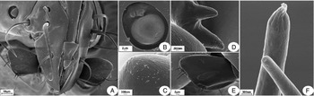
SEM photographs show that M. ruficauda larva has different sensorial structures mainly located in the antennae and maxillary palps (fig. 1A). We found that the antenna has three distinctive sensilla (fig. 1B). The bigger structure is a dome-like sensillum that, according to different authors, is a multiporous chemosensillum (fig. 1C; Zacharuk et al., Reference Zacharuk, Yin and Blue1971; Nicastro et al., Reference Nicastro, Melzer, Hruschka and Smola1998). The other two sensilla are smaller in size and different in morphology. One of the sensillum is a peg-like sensillum with finger-like processes (fig. 1D, upper). This sensillum has channels and could also have a chemosensorial function according to different authors (Bay & Pitts, Reference Bay and Pitts1976; White & Bay, Reference White and Bay1980; Henderson & Wellington, Reference Henderson and Wellington1982; Mayo et al., Reference Mayo, Anderson, Burguete and Chillida1987; Nicastro et al., Reference Nicastro, Melzer, Hruschka and Smola1998). Finally, the third sensillum is a peg-like sensillum, but instead of channels it has a coarse surface and unlike the other sensilla, its function is unknown (fig, 1D, lower).
External morphology of the cephalic capsule of the second-instar larva of M. ruficauda. (A) General view showing the disposition of antennae, maxillary palps and mouthparts. (B) Detail of the antennae showing three distinct sensilla. (C) Detail of the sensorial cone with probable chemosensorial function. (D) Detail of the peg-like sensillum with finger-like processes and the peg-like sensillum with coarse surface. (E) Detail of a maxillary palp showing five chemosensorial sensilla and the digitiform sensillum. (F) Detail of the two types of sensilla found in the maxillary palps. One sensillum has finger-like processes and the other one has an apical pore typical of chemosensors.

Regarding the maxillary palps, we could identify five sensilla distributed in two groups in the tip and other sensillum in the frontal surface of the palps (fig. 1E). The distal group in the palp tip has two styloconic sensilla with finger-like processes at the tip and a uniporous chaetic sensillum. The other group of sensilla is found proximate to the body and consists of one styloconic sensillum with finger-like processes and a uniporous chaetic sensillum (fig. 1F). Based on the described morphology, the proposed function of these types of structures is olfaction and gustatory-tactile, respectively (Zacharuk, Reference Zacharuk1980; Ryan, Reference Ryan2002). We also found on the dorsal view of the maxillary palp a digitiform sensillum. According to the available literature, it serves as a chemoreceptor but also as a mechanoreceptor and a thermo-hygroreceptor (Zacharuk et al., Reference Zacharuk, Albert and Bellamy1977; Honomichl & Guse, Reference Honomichl and Guse1981; Giglio et al., Reference Giglio, Ferrero, Perrotta, Tripepi and Brandmayr2003, Reference Giglio, Perrotta, Talarico, Zetto Brandmayr and Ferrero2013).
Function of sensory structures
The occlusion of antennae and maxillary palps influenced the orientation response of larvae to hosts. In general, we found that when larvae had either both the antennae or the maxillary palps occluded, they oriented at random in the experimental arena (table 2).
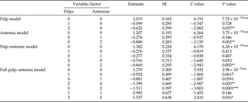
Estimated regression coefficients and standard errors (SE) of treatments that affect orientation to hosts.

Variable/factor: palps, number of palps occluded; antennae, number of antennae occluded.
* P < 0.05, ** P < 0.01.
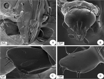
Regarding the control series, when larvae were not treated and no host was offered (negative control), as expected, they distributed randomly in the arena (χ2 (1;0.05) = 1.125, P = 0.289). The treatment of occlusion with the Kwik-seal silicon resulted in a very efficient non-invasive method for studying the function of the sensory structures. We were able to selectively occlude either the antennae or maxillary palps (fig. 2B, D).
Detail of the occlusion procedure of the antennae and maxillary palps for behavioural experiments. (A) Cephalic capsule showing the position of the antennae. (B) Cephalic capsule showing both antennae occluded with the silicone. (C) Maxillary palp without occlusion. (D) Maxillary palp occluded by the silicone.

In the case of the treatments, the use of silicone resulted innocuous since larvae from the sham control oriented to the host in similar levels as the positive control (χ2 (1;0.05) = 13.333, P < 0.001).
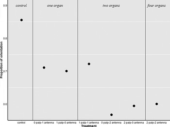
Both palp-model and antenna-model showed high levels of residual deviance (9.94 and 15.66 on 4 degrees of freedom). Although some factors showed differences with the intercept, these models performed badly so they were discarded (table 2). The palp–antenna model unified the information of previous models, including the information simultaneously. This model showed a residual deviance of 6.90 on 2 degrees of freedom, which is still very high and hence discarded. Finally the full palp–antenna model included the interaction between the two factors (occlusion of antennae and maxillary palps). As expected, when both antennae and maxillary palps were occluded the proportion of larvae that orient to the host was significantly lower than the intercept (table 2, fig. 3). However, when only one structure (an antenna or a maxillary palp) or two structures but of different type (an antenna and a maxillary palp) were occluded the proportion of larva orienting to the host was not significantly different from the intercept (table 2, fig. 3).
Proportion of larvae the oriented to a host for every treatment. Treatments: control, larvae had all sensorial structures functional (n = 60); one organ, larvae with either one antenna (n = 38) or one maxillary palp occluded (n = 30); two organs, larvae with one maxillary palp and one antenna occluded (n = 61) or with either two antennae (n = 28) or two maxillary palps occluded (n = 32); four organs, larvae with both antennae and both maxillary palps occluded (n = 35).

The final model retained was the full palp–antenna model which was supported by the ANOVA analysis on the models. The difference between the palp and antenna models was not statistically significant indicating that both models perform equally bad and should be discarded. The palp–antenna model reduced the residual deviance significantly (Deviance: 8.762, P < 0.013) indicating that the inclusion of both factors (palp and antenna) is necessary. The inclusion of the interaction between the factors was further supported since the full palp–antenna model significantly reduced the residual deviance (Deviance: 6.897, P < 0.032). In light of all the analysis, we retained the full palp–antenna model as the model that best explains the data.
Host acceptance
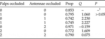
Regarding host acceptance, the experiments showed in general that the occlusion of the antennae or maxillary palps do not influence the acceptance of a host. A high proportion of larvae from every treatment attacked the host compared with the non-treated group (control series, table 3).
Proportion of treated larvae that accepted hosts compared with the proportion of accepted healthy third instar C. signaticollis (CS3) hosts.

Palps occluded, number of palps occluded. Antennae occluded, number of antennae occluded. Prop, proportion of larvae that accepted hosts. Q, statistic analogous to Dunnett's test (critical value: Q 0.05(1),∞,7 = 2.29). P > 0.05 denotes not statistically significant differences.
1 Since the response of untreated larvae to hosts was used as a control, there is no comparison against itself.
Discussion
In this work, we realized a morpho-functional study of the sensory structures that mediate orientation and acceptance of the host in the larva of M. ruficauda. We found conspicuous sensory structures located in the antennae and the maxillary palps. Regarding the influence of the different sensory structures on the location to the host, we were able to determine that orientation to the host requires of at least one functional antenna and one maxillary palp at the same time. Finally, regarding host acceptance, we found that neither the sensory structures located in the antennae nor in the maxillary palps are needed during the process of accepting the host.
Regarding the morphology, we were able to characterize the sensory structures in the second-instar larva of M. ruficauda. We found that the antennae have three different sensilla. Two of those sensilla have clearly chemosensorial function while the remaining one has an unknown function. The most prominent structure, the sensorial cone, is located at the distal tip of the last antennal article in all taxa where articles are externally visible. Ultrastructural analysis in other taxa revealed features indicating that it represents a multiporous chemosensillum. Another structure found in the antenna is an aporous grooved like sensillum with fingertip projections. These structures have been found in different larvae of Diptera species, but the channels have an electronic dense material showing that this structure is chemoreceptive (Nicastro et al., Reference Nicastro, Melzer, Hruschka and Smola1998). It should be noted that in M. ruficauda the adult has many multiporous sensilla, some uniporous sensilla and sensory pits with multiporous sensilla inside (Groba et al., Reference Groba, de Cidre and Castelo2013). Hence, sensory structures found in the larva may be the precursors of the ones that adult show and could have similar functions.
Regarding the maxillary palps we found two distinct groups of sensilla but with only two types of sensillum. Both types of sensilla are possibly chemosensorial. In this case, the first type of sensilla is a fingertip-like sensilla as the one found in the antennae but more elongated with olfactive function. The other type of sensillum is a uniporous sensillum with possible gustatory–tactile function, but there are some studies in adult arthropods showing that this type of sensillum can detect volatiles (Slifer, Reference Slifer1970). Finally, we found a digitiform sensillum that has been described in other species as a thermohygro receptor. If this digitiform sensillum has the same function in M. ruficauda then it would be very useful for the larva to avoid the surface after they bury in the soil. The fact that M. ruficauda larvae show many different kinds of chemosensorial sensilla could indicate the importance of olfaction in this species. In fact, maxillary palps show to be extremely important since they bear olfactive, gustative and thermohygro sensors. It should be noted that these larvae must locate the best available host while moving through the soil structure that is composed of a complex matrix of elements. We did found also other minor sensilla distributed along the head capsule, but whose functions are unknown. Finally, we were able to eliminate the functionality of different structures using a non-invasive method that probed to be as effective as the direct ablation of the structures.
Regarding the function of the sensory structures we were able to clearly show the chemosensorial nature of at least the sensilla of the main head sensory organs. We found that at least sensilla from one antennae and one maxillary palp should be functional for the parasitoid larva to be able to orient to its host. Previous studies (Crespo et al., Reference Crespo, Lazzari and Castelo2011) have shown that only sensilla from the maxillary palps were needed for locating a host. Although our results may show to be contradictory, it should be noted that Crespo et al. (Reference Crespo, Lazzari and Castelo2011) only ablated maxillary palps leaving antennae untouched. This way, their results showed that when only one maxillary palp was functional, orientation to the host was positive but those larvae also had both antennae functional. In our work, we were able to establish the importance of both structures. Our results also reinforce the idea that M. ruficauda larvae do not need bilateral information in order to locate a host suggesting that a sequential sampling of the environment is the way in which this species acquires information.
Finally, regarding host acceptance we found that, regardless of the specific treatment, larvae accepted the host in high proportion. This result shows clearly that at least sensilla from the antennae and maxillary palps are not involved in accepting the host. When larva had both structures occluded they do accepted the host indicating that M. ruficauda has a different mechanism for deciding to attach to a specific host. When analysing the SEM pictures there are other sensilla in the cephalic capsule and mandibles that could be involved in the acceptance of the host.
In this work, we were able to develop a novel and non-invasive methodology for studying the function of diverse sensilla in M. ruficauda larvae. This methodology allows the occlusion of the structure of interest and eventually it can be reversed recovering full functionality. However, in our experiments we were unable to remove the silicone because of the small size of the structure treated. Nonetheless, this approach can allow for many behavioural studies to be complemented with morphological information of sensilla that are available from different species and shed some light in their function.
Acknowledgements
We thank local beekeepers and farmers form Pampas region of Argentina for allowing us to work on their farms. We also thank Christoph Kleineidam for providing us with the Kwik-sil silicone to perform the experiments. The Advanced microscopy centre kindly helped us with the photographs. J. E. Crespo and M. K. Castelo are researchers of CONICET. All experiments performed conform to the legal requirements of Argentina and to accepted international ethical standards, including those relating to conservation and animal welfare.
Funding
This work has been funded through the grants PICT n° 2614-2014, UBACyT 2014 n° 0059 to J. Crespo, PIP-CONICET 2014 N° 11220130100368CO to M. Castelo and the Programme Marie Curie Actions FP7-PEOPLE-2012-IRSES n° 319015.