Catheter ablation for the treatment of supraventricular tachycardia has emerged over the last 3 decades as a safe and effective therapy in both children and adults. It is now offered as a standard elective treatment option for supraventricular tachycardia in children as young as 5 years of age, and in this population is associated with a high success rate and a low complication rate. The Prospective Assessment after Pediatric Cardiac Ablation study, published in 2004, established benchmarks for expected outcomes of radiofrequency catheter ablation based upon multiple centres’ outcomes in a paediatric population. Reference Van Hare, Javitz and Carmelli1
Imaging in the Prospective Assessment after Pediatric Cardiac Ablation study was limited to fluoroscopy and standard electrophysiology recording methods. Since that time, technology has advanced significantly, with the most important advancements being cryoablation Reference Collins, Dubin, Chiesa, McDaniel and Van Hare2–Reference Drago, Russo, Silvetti, De Santis, Iodice and Onofrio4 and three-dimensional mapping systems. A contemporary comparison of outcomes between Prospective Assessment after Pediatric Cardiac Ablation and more recent data from the Multicenter Pediatric and Congenital EP Quality Initiative registry, published by Dubin et al, demonstrated significant improvement in acute success rates and fluoroscopy and procedure times. Reference Dubin, Jorgensen and Radbill5
The use of three-dimensional mapping has been shown in single centre studies to reduce the amount of fluoroscopy usage, or eliminate it altogether, and therefore catheter ablation-associated radiation exposure to the patient and electrophysiology team is also reduced or eliminated. Reference Kirchhof, Loh and Eckardt6–Reference Razminia, Willoughby and Demo13 As the use of three-dimensional mapping systems has expanded, in order to assess the impact of these systems on outcomes in catheter ablation, the Catheter Ablation with Reduction or Elimination of Fluoroscopy registry was established in both the paediatric and adult population. Using the Prospective Assessment after Pediatric Cardiac Ablation study as a standard, we queried the Catheter Ablation with Reduction or Elimination of Fluoroscopy database for comparable procedures to determine if there are important differences in procedural parameters and outcomes. This study reports acute outcomes of ablation of common supraventricular tachycardia substrates in the paediatric population from the Catheter Ablation with Reduction or Elimination of Fluoroscopy registry, using the early results from the Prospective Assessment after Pediatric Cardiac Ablation study for historical comparison.
Materials and methods
Catheter Ablation with Reduction or Elimination of Fluoroscopy registry
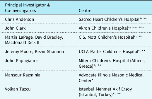
The Catheter Ablation with Reduction or Elimination of Fluoroscopy registry is a multicentre international registry that retrospectively and prospectively collects data from adult and paediatric centres that utilise a three-dimensional mapping system (CARTO, Biosense Webster, Inc or Ensite, St. Jude Medical/Abbott) to perform cardiac catheter ablation procedures. Participating centres and investigators are listed in Table 1. This investigator-initiated registry, implemented in 2012, stores data from male and female patients of any age and diagnosis who have had cardiac catheter ablation procedures performed with three-dimensional mapping systems utilised for the primary or sole purpose of three-dimensional non-fluoroscopic imaging in order to reduce or eliminate fluoroscopy usage and radiation exposure. At the time of this study, there were 2038 patients enrolled in the Catheter Ablation with Reduction or Elimination of Fluoroscopy registry. The registry includes patients undergoing ablation for any tachycardia mechanism, including all forms of both supraventricular and ventricular tachycardia. All registry data have been securely stored and maintained electronically using Research Electronic Data Capture by Vanderbilt University, which is managed and overseen by Akron Children’s Hospital.
CAREFL registry participating centres

* Centres that have entered data into the registry by 9/30/18.
** Paediatric Centres that contributed data to the present study.
*** Lead centre.
Study design
This international, multicentre, retrospective cohort study utilised existing Catheter Ablation with Reduction or Elimination of Fluoroscopy registry data from six paediatric centres (listed in Table 1) to determine acute safety and effectiveness of catheter ablations in children undergoing the procedure with three-dimensional non-fluoroscopic imaging. Results of this study were compared to historical results previously reported in the cohort of the Prospective Assessment after Pediatric Cardiac Ablation study in order to evaluate whether new benchmarks for safety and effectiveness of paediatric cardiac catheter ablations could be established. All data management, statistical analyses, and Institutional Review Board approvals occurred at the coordinating site, Akron Children’s Hospital.
Data were collected for each catheter ablation performed, including patient demographics, clinical characteristics, medical history, procedural details (duration, fluoroscopy use, complications), and clinical outcomes.
Study population
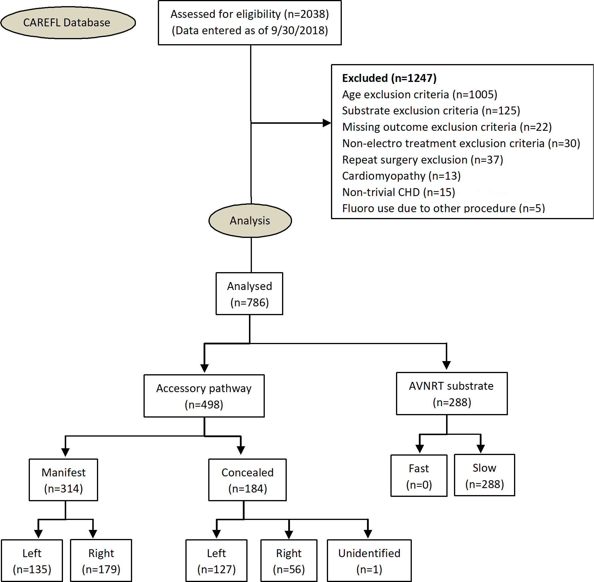
Patients included in the Catheter Ablation with Reduction or Elimination of Fluoroscopy cohort, the “study group”, were selected by matching the enrollment criteria of the Prospective Assessment after Pediatric Cardiac Ablation study, as closely as possible. Entry criteria for the Prospective Assessment after Pediatric Cardiac Ablation cohort, the “comparison cohort”, included children, aged 0–16 years, undergoing catheter ablation for supraventricular tachycardia due to either atrioventricular nodal reentry or an accessory pathway. Exclusion criteria included patients with more than trivial CHD, patients with cardiomyopathy, and patients undergoing any simultaneous non-electrophysiologic procedures. These same criteria were then used to filter the Catheter Ablation with Reduction or Elimination of Fluoroscopy database for patients from September 2006 through September 2018. Figure 1 shows the patient selection flow chart.
CAREFL flow of patients from database to analysis.

Variables and endpoints
Data from all variables and endpoints were extracted from the Catheter Ablation with Reduction or Elimination of Fluoroscopy registry. Demographic data and basic patient characteristics collected included gender, age, height, weight, body mass index, and symptoms. Medical history gathered included major non-cardiac co-morbidities, prior arrhythmias, and treatment with antiarrhythmic drugs. Procedural data included number of radiofrequency and cryoablation lesions placed, ablation substrate type, location of pathway, procedure length, success of ablation, minor complications, major complications, and total fluoroscopy time for each procedure. Not all contributing centres record fluoroscopy dose, so it was not assessed in this study. Decisions regarding timing of discontinuation of medications, sedation or anesthesia, and radiofrequency versus cryoablation energy were at the discretion of the performing physician. The database does not have robust means of tracking such information.
Statistical analysis
Baseline demographic and clinical characteristics, along with measures of safety and effectiveness, were summarised using descriptive statistics. Comparability of groups in the study group and the comparison cohort was established as described above in the inclusion/exclusion criteria, as well as via comparison of cohort composition (age, gender, structural heart disease, and tachycardia mechanism) via use of Chi-Square Goodness of Fit Test for binomial data and the One Sample T-Test for continuous data, prior to conduct of any inferential statistics.
To assess safety and effectiveness of the three-dimensional non-fluoroscopic imaging procedures, evaluation of the study group complication rates, fluoroscopy time, procedure length, and success were compared to those reported by the comparison study by application of the Chi-Square Goodness of Fit Test for binomial data or the One Sample T-Test for continuous data. Success in this data was defined as lack of inducibility of tachycardia or elimination of substrate.
Results
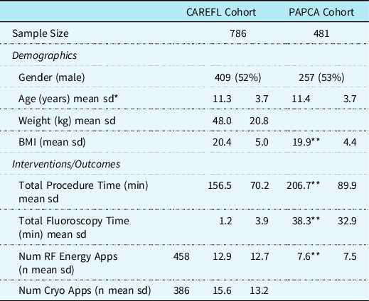
A total of 786 ablation procedures were performed which met the inclusion and exclusion criteria for this study. Forty-one (5%) of these procedures had more than one tachycardia mechanism ablated. Table 2 lists the demographic and procedural characteristics of the study group. Mean (standard deviation) age was 11.3 (3.7) years, and mean body weight was 48.0 (20.8) kg with a mean BMI of 20.4 (5.0). The mean total procedure time was 156.5 (70.2) minutes. Mean fluoroscopy time was 1.2 (3.9) minutes, with a range of 0.0 – 61.0 minutes, while 77.3% of procedures were completed without the use of fluoroscopy. There was no evidence in the sample of significant baseline differences between the cohorts by age (p = 0.49), gender (p = 0.59), or accessory pathway (p = 0.56). The mean total procedure time was significantly shorter in the Catheter Ablation with Reduction or Elimination of Fluoroscopy cohort compared to the Prospective Assessment after Pediatric Cardiac Ablation cohort (156.5 versus 206.7 minutes, p < 0.01). The mean total fluoroscopy time was also significantly shorter in the study group (1.2 versus 38.3 minutes, p < 0.01). Table 3 presents the average number of radiofrequency versus cryoablation applications by substrate.
Demographic and procedural characteristics

* Standard deviation.
** Statistically significant (p < 0.01)
RF versus Cryo applications by substrate

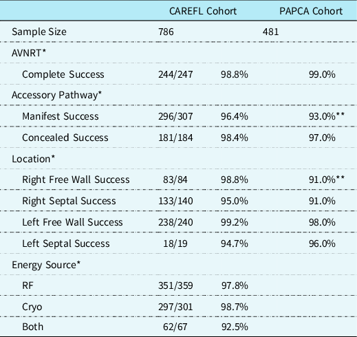
Table 4 lists the immediate results of ablation procedures. The overall success rate was 97.7% for the entire study group. The success rate was 98.7% for cryoablation and 97.8% for radiofrequency. For those who received both, the success rate was 92.5%. The success rate for manifest accessory pathway ablation was 96.4%, which was significantly different from the comparison cohort (93%, p = 0.02). The success rate for concealed accessory pathways was 98.4%, which was not significantly different from the comparison cohort (97%, p = 0.28). Atrioventricular nodal reentrant tachycardia ablation success was 98.8%, which was not significantly different from the comparison cohort (99%, p = 0.74). There were 26 (3.0%) minor complications reported in 23 patients. Various forms of transient heart block were the most common, seen in 20 total patients. Of these 20 patients, there were 5 with transient 3rd degree heart block, 6 with 2nd degree block, 5 with 1st degree block, and 4 with bundle branch block. There were three haematomas, and three complications listed as “other”. There were two (0.3%) major complications in the study group. One was a severe medication reaction and the other was bleeding requiring transfusion. There was no permanent atrioventricular block, radiation-induced skin burns, or deaths. In the comparison study, there were 8 (1.6%) major complications, consisting of 6 episodes of permanent heart block, one skin burn, and one intracardiac thrombus. Occurrence of a major complication was significantly less in the study group compared to the control cohort (p < 0.01).
Procedural Outcomes

* Excluding partial success/partial failure.
** Statistically Significant (p < 0.01).
Discussion
The current study highlights the continued improvements occurring for catheter ablation of supraventricular tachycardia in children. In 2004, the Prospective Assessment after Pediatric Cardiac Ablation study became the benchmark for catheter ablation procedures performed in pediatric patients. As the practice of paediatric electrophysiology has matured and technological enhancements have provided new tools, new standards for safety and efficacy should be defined. The community of practicing paediatric electrophysiologists is keenly aware of this need, especially as it relates to the need to minimise the exposure of ionising radiation from fluoroscopy to our patients and to our electrophysiology staff. Between 1982 and 2006, there was a greater than 600% increase in the average annual medical radiation exposure to United States citizens. Reference Schauer and Linton14 Early awareness of this problem led to improved protocols for fluoroscopy use. Several measures incorporating the “as low as reasonably achievable” principle, including use of collimation, reducing the distance of the image intensifier to the patient, use of magnification and frame acquisition rates, as well as shielding of the patient and operator, helped to reduce the dose of ionising radiation delivered to the patient (and in many cases, to electrophysiology staff). However, the amount of fluoroscopy usage, especially for difficult ablation procedures, was still considerable, as seen in the comparison study results. One can make a strong argument that there is no safe minimum dose of ionising radiation from fluoroscopy usage. Reference Brenner, Doll and Goodhead15,Reference Beels, Bacher, De Wolf, Werbrouck and Thierens16 This need to reduce or eliminate the exposure to ionising radiation from fluoroscopy usage makes the implementation of three-dimensional non-fluoroscopic imaging all the more relevant, especially in the paediatric population.
In the current study, utilisation of three-dimensional non-fluoroscopic imaging achieved a 97.2% reduction in fluoroscopy times compared to previous benchmarks. In 77% of the procedures, fluoroscopy use was completely avoided. This was accomplished while simultaneously shortening procedure times and reducing the complication rate, without negatively impacting success. Although it appears that most paediatric electrophysiology labs in the United States, if not worldwide, are now equipped with three-dimensional mapping systems, there remains marked variation in experience with, and usage of these systems. Factors contributing to variability in usage are likely many and complex and may include cost concerns, variability in technical support, generally excellent outcomes with catheter ablation using standard fluoroscopic imaging, and the learning curve. Concerns about safety and efficacy of ablation with three-dimensional non-fluoroscopic imaging were undoubtedly a past concern as well, but the data presented herein make a strong argument that use of three-dimensional non-fluoroscopic imaging is at least as safe and effective as fluoroscopic imaging for ablation of the common types of supraventricular tachycardia encountered in children. Success rates were not worse, and complication rates were significantly lower in the study group. The ability to mark an area of interest, and return to that location later, is one of the benefits of three-dimensional mapping systems that may improve procedural success. As experience increases, and as three-dimensional mapping technology improves, the percentage of ablation procedures employing entirely three-dimensional non-fluoroscopic imaging will undoubtedly increase.
Another concern that may have contributed to reduced implementation of the use of three-dimensional non-fluoroscopic imaging in the past was concern about the length of the procedure. Procedure times in the study group turned out to be significantly shorter than in the comparison cohort, similar to the recent Multicenter Pediatric and Congenital EP Quality Initiative cohort. Reference Dubin, Jorgensen and Radbill5 Three-dimensional mapping procedures, in the context of performing catheter ablation, were expected to take longer than using traditional fluoroscopic imaging, where the image is acquired with a simple step on the pedal. Acquiring a three-dimensional geometry is an extra step not required in a traditional fluoroscopically guided procedure. However, there may be a secondary benefit of the use of three-dimensional non-fluoroscopic imaging due to the ability to continuously image the catheters, and their position relative to the cardiac chambers throughout the remainder of the procedure, without the need to step on the fluoroscopy pedal to reevaluate the position of the catheters. An additional way to reduce duration of the procedure is that a complete geometry of the chamber mapped is not always necessary, and this step can be eliminated. It is interesting to note that patients in the study group received more radiofrequency lesions, on average, than in the control group (12.9 versus 7.6). This difference could represent a lack of confidence on the physician’s part in the three-dimensional mapping system. It could, therefore, be a potential downside of fluoroless navigation. Other possibilities would be a difference in style of practice, a goal to decrease recurrence, or in some other unmeasured variable. The difference did not result in higher procedure times or increased complication rates. It may have influenced the higher success rates seen in the study group with manifest accessory pathways and right free wall pathways. It will bear following over time. There was also a trend toward higher success rates for septal accessory pathways, but it did not reach statistical significance. If this trend is real, it will require data to determine if this is an effect of cryoablation, three-dimensional mapping, or some other undefined factor. There were insufficient patients with radiofrequency of septal pathways to allow statistical comparison between the study group and control group.
Finally, an important finding in this study is the notable absence of atrioventricular block in the study group. The authors feel that this reduced incidence of atrioventricular block is likely due to the increased utilisation of cryoablation, which now has an abundance of literature to support its benefit in reducing the occurrence of atrioventricular block for ablation of substrates in proximity to the atrioventricular node. Reference Skanes, Dubuc and Klein17–Reference Law, Von Bergen, Gingerich, Saarel, Fischbach and Dick19 However, we speculate that three-dimensional non-fluoroscopic imaging may also provide some benefit in avoiding the complication of atrioventricular block by providing continuous visualisation of the catheters during the procedure and providing tools for marking crucial conduction system landmarks that remain on the image even if catheters have moved.
Our study largely validates the contemporary data published from the Multicenter Pediatric and Congenital EP Quality Initiative registry study authored by Dubin and colleagues, Reference Dubin, Jorgensen and Radbill5 demonstrating improved outcomes in terms of acute procedural success, procedure times, and fluoroscopy exposure times, although there are important differences in study design and clinical sites. Most important of these differences is that the study but Dubin et al includes patients with CHD and was largely carried out in academic centres, with higher likelihood of trainees participating in procedures. While it should always be the goal, in every case, to minimise or eliminate radiation exposure, it should be done so according to the as low as reasonably achievable principle. Fluoroscopy presently remains an important tool to have available for every case, if needed.
Limitations
The major confounding factor within this study is the use of cryoablation. Cryoablation was not utilised in the comparison study but was heavily utilised in the current study. There are insufficient cases in the database utilising radiofrequency energy for atrioventricular nodal reentrant tachycardia or septal accessory pathways to make a comparison of radiofrequency with the comparison group. However, the data from the study group represent current practice in catheter ablation in children and therefore remains useful as the new benchmark. This cohort study is neither randomised nor blinded. Additionally, contributing centres have a significant degree of experience with use of three-dimensional non-fluoroscopic imaging (average of 9 years, with a range of 3–13 years). Also, two of the contributing centres in the study database also participated in the comparison study. There may be an influence of physician experience benefitting the cohort study outcomes. While this study is intended to evaluate the effect of three-dimensional mapping on ablation outcomes, other confounding influences cannot be eliminated. The study database did not track irrigated tip catheters or contact force sensing until recently. In addition, the database does not gather information regarding CT or MRI imaging import. The use of ultrasound is noted in the database but the indication for it and extent of its utility is not tracked. The influence of these technologies on the current study cannot be assessed and represent a limitation of comparing different time periods. Finally, the majority of the procedures were performed utilizing the Ensite system, with proportionally fewer procedures utilising the CARTO system (only 16 patients in the cohort, all of which also utilised fluoroscopy). Therefore conclusions regarding the amount of fluoroscopy usage when CARTO (Biosense – Webster) systems are utilised could not be adequately evaluated in this study.
Conclusions
This international, multicentre study demonstrates that catheter ablation with utilisation of three-dimensional non-fluoroscopic imaging for supraventricular tachycardia caused by accessory pathways or atrioventricular nodal reentrant tachycardia remains a highly safe and effective procedure in the paediatric population. More importantly, this study provides evidence that three-dimensional non-fluoroscopic imaging can dramatically decrease or eliminate fluoroscopy exposure in children undergoing catheter ablation. While these results represent the current best benchmark for procedural outcomes and expected radiation exposure, it is anticipated that, with continued technological improvements and clinical experience, outcomes will continue to improve.
Acknowledgements
The authors would like to thank the Rebecca D. Considine Research Institute for their administrative support.
Financial support
This research received no specific grant from any funding agency, commercial, or not-for-profit sectors.
Conflicts of interest
Dr Razminia is a consultant for Abbott. The other authors have no conflicts of interest to declare.