Introduction
The aetiology of male infertility remains a matter of debate. For many years male fertility has been defined as the ability of sperm to fertilize an oocyte: for human in vitro fertilization (IVF). Obtaining 2- to 4-cell embryos was considered a test of sperm fertilizing ability, with the assumption that all of the resulting embryos had the same developmental potential, irrespective of sperm and oocyte quality. Morphological aspects of spermatozoa have been considered. Several authors (Ron-El et al., Reference Ron-El, Nachum, Herman, Raziel, Nachum and Caspi1991) observed that, even in the presence of an acceptable fertilization rate, severe defects in sperm morphology and oligo-asthenospermia are associated with poor human embryo development and abnormal morphology. The same negative relationship was observed between sperm quality and the ability of embryos to develop to blastocyst stage (Janny & Menezo Reference Janny and Menezo1994; Menezo & Dale Reference Menezo and Dale1995). It is now, therefore, admitted that differences in male fertility are not related simply to failure of sperm to penetrate the oocyte. Intracytoplasmic sperm injection (ICSI) allows better embryo development in some cases, but this technique also has its negative aspects (Morozumi & Yanagimachi Reference Morozumi and Yanagimachi2005). Attention has recently been focused more carefully on the quality of sperm DNA. Several methods have also been developed to evaluate paternal DNA integrity independent of all semen parameters, including sperm morphology, concentration and motility. The different technologies that have been used yield the same conclusions: sperm DNA fragmentation leads to reduced fertility (Henkel et al., Reference Henkel, Hajimohammad, Stalf, Hoogendijk, Mehnert, Menkveld, Gips, Schill and Kruger2004), especially when the treatment performed is artificial insemination with sperm from the husband or regular IVF (Evenson et al., Reference Evenson, Larson and Jost2002). For this type of sperm decay, ICSI may improve embryo developmental potential (Lopes et al., Reference Lopes, Sun, Jurisicova and Casper1998a). In order to gain insight into the role of some precise DNA lesions, we decided to establish the background level of several oxidative DNA lesions in spermatozoa. Reactive oxygen species have a heavily deleterious impact on sperm DNA, leading potentially to the formation of 8-oxo-7,8-dihydro-2′-deoxyguanosine (8-oxo-dGuo), the well-known oxidative product of DNA. This compound seems to be related not only to DNA fragmentation (Lopes et al., Reference Lopes, Jurisicova, Sun, Meriano and Casper1998b; Oger et al., Reference Oger, Da Cruz, Panteix and Menezo2003), but to also have a mutagenic effect per se. Oxidative stress can directly lead to changes in DNA bases and generates ethenonucleosides, leading to one type of lesion that occurs in human sperm that is related to lipid peroxidation (Aitken et al., Reference Aitken, Harkiss and Buckingham1993). Among these lesions, the compounds 1,N2-etheno-2′-deoxyguanosine (ɛdGuo) and 1,N6-etheno-2′-deoxyadenosine (ɛdAdo) probably arise from a reaction with 4-hydroxy-2-nonenal, the main aldehyde compound released during lipid peroxidation (Douki et al., Reference Douki, Odin, Caillat, Favier and Cadet2004). However, they could be also be formed after occupational exposure to vinyl chloride or urethane. Chlorinated nucleosides such as 5-chloro-2′-deoxycytidine, 8-chloro-2′-deoxyguanosine and 8-chloro-2′-deoxyadenosine are another type of DNA adducts that is generated by hypochlorite during processes that involve inflammation (Badouard et al., Reference Badouard, Douki, Faure, Cadet and Favier2005). Semen contains polymorphonuclear cells that when stimulated are susceptible to the generation of large amounts of hypochlorite. Fortunately, mammalian cells, including oocytes, have systems that allow DNA repair (Bessho et al., Reference Bessho, Tano, Kasai, Ohtsuka and Nishimura1996; Wood et al., Reference Wood, Mitchell, Sgouros and Lindhal2001; Zeng et al., Reference Zeng, Baldwin and Shultz2004): it is obvious that the quality of the oocytes is of major importance in early embryogenesis. If left unrepaired, these sperm DNA lesions have profound effects on cell viability, leading to apoptosis or oncogenic transformations (Boiteux & Radicella 1999; Cline & Hanawalt 2005).
Materials and Methods
Isolation of spermatozoa
Thirty-nine patients who attended for a sperm migration test before IVF or artificial insemination at a fertility centre in Lyon (France) at the ‘Institut Rhonalpin pour l'étude de la reproduction humaine’ donated semen samples.The sperm was processed with a two-phase (45–90%) Percoll gradient (SupraSperm: Medicult), diluted with flushing medium (Medicult). The sperm count and quality of the preparation were checked under a microscope with a heated stage. The minimum count requested after migration was 1.2 millions live motile cells in the sperm suspension (0.5 ml). This value corresponds to our optimum sperm samples, irrespective of the assisted reproductive technology procedure requested. Samples were diluted in culture medium (EARLE) containing 0.4% human serum albumin and frozen in liquid nitrogen until DNA extraction.
Sperm DNA extraction
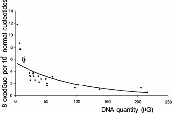
After thawing the samples at room temperature, spermatozoa were centrifuged 10 min at 3000 g (+4°C), washed in PBS buffer, centrifuged a second time and the supernatant was removed. DNA extraction was performed on the pellet. DNA from spermatozoa is hypercondensed and therefore a special protocol was optimized in order to extract the cellular DNA. Our previously described protocol for DNA extraction and digestion was optimized in order to minimize adventitious DNA oxidation during the work-up (Ravanat et al., Reference Ravanat, Douki, Duez, Gremaud, Herbert, Hofer, Lasserre, Saint Pierre, Favier and Cadet2002) by adding 10 mM dithiothreitol (DTT) to the two lysis buffers A and B (see Oger et al., Reference Oger, Da Cruz, Panteix and Menezo2003). The highly compacted nature of DNA in sperm cells caused difficulties in the first step of our study using our chaotropic technique with NI for extraction (Ravanat et al., Reference Ravanat, Douki, Duez, Gremaud, Herbert, Hofer, Lasserre, Saint Pierre, Favier and Cadet2002). Dithiothreitol allows destabilization of the nuclear matrix (Ward et al., Reference Ward, Kishikawa, Yanagimachi and Yanagimachi2000) possibly through the opening of disulphide bounds (Bedford & Calvin Reference Bedford and Calvin1974). In order to avoid these peroxidation side-effects, related to the low quantities of DNA extracted, we used only those samples for which the quantity of DNA extracted was over 30 µg (Fig. 1). We had previously observed similar effects of DNA quantity using other biological media and a sufficient amount of DNA remains a critical point in the technique, even with the addition of iron chelators to the buffer. The DNA samples were digested enzymatically with a mixture of nuclease P1 (Roche), alkaline phosphatase (Roche) and phosphodiesterase (Sigma), (Ravanat et al., Reference Ravanat, Douki, Duez, Gremaud, Herbert, Hofer, Lasserre, Saint Pierre, Favier and Cadet2002).
Relationship between the quantity of DNA extracted and its spontaneous oxidation.

HPLC – MS/MS measurements
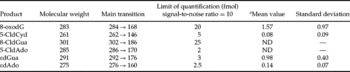
After hydrolysis, different lesions were measured using an Agilent (Massy, France) HPLC system, equipped with a thermostated auto sampler, a binary HPLC pumping system, an oven and an UV detector. The different lesions were separated using a reversed phase Lichrocart-RP 18e (3 µm, 0.2 cm × 12.5 cm) column from Merck®. The column was maintained at 28 °C and eluted with a flow rate of 0.2 ml/min in two different gradients, starting with 100% ammonium formate 5 mM (pH 6.5). The proportion of acetonitrile (AcN) added during the gradient depended upon the group of DNA lesions analysed. For the analysis of 8-oxo-dGuo and chlorinated deoxynucleosides, the percentage of AcN reached 25% in 25 min, whereas it reached 20% in 28 min for the separation of ethenonucleosides. The column was then washed for 5 min with 50% of AcN and equilibrated in starting conditions for 15 min before the next sample injection. The eluent was then passed through the UV detector set at 260 nm in order to monitor the normal nucleosides. After the addition of methanol (MeOH: 0.1 ml/min), the eluent was directed onto an API3000 tandem mass spectrometer (Applied Biosystems) through a ‘Turbospray’ electrospray source. The system was controlled using Analyst software 1.4.1. The different parameters of ionization were optimized as previously described (Douki et al., Reference Douki, Odin, Caillat, Favier and Cadet2004; Badouard et al., 2005) and the positive mode was determined to give optimal sensitivity for detecting the lesions. The different lesions were quantified by external calibration, apart from the ethenonucleosides, which could calibrated internally. The limit of quantification of the different lesions, corresponding to a signal-to-noise ratio of 10, is summarized in Table 1 and varies from 2–25 fmol injected.
Limit of quantification and basal level of the different DNA lesions in good quality sperm.

aNumber/106 normal bases.
Results
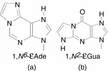
As shown in Table 1, the formation of 8-oxo-deoxyguanosine is the most abundant DNA lesion observed in our sperm samples. Ethenoguanosine (εGua: Fig. 2b) formation is observed at a very similar level, close to one lesion for one million bases, nearly 10 times higher than the level observed for ethenoadenosine (εAde, Fig. 2a). We were able to detect 5-chlorocytosine, but not 8-chloroguanosine or 5-chloroadenosine.
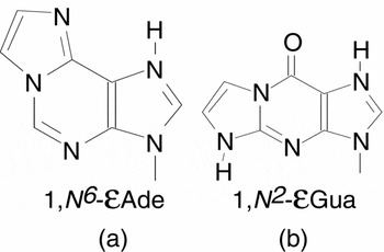
(a) Structure of 1,N6-ethenoadenosine. (b) Structure of 1,N2-ethenoguanosine.
Discussion
When performing DNA lesion studies, care has to be taken to assess the amount of extracted DNA, as this may have a negative impact on the significance of the results observed. For this reason, we used only our best sperm samples for these experiments. It was not surprising to detect 8-oxo-dGuo, which has been described in numerous papers and at higher levels than described here. This is probably related to the quality of the samples analysed here, but also to side oxidation reactions observed, due to inappropriate preparation conditions. This problem was solved by adding the reducing agent dithiothreitol (DTT) to break protamine disulphide bridges. The level of 8-OHdG is similar to that observed in leukocytes, using the same technology (Badouard et al., Reference Badouard, Masuda, Nishino, Cadet, Favier and Ravanat2005a, b). DNA in spermatozoa is highly condensed, due to protamine disulphide bridges and the presence of the antioxidant enzyme glutathione peroxidase HPGPx (Ursini et al., Reference Ursini, Heim, Kiess, Maiorino, Roveri, Wissing and Flohe1999); this should favour a lower level of oxidative decay in sperm. However, these protective systems are counterbalanced by the higher oxygen pressure in sperm when it is directly exposed to air compared to blood, especially in in vitro conditions.
More interestingly, we have detected high levels of two ethenonucleosides: ethenoguanosine and ethenoadenosine. These compounds arise from 4-hydroxy-2-nonenal, the main aldehyde compound released during lipid peroxidation (Douki et al., Reference Douki, Odin, Caillat, Favier and Cadet2004). These compounds have never been previously described in sperm; the quantity of etheno adducts to guanosine is similar to that observed in leukocytes, but is lower for ethenoadenosine (Badouard et al., Reference Badouard, Masuda, Nishino, Cadet, Favier and Ravanat2005a, b).
The quantity of chlorinated bases is low, but this could be a consequence of the small amount of infiltrated polymorphonuclear cells (PMN) in good quality sperm. Sperm cells contain no enzymes that can produce chlorine derivatives such as myeloperoxidase, abundant in PMN. We could expect a higher level of such derivatives in bad quality or infected sperm. 5-Chlorodeoxycytosine is the only chlorinated derivative detected in our conditions. Compared to leucocytes, we observed a similar level of etheno adducts to those of guanosine, but a lower level of adenosine adducts (Badouard et al., Reference Badouard, Masuda, Nishino, Cadet, Favier and Ravanat2005a, b).
During the process of fertilization, the oocyte cytoplasm should theoretically repair any decays in the integrity of sperm DNA (Hamatani et al., 2004; Zeng et al., Reference Zeng, Baldwin and Shultz2004). Nucleotide excision repair and base excision repair are good candidates for this mechanism. APEX (multifunctional DNA repair enzyme) and OGG1 (DNA glycosylase) are present in the human oocyte (El Mouatassim et al., unpublished). However, trans-4-hydroxy-2-nonenal, involved in ethenonucleotide synthesis, inhibits nucleotide excision repair in human cells (Cline et al., Reference Cline, Riggins, Tornaletti, Marnett and Hanawalt2004; Feng et al., Reference Feng, Hu and Tang2004), thus increasing the risk of cancer. Oocyte DNA repair capacity is decreasing with age (Hamatani et al., 2004). In case of weak, or inefficient repair capacity, such DNA lesions have profound effects on developmental arrests (Henkel et al., Reference Henkel, Hajimohammad, Stalf, Hoogendijk, Mehnert, Menkveld, Gips, Schill and Kruger2004) and/or cell viability. It is now well documented that transmission of altered DNA is associated with an increase in childhood cancer (Boiteux & Radicella, Reference Boiteux and Radicella2000; Zenzes et al., Reference Zenzes2000; Cline & Hanawalt, Reference Cline and Hanawalt2003).
It has been demonstrated that food and environment interact with each other and affect DNA quality (Talaska et al., Reference Talaska, Al-Zoughool, Malaveille, Fiorini, Schumann, Vietas, Peluso, Munnia, Bianchini and Allegro2006). It has therefore been tempting to reduce or avoid the generation of DNA decays related to free radical exposure. Numerous controversial studies have evaluated the efficacy of antioxidants in the treatment of male fertility (Agarwal et al., Reference Agarwal, Nallela, Allamaneni and Said2004). The majority of investigators agree that this treatment results in only partial success. In a recent study, we have analysed the impact of antioxidant treatments on sperm DNA fragmentation. It was found that they reduce strand breaks but increase DNA decondensation (Ménézo et al., 2007), opening the gate for new and more sophisticated antioxidant treatments.
In conclusion, the impact of ROS stress can be detected sensitively and accurately at a molecular level, not only for 8-OHdG but also for a new class of DNA adducts. We have established the references values, but further work is needed for clinical assays on sperm with low counts and high morphological anomalies, in which an increase of such decay should be expected. This aspect should not be overlooked in assisted reproductive technology, in order to understand the limitations observed in the results and in the progress that could be made.
This paper was presented in part at the 2006 ESHRE meeting in Prague (oral presentation).