Introduction
Trichinellosis is a cosmopolitan foodborne zoonosis caused by a parasitic tissue-dwelling nematode of the genus Trichinella (Murrell & Pozio, Reference Murrell and Pozio2011). The disease has been reported to occur in 66 countries and infects an estimated 11 million people (Dupouy-Camet, Reference Dupouy-Camet2009; Yang et al., Reference Yang, Gu, Yang, Wei, Wang, Cui, Pan, Li and Zhu2010). Scattered outbreaks of human trichinellosis have been reported over time in Asia and Europe (Ranque et al., Reference Ranque, Faugère, Pozio, La Rosa, Tamburrini, Pellissier and Brouqui2000; Khumjui et al., Reference Khumjui, Choomkasien, Dekumyoy, Kusolsuk, Kongkaew, Chalamaat and Jones2008; European Food Safety Authority, 2011; Murrell & Pozio, Reference Murrell and Pozio2011). Trichinella infections are normally acquired through ingestion of raw or undercooked infected meat (Chai et al., Reference Chai, Murrell and Lymbery2005; Murrell et al., Reference Murrell, Dorny, Flisser, Nash and Pawlowski2005) or food contaminated with infective muscle larvae (ML) (Slifko et al., Reference Slifko, Smith and Rose2000; Dabanch, Reference Dabanch2003; Pozio & Rossi, Reference Pozio and Rossi2008). The ingested ML develop into adults that, in the small intestines, release newborn larvae (NBL) that migrate to the muscles (Gao et al., Reference Gao, Wang and Liu2014).
The risk of future human T. zimbabwensis infection in sub-Saharan Africa is increasing due to poverty, food insecurity, climate change and failure of veterinary controls and surveillance (De Merode et al., Reference De Merode, Homewood and Cowlishaw2004; Pozio & Murrell, Reference Pozio and Murrell2006; Gottstein et al., Reference Gottstein, Pozio and Nöckler2009; Magwedere et al., Reference Magwedere, Hemberger, Hoffman and Dziva2012; Mukaratirwa et al., Reference Mukaratirwa, La Grange and Pfukenyi2013). Furthermore, globalization has exacerbated the risk through increased movement of people, wildlife and livestock in and out of T. zimbabwensis endemic zones of southern Africa (Mukaratirwa et al., Reference Mukaratirwa, La Grange and Pfukenyi2013). Natural T. zimbabwensis infections have been reported in a variety of vertebrates (Pozio et al., Reference Pozio, Foggin, Gelanew, Marucci, Hailu, Rossi and Morales2007; La Grange et al., Reference La Grange, Marucci and Pozio2009, Reference La Grange, Marucci and Pozio2010, Reference La Grange, Govender, Mukaratirwa and Hughes2012). In addition, experimental infections have shown that the parasite can infect non-human primates, pigs and rodents (Mukaratirwa & Foggin, Reference Mukaratirwa and Foggin1999; Pozio et al., Reference Pozio, Marucci, Casulli, Sacchi, Mukaratirwa, Foggin and La Rosa2004; Mukaratirwa et al., Reference Mukaratirwa, Dzoma, Matenga, Ruziwa, Sacchi and Pozio2008). Although there have been no cases of human infections due to T. zimbabwensis in sub-Saharan Africa, the parasite remains a public health risk.
Trichinella infection surveillance, control and treatments have been hampered by wide distributions of domestic, synanthropic and sylvatic reservoirs, the lack of a licensed vaccine and efficacious drugs against encysted ML, and lack of reliable diagnostic tools for screening early cases of infection (Yépez-Mulia et al., Reference Yépez-Mulia, Hernández-Bello, Arizmendi-Puga, Fonseca-Liñán and Ortega-Pierres2007; Gruden-Movsesijan et al., Reference Gruden-Movsesijan, Ilic, Mostarica-Stojkovic, Stosic-Grujicic, Milic and Sofronic-Milosavljevic2008; Feng et al., Reference Feng, Wu, Wang, Bai, Shi, Tang, Liu, Song, Boireau, Wang, Zhao and Liu2013). However, research continues into the testing of parasite-derived somatic antigens and crude extracts in an effort to develop vaccines, identifying putative molecules to be used in the development of diagnostic tools, vaccines and immunotherapies (Ruangkunaporn et al., Reference Ruangkunaporn, Watt, Karnasuta, Jongsakul, Mahannop, Chongsa-nguan and Chaicumpa1994; Pozio et al., Reference Pozio, Sofronic-Milosavljevic, Gomez Morales, Boireau and Nöckler2002; Deville et al., Reference Deville, De Pooter, Aucouturier, Lainé-Prade, Cote, Boireau and Vallée2005; Bień, Reference Bień2007; Frey et al., Reference Frey, Schuppers, Nöckler, Marinculić, Pozio, Kihm and Gottstein2009; Nagano et al., Reference Nagano, Wu, Asano and Takahashi2011; Li et al., Reference Li, Yao, Pan, Liu, Hu, Wu and Zhou2013). The present study was undertaken to determine differential immune responses induced in mice infected with T. zimbabwensis. Trichinella zimbabwensis is the most prevalent Trichinella species in southern Africa, and its infectivity (Hurnikova et al., Reference Hurnikova, Dubinsky, Mukaratirwa, Foggin and Kapel2004; Pozio & La Rosa, Reference Pozio and La Rosa2005; Matenga et al., Reference Matenga, Mukaratirwa, Bhebhe and Willingham2006), host range (Mukaratirwa et al., Reference Mukaratirwa, Nkulungo, Matenga and Bhebhe2003, Reference Mukaratirwa, Dzoma, Matenga, Ruziwa, Sacchi and Pozio2008; Matenga et al., Reference Matenga, Mukaratirwa, Bhebhe and Willingham2006), biochemistry (La Rosa et al., Reference La Rosa, Marucci and Pozio2003), diagnosis (Ludovisi et al., Reference Ludovisi, La Grange, Gómez Morales and Pozio2013) and treatment (Mukaratirwa et al., Reference Mukaratirwa, Gcanga and Kamau2015) have been studied extensively. However, there is a paucity of information on the immune responses induced during the phases of its life cycle.
Materials and methods
Experimental infections in mice
Sixty female BALB/c mice aged 6–8 weeks were sourced from the University of Cape Town, South Africa, and maintained in pathogen-free individual ventilated cages at the Biomedical Resources Unit of the University of KwaZulu-Natal (UKZN), Westville Campus, South Africa. The mice were fed with heat-sterilized rodent pellets (Meadow feeds, Durban, Republic of South Africa) and clean water ad libitum. The experimental mice were randomly assigned to two groups; Trichinella-infected (n= 30) and the non-infected control group (n= 30).
A crocodile-derived T. zimbabwensis (ISS1209) parasite strain was used. The isolate was maintained in our laboratory by serial passage in Sprague–Dawley rats. Muscle larvae were obtained from infected stock rats: whole carcasses were digested at 42 days post infection (dpi) according to the digestion method previously described by Kapel & Gamble (Reference Kapel and Gamble2000). Each mouse was orally infected with 500 ML and, at 0, 7, 14, 21, 28 and 35 dpi, groups of six mice were sacrificed humanely and blood was collected for sera.
Immunological protocols
Preparation of T. zimbabwensis antigen was performed as described previously by Escalante et al. (Reference Escalante, Romarís, Rodríguez, Rodríguez, Ga and Ubeira2004). The ML obtained as described earlier were sonicated at five pulses of 100 W for 30 s each and centrifuged at 40,000 × g at 4°C for 60 min. The supernatant was collected. The protein concentration was determined by Bradford assay (Bradford, Reference Bradford1976) then an anti-protease cocktail (Sigma-Aldrich, St. Louis, Missouri, USA) was added. The antigen was diluted to working concentration and stored at − 80°C until use.
Trichinella-specific IgG, IgM, IgG1, IgG2a, IgG2b and IgG3 antibodies were measured in sera collected on 0, 7, 14, 21, 28, 35 dpi by enzyme-linked immunosorbent assay (ELISA) in both experimental and control groups. Microplates were coated with 6.0 μg/ml of crude T. zimbabwensis larval antigen in 100 μl bicarbonate buffer (4 mm Na2CO3, 8 mm NaHCO3, pH 9.6) and incubated overnight at 4°C. The microtitre plates were blocked with 0.05% Tween 20 in phosphate-buffered saline (PBS) containing 5% bovine serum albumin (BSA) and incubated at 37°C for 2 h. Serum samples at a dilution of 1:200 were added in triplicate wells and incubated at 37°C for 2 h. Horseradish peroxidase-conjugated goat anti-mouse IgG and IgM (Santa Cruz Biotechnology, California, USA) antibodies were added at a dilution of 1:2000, and IgG1, Ig2a, IgG2b and IgG3 at a dilution of 1:1000. After 2 h of incubation at 37°C, 100 μl substrate (TMB substrate; KPL, Gaithersburg, Maryland, USA) was added. Optical density (OD) values were measured at 630 nm using a microplate reader (BioTek, Winooski, Vermont, USA).
Concentrations of interferon-γ (IFN-γ), tumour necrosis factor-α (TNF-α), interleukin-4 (IL-4) and IL-10 were measured in sera by mouse cytokine-specific ELISA kits (RnD systems, Minneapolis, Minnesota, USA) according to the manufacturer's guidelines. Cytokine concentrations were obtained from standard curves generated by recombinant cytokines.
Data analysis
Antibody responses and cytokine concentrations at each day of sacrifice were expressed as means ± standard error (SE) and analysed using a two-way analysis of variance (ANOVA); the levels of significance were determined by Bonferroni post-hoc test analyses using Graph pad PRISM version 5.04 for windows (Graphpad software, San Diego, California, USA) and a P value of < 0.05 was considered to be significant.
Results
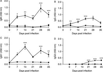
Following infection, the mice were found to be seropositive for parasite-specific IgM (fig. 1A), IgG (fig. 1B) and IgG1 (fig. 1C) antibodies. A gradual increase in IgM levels was observed at day 7 pi, which reached peak levels on days 14 and 28 pi (fig. 1A). Significant levels of IgG were observed compared to the control group (ANOVA, F 3,29= 5.01, P= 0.001; Bonferroni post-hoc P< 0.01) (fig. 1B). High levels of anti-T. zimbabwensis-specific IgG1 levels were observed at day 14 pi (fig. 1C) and declined at day 21 pi (ANOVA, F 5,27= 5.01, P= 0.0007; Bonferroni post-hoc P < 0.001). Trichinella-specific IgG3 antibody levels were detected from day 21 pi and remained elevated throughout the course of infection (fig. 1D). A decrease in IgG, IgM and IgG1 antibody responses was noted after day 28 pi. Overall, the Trichinella-infected group had significantly higher antibody responses as compared to the control group (P < 0.001).
Optical densities (mean ± SE) of Trichinella-specific IgM (A), IgG (B), IgG1(C) and IgG3 (D) antibodies in Trichinella-infected mice (●) and control mice (■). Levels of significance: *, P < 0.05; **, P < 0.01; ***, P < 0.001.

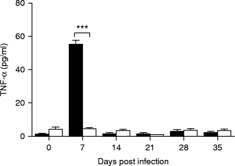
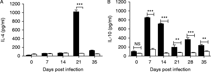
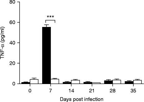
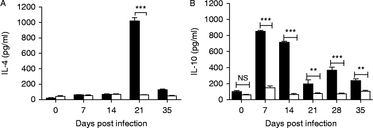
At day 7 pi, significant levels of TNF-α (ANOVA, F 5,43= 181.87, P < 0.0001; Bonferroni post-hoc P < 0.001) (fig. 2) and IL-10 (ANOVA, F 5,34= 98.02, P < 0.0001; Bonferroni post-hoc P < 0.001) (fig. 3B) were observed in the infected group compared to the control group. Interleukin-10 concentrations showed a gradual decline, attaining low levels at day 21 pi (ANOVA, F 5,34= 98.02, P < 0.0001; Bonferroni post-hoc P < 0.01). At days 28 and 35 pi, a gradual increase and decline were observed, respectively. Significantly higher levels of IL-4 were detected at day 21 pi (ANOVA, F 4,24= 382.91, P < 0.0001; Bonferroni post-hoc P < 0.001) (fig. 3A), which declined at day 35 pi to the level of the control group. Overall, there were significant differences in the levels of variation amongst infection groups over the infection period for all the parameters.
Tumour necrosis factor alpha (TNF-α) concentrations (mean ± SE) in Trichinella-infected (black bars) and control mice (white bars). Levels of significance: *, P < 0.05; **, P < 0.01; ***, P < 0.001.
The concentrations (mean ± SE) of IL-4 (A) and IL-10 (B) of Trichinella-infected (black bars) and control mice (white bars). Levels of significance: *, P < 0.05; **, P < 0.01; ***, P < 0.001.
Discussion
Our findings show that the intestinal stage of T. zimbabwensis infection was characterized by the early production of T. zimbabwensis-specific IgM and IgG antibodies. This implies that the host intestinal epithelial mucosa elicits a protective innate immune response against the NBL and adult worms (AW) in the intestinal tract (Picherot et al., Reference Picherot, Oswald, Cote, Noeckler, Le Guerhier, Boireau and Vallée2007). Therefore, it is plausible to deduce that parasite-derived antigens are released by NBL and AW to interact with enterocytes and immune cells influencing, in the long run, T-cell polarization and production of parasite-specific antibodies (Yépez-Mulia et al., Reference Yépez-Mulia, Montaño-Escalona, Fonseca-Liñán, Muñoz-Cruz, Arizmendi-Puga, Boireau and Ortega-Pierres2009). The resultant molecular dialogue between the host and the parasite results in the induction of a mixed T-helper 1 (Th1)- and Th2-type immune response, similar to that induced in mice with chronic T. spiralis infection (V. Fabre et al., Reference Fabre, Beiting, Bliss and Appleton2009). Our findings also show that there was markedly enhanced production of the Th1-type cytokine, TNF-α, and parasite-specific IgG1 (Th2-type dependent) antibody as early as 7 dpi, which is not the case with T. spiralis infection (Beiting et al., Reference Beiting, Gagliardo, Hesse, Bliss, Meskill and Appleton2007; M.V. Fabre et al., Reference Fabre, Beiting, Bliss, Gebreselassie, Gagliardo, Lee, Lee and Appleton2009). In chronic T. spiralis infection, antibodies were measurable at 15 dpi, with low IgG2a levels. Therefore, the enteric phase of T. zimbabwensis infection majorly favoured a mixed Th1- and Th2-type response, as opposed to T. spiralis and T. pseudospiralis infections which favoured a low-level Th1-type response with an earlier Th2-type response (Wakelin et al., Reference Wakelin, Goyal, Dehlawi and Hermanek1994). The role of T cells in both parasites has been fully elucidated (V. Fabre et al., Reference Fabre, Beiting, Bliss and Appleton2009) and the differences in immune responses observed between the encapsulating and the non-encapsulating Trichinella species may be attributed to the challenge dose, parasite antigenic diversity, animal-host sex and the rate of worm expulsion (Furze & Selkirk, Reference Furze and Selkirk2005; Fu et al., Reference Fu, Wang, Tong, Pan, Long, Qian and Hou2009; Hlaka et al., Reference Hlaka, Chitanga and Mukaratirwa2015). The mixed Th1- and Th2-type responses elicited create a pro-inflammatory and anti-inflammatory environment that favours parasite immune evasion and establishment (Hewitson et al., Reference Hewitson, Grainger and Maizels2009; Gruden-Movsesijan et al., Reference Gruden-Movsesijan, Ilic, Colic, Majstorovic, Vasilev, Radovic and Sofronic-Milosavljevic2011; Ilic et al., Reference Ilic, Worthington, Gruden-Movsesijan, Travis, Sofronic-Milosavljevic and Grencis2011; Sofronic-Milosavljevic et al., Reference Sofronic-Milosavljevic, Ilic, Pinelli and Gruden-Movsesijan2015). The parasite-driven immunosuppression of the host regulatory response offers mutual benefit to the parasite and the host (Ilic et al., Reference Ilic, Gruden-Movsesijan and Sofronic-Milosavljevic2012; Ashour, Reference Ashour2013). This phenomenon has also been reported during chronic T. spiralis infection in mice, where there has been evidence of immune regulatory induction enabling the parasite to survive longer, limiting host pathology and overt inflammatory responses (Beiting et al., Reference Beiting, Gagliardo, Hesse, Bliss, Meskill and Appleton2007). These findings are corroborated by our previous findings, where we established that T. zimbabwensis AW persist in the intestinal tract for up to 21 days post infection (Onkoba et al., Reference Onkoba, Kamau, Chimbari and Mukaratirwa2015) as compared to other Trichinella parasites that are eliminated in 7–18 dpi (Sadaow et al., Reference Sadaow, Intapan, Boonmars, Morakote and Maleewong2013). The differences in the persistence of AW suggest that suppressive parasite or host factors may be at work, or that antigenic diversity exists between the non-encapsulating T. zimbabwensis and the encapsulating T. spiralis parasites (Gao et al., Reference Gao, Liu, Wu, Wang, Gong, Lu, Xia, Song, Wang, Du, Liu, Han, Tang, Yang, Jin, Zhang and Liu2012, Reference Gao, Wang and Liu2014). In addition, parasite-induced immunomodulatory responses may also be slowing the rate at which the hosts generate immune-mediated worm expulsion and parasite killing (Goyal & Wakelin, Reference Goyal and Wakelin1993; Wakelin et al., Reference Wakelin, Goyal, Dehlawi and Hermanek1994; Wakelin & Goyal, Reference Wakelin and Goyal1996). Levels of the TNF-α cytokine at 7 dpi were elevated, suggesting that a protective immune response is initiated against T. zimbabwensis AW and NBL, which is geared towards AW expulsion, abrogation of inflammation caused by NBL or parasite killing (Harnett & Harnett, Reference Harnett and Harnett2010) and eventual tissue healing (Artis et al., Reference Artis, Humphreys, Bancroft, Rothwell, Potten and Grencis1999; Maizels et al., Reference Maizels, Pearce, Artis, Yazdanbakhsh and Wynn2009).
In the course of infection, we observed that the elevated levels of TNF-α at 7 dpi corresponded with a decrease of IL-4, and vice versa at 21 dpi. This implies that the infection orchestrates down-regulation of IL-4, which is a primary driver of the Th2-type response essential for parasite establishment (Wakelin et al., Reference Wakelin, Goyal, Dehlawi and Hermanek1994; Wu et al., Reference Wu, Nagano, Asano and Takahashi2010). In the migrating phase of larval stages, the levels of IL-10 and parasite-specific IgG1 and IgG3 antibodies were significantly elevated. During T. spiralis infection, IL-10 levels have also been shown to coincide with IgG1 levels (Beiting et al., Reference Beiting, Bliss, Schlafer, Roberts and Appleton2004). In both wild-type and IL-10 deficient mice, it has been established that the inflammatory response that occurs as a result of migrating larvae is attenuated independently of IL-10 production (V. Fabre et al., Reference Fabre, Beiting, Bliss and Appleton2009). Similarly, during T. spiralis infection, IL-10 and transforming growth factor-β (TGF-β) have been indicated to control inflammatory responses (Hübner et al., Reference Hübner, Shi, Torrero, Mueller, Larson, Soloviova, Gondorf, Hoerauf, Killoran, Stocker, Davies, Tarbell and Mitre2012). However, Beiting et al. (Reference Beiting, Gagliardo, Hesse, Bliss, Meskill and Appleton2007) have shown that IL-10 alone does not affect parasite survival, but that in combination with TGF-β parasite death occurs. Thus, the decline in the levels of IgM and IgG1 antibodies with significant IgG3 antibody elevation shows that IL-10 in combination with other immune cells down-regulates the production of parasite-specific antibodies.
The mixed cytokine and antibody response shows that the T. zimbabwensis parasite does not fully manage to shut off the effects of innate effector cells, evident by the presence of initial parasite-specific antibodies that co-exist with either Th1- or Th2-type responses. Therefore, based on our observations, T. zimbabwensis does not seem to induce solely either Th1- or Th2-type immune responses (Ilic et al., Reference Ilic, Worthington, Gruden-Movsesijan, Travis, Sofronic-Milosavljevic and Grencis2011), similar to a report of soil-transmitted helminth (STH) infections (Anthony et al., Reference Anthony, Rutitzky, Urban, Stadecker and Gause2007). STHs provoke a strong CD4+ Th2-type-mediated resistance that limits the establishment of helminthic infections in the host and the resultant pathologies (Yazdanbakhsh et al., Reference Yazdanbakhsh, Van Den Biggelaar and Maizels2001; Maizels et al., Reference Maizels, Hewitson and Smith2012).
In the present study, we demonstrate for the first time that T. zimbabwensis orchestrates an immunomodulatory response that encompasses mixed Th1- and Th2-type and T-regulatory responses, which are similar to those of other intestinal nematodes during chronic infection (Else & Finkelman, Reference Else and Finkelman1998). Our study was not able to elucidate the actual role of Trichinella-derived excretory and/or secretory products in T-cell priming, activation and expansion. However, T. spiralis secreted products have been shown to have an ability to induce T-cell priming that results in expansion of regulatory T and B cells, and dendritic cell (DC) stimulation in in vitro and murine studies (Ilic et al., Reference Ilic, Colic, Gruden-Movsesijan, Majstorovic, Vasilev and Sofronic-Milosavljevic2008; Aranzamendi et al., Reference Aranzamendi, Fransen, Langelaar, Franssen, van der Ley, van Putten, Rutten and Pinelli2012).
In the parasite establishment phase in the striated muscles, our results showed that the presence of ML in the predilection site is associated with high levels of Trichinella-specific IgM, IgG, IgG1 and IgG3 antibodies and regulatory cytokine (IL-10). This may be as a result of the muscular inflammation caused by encysting ML and migrating NBL. Studies have shown that the resolution of muscular inflammation is dependent on how successful the enteric-phase immunoregulation was (Helmby & Grencis, Reference Helmby and Grencis2003). Therefore, we observed that as the number of ML increased in striated muscles, the levels of Th1- and Th2-type cytokines dropped. This is an indication that the successful parasite establishes itself and initiates induction and modulation of host effector mechanisms (Artavanis-Tsakonas et al., Reference Artavanis-Tsakonas, Tongren and Riley2003; Hewitson et al., Reference Hewitson, Grainger and Maizels2009). V. Fabre et al. (Reference Fabre, Beiting, Bliss and Appleton2009) established that nitric oxide is involved in T. spiralis parasite killing during the chronic phase of infection, but in the muscle phase the ML are capable of inhibiting inducible nitric oxide synthase (iNOS) to improve their survival and establishment.
We have shown, for the first time, that the non-encapsulated T. zimbabwensis parasite induces a mixed Th1- and Th2-type response with a Th2-biased immune response in the parasite establishment phase. Therefore, we can speculate that the NBL may be responsible for the mixed Th1- and Th2-type and regulatory responses in the enteric and migrating phases of the parasite life cycle. The study was not able to elucidate extensively the manner in which the different short-lived immune responses are induced in the body compartments during NBL migration. Further studies are needed to establish the actual immune cells that are involved in anti-T. zimbabwensis immunity, immunomodulation and establishment of parasitism.
Acknowledgements
Our utmost gratitude to Mr David Buti, Dr Linda Bester and Dr Sanil Singh for providing care and housing for study animals at the Biomedical Resources Unit.
Financial support
This study received financial support from the College of Health Sciences of the University of KwaZulu-Natal through a PhD studentship bursary awarded to W.N.O.
Conflict of interest
None.
Ethical statement
All experimental protocols and procedures of the study were reviewed and approved by the Animals Ethics Committee of the University of KwaZulu-Natal (UKZN) (ref no: 114/13/Animal) in accordance with the South African national guidelines on animal care, handling and use for biomedical research. The experiments are reported in accordance with ARRIVE guidelines (Kilkenny et al., Reference Kilkenny, Browne, Cuthill, Emerson and Altman2013).