Hydatid cyst is a zoonosis caused by larvae of the tapeworm Echinococcus granulosus. It is endemic in Xinjiang, China. Cardiac hydatid cyst disease is uncommon and represents only approximately 0.5–2% of all hydatid cyst cases. Reference Kahlfuß, Flieger and Roepke1 The common locations of cardiac hydatid cysts are the left and right ventricles and interventricular septum. In 50% of such cardiac cases, multiple organ inclusion can be found. Primary pericardial hydatid cyst is rare. Reference Padmanabhan, Kumar and Mohammed2,Reference Epçaçan, Ramoğlu and Özdemir3 In this study, we report on an asymptomatic patient who was incidentally diagnosed with primary pericardial hydatid cyst.
Case report
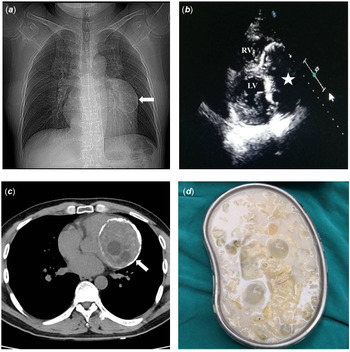
A 39-year-old male was admitted to a hospital with a space-occupying lesion of the lung. He lived in a small county and was a butcher. In addition, he had no symptoms, and his past medical history was unremarkable. His physical examination was not notable, and his routine laboratory test results were normal. An enzyme-linked immunosorbent assay was positive for Echinococcus antibodies. Electrocardiogram showed sinus rhythm and non-specific ST-T changes. Chest X-ray showed a mixed density lump near the left upper hilum, and discontinuous arc calcification could be observed at the edge of the mass, which measured 8 cm in diameter, indicating the possibility of a hydatid cyst (Fig 1a). Thoracic CT confirmed the presence of a 7.2 cm × 8.6 cm cystic lesion in the pericardium adjacent to the left ventricle. The lesion had a complete, thick wall, and some edges presented eggshell-like calcification (Fig 1b). No other cystic lesion in the mediastinal or lung areas was observed. Echocardiography revealed an 8.5 cm × 7.5 cm echolucent cystic mass that was suspected to be a hydatid cyst within the pericardium adjacent to the left and right ventricles (Fig 1c). The overall left ventricular size and systolic function of the patient were normal, and no pericardial effusion was observed. Abdominal ultrasound showed normal findings.
Chest radiograph showed a lump near the left upper hilum. Peripheral wall calcification could be seen (a); echocardiography confirmed the presence of a huge cyst in the pericardial cavity near the left and right ventricular walls. LV = left ventricle; RV = right ventricle (b); CT showed a pericardial hydatid cyst with peripheral wall calcification and multiple daughter cysts adjacent to the left ventricle (c); removed endocyst and daughter cysts (d).

The patient was operated on with a beating heart with cystectomy and partial pericardiectomy (Fig 1d). Preoperatively, he received albendazole for 12 weeks. At the routine follow-up examination 12 months post-operatively, he showed no evidence of recurrence.
Discussion
Hydatid disease is a zoonotic disease transmitted from animals to humans. It is a parasitic infestation by Echinococcus, and the frequently affected organs are the liver and lungs, but multiple organ involvement can also occur. Cardiac involvement is rarely encountered (0.5–2%). Reference Kahlfuß, Flieger and Roepke1 Cysts may reach the heart through the lymphatic system or by escaping the liver and lung filters during primary infection. The myocardium becomes involved when the cysts travel through the heart and reach the myocardium by coronary circulation. Reference Yasim, Ustunsoy and Gokaslan4 The distribution of cardiac echinococcosis is similar to that of coronary blood flow. Cardiac hydatid cysts are frequently found in the left ventricle (55–60%), right ventricle (15%), left atrium (8%), interventricular septum (5–9%), pericardium (8%), and pulmonary arteries (7%). Reference Kahlfuß, Flieger and Roepke1 However, our patient presented with primary pericardial hydatid cyst, which is rare. He was asymptomatic, and his diagnosis was made on incidental findings. Notably, he lived in endemic regions and was a butcher. His family lived in close proximity with flocks and dogs. He could be infected through the ingestion of food or water contaminated with parasite’s eggs or via direct handling of animal hosts.
The hydatid cyst grows slowly and unless located in a critical anatomic site, it takes many years to evolve. Patients with cardiac hydatid cysts may remain asymptomatic for years or present minor non-specific symptoms. The clinical signs and symptoms a patient presents depend on the size, location, and evolutionary status of the cysts. Reference Jamli, Cherif and Ajmi5 Although it is a benign disease, sometimes it can cause serious morbidity and mortality. In other patients, the cysts can induce chest pain, palpitations, cough, dyspnoea, fever, valvular dysfunctions, pulmonary hypertension, or atrioventricular blocks. The rare complications of cardiac hydatid cysts include generalised allergic–anaphylactic phenomena, ventricular tachycardia, cardiac tamponade, and pulmonary or systemic embolism with lower limb ischaemia or brain infarction. Complications may cause sudden death.
Early diagnosis of cardiac hydatid cysts is important to avoid these complications. However, clinical symptoms are usually non-specific; thus, diagnoses are difficult to obtain on the basis of clinical symptoms only. In endemic areas, cardiac disease must be kept in mind even in the absence of past medical history of hydatid disease. Chest radiographs usually show abnormal heart shadows, but calcified lobular masses may be a sign of hydatid disease. Diagnosis of hydatid disease of the heart depends on a series of tests, including hydatid serology, radiography, echocardiography, CT, and MRI. CT and MRI are important to locate and define the anatomical relationships of hydatid cysts. Reference Dursun, Terzibasioglu and Yilmaz6 They are essential for the exact surgical removal of the cysts.
The best treatment method for a cardiac hydatid cyst is surgical intervention considering the progressive and potentially fatal complications in its natural course. Reference Brunetti, Kern and Vuitton7 Approximately 10% of all hydatid cysts tend to recur after surgery. Reference Ben-Hamda, Maatouk and Ben-Farhat8 Chemotherapy with albendazole or mebendazole after surgery substantially decreases recurrence. The duration of chemotherapy is variable.
Primary pericardial hydatid cysts are rare and often asymptomatic in the early stages but are potentially lethal. It can be suspected in patients coming from endemic areas. Transthoracic echocardiography, CT, and MRI are important in diagnosing and locating cardiac hydatid cysts. Furthermore, surgical excision is deemed necessary.
Acknowledgements
None.
Financial support
This research received no specific grant from any funding agency, commercial, or not-for-profit sectors.
Conflicts of interest
None.
Ethical standards
Not applicable.