Introduction
In vitro production (IVP) and transfer of embryos have negligible contribution to the genetic management of wildlife species including marine mammals (Pukazhenthi et al., Reference Pukazhenthi, Comizzoli, Travis and Wildt2006). Research on in vitro maturation (IVM), fertilization (IVF) and culture (IVC) of whale embryos will greatly contribute to our basic understanding of reproductive physiology of cetaceans resulting in application of various assisted reproductive technologies (ARTs) to increase the population and aid in management of cetaceans. Accordingly, several studies on ARTs with respect to IVM (Fukui et al. Reference Fukui, Mogoe, Ishikawa and Ohsumi1997a, Reference Fukui, Iwayama, Matsuoka, Nagai, Koma, Mogoe, Ishikawa, Fujise, Hirabayashi, Hochi, Kato and Ohsumi2007; Asada et al., Reference Asada, Horii, Mogoe, Fukui, Ishikawa and Ohsumi2000, Reference Asada, Tetsuka, Ishikawa, Ohsumi and Fukui2001a; Iwayama et al., Reference Iwayama, Ishikawa, Ohsumi and Fukui2005; Fujihira et al., Reference Fujihira, Kobayashi, Hochi, Hirabayashi, Ishikawa, Ohsumi and Fukui2006), cryopreservation of oocytes (Asada et al., Reference Asada, Horii, Mogoe, Fukui, Ishikawa and Ohsumi2000; Iwayama et al., Reference Iwayama, Hochi, Kato, Hirabayashi, Kuwayama, Ishikawa, Ohsumi and Fukui2004; Fujihira et al., Reference Fujihira, Kobayashi, Hochi, Hirabayashi, Ishikawa, Ohsumi and Fukui2006), IVF (Fukui et al., Reference Fukui, Mogoe, Ishikawa and Ohsumi1997b; Asada et al., Reference Asada, Tetsuka, Ishikawa, Ohsumi and Fukui2001a), intracytoplasmic sperm injection (ICSI) (Asada et al., Reference Asada, Wei, Nagayama, Tetsuka, Ishikawa, Ohsumi and Fukui2001b; Fujihira et al., Reference Fujihira, Kobayashi, Hochi, Hirabayashi, Ishikawa, Ohsumi and Fukui2006; Fukui et al., Reference Fukui, Iwayama, Matsuoka, Nagai, Koma, Mogoe, Ishikawa, Fujise, Hirabayashi, Hochi, Kato and Ohsumi2007) and inter-species somatic cell nuclear transfer (Ikumi et al., Reference Ikumi, Sawai, Takeuchi, Iwayama, Ishikawa, Ohsumi and Fukui2004) have been conducted in common minke (Balaenoptera acutorostrata) and Antarctic minke (B. bonaerensis) whales. However, still there is no information on IVM, IVF and embryo development of oocytes derived from other whales. The authors had the opportunity to board a vessel in the North Pacific Ocean to collect and use follicular oocytes as the first attempt for IVM, IVF and IVC in other whales namely sei (B. borealis) and Bryde's (B. edeni) whales.
It is desirable to retrieve as many follicular oocytes as possible from an animal at any IVP programme. The number of oocytes retrieved per animal is very important to maximize the utilization of oocytes at IVP if the sampling of ovaries is very difficult particularly in cetaceans. The quality and quantity of oocytes retrieval per ovary or animal may vary depending on the reproductive status and age of animals and on oocyte harvesting technique (Vazquez et al., Reference Vazquez, Moreno, Hanneman, Evans and Kraemer1993; Wani et al., Reference Wani, Wani, Khan and Sidiqi1999; Rocha et al., Reference Rocha, Bastos, Cunha, Adona and Santos2006). Accordingly, Iwayama et al. (Reference Iwayama, Hochi, Kato, Hirabayashi, Kuwayama, Ishikawa, Ohsumi and Fukui2004) retrieved numerically higher number of oocyte in prepubertal minke whales than that of pubertal counterparts. However, there is no information on the number of oocytes retrieval from individual sei and Bryde's whales at capture.
It has been demonstrated that bovine oocytes appear to acquire developmental competence in vitro only when the cumulus cells tightly surround the oocytes (Leibfried-Rutledge et al., Reference Leibfried-Rutledge, Crister, Parrish and First1989). Further, the presence of cumulus cell layers during oocytes maturation showed high frequency of embryonic development in mice (Cross & Brinster, Reference Cross and Brinster1970) and rat (Vanderhyden & Armstrong, Reference Vanderhyden and Armstrong1989). Moreover, there is a report that oocyte grade with respect to number of cumulus cells influences the developmental competence of IVF embryos in Antarctic minke whales (Asada et al., Reference Asada, Tetsuka, Ishikawa, Ohsumi and Fukui2001a). However, until now, there is no study on the effects of oocyte grade on developmental competence of embryos of sei and Bryde's whales.
Protein is widely used as a supplement in culture media even though preimplantation development of embryos can be obtained in protein-free media. Embryos cultured in protein-supplemented media are superior to embryos cultured in protein-free media in terms of developmental potential (Krisher et al., Reference Krisher, Lane and Bavister1999; Wrenzycki et al., Reference Wrenzycki, Hermann, Carnwath and Niemann1999; Choi et al., Reference Choi, Lee, Lim, Kang and Hwang2002). Asada et al. (Reference Asada, Tetsuka, Ishikawa, Ohsumi and Fukui2001a) also obtained higher rate of development of IVF embryos cultured in medium supplemented with FWS than that of no serum supplementation in Antarctic minke whales. Serum is commonly used as protein source in many embryo culture media although it may negatively affect embryo development (Bavister, Reference Bavister1995). When serum-free media are desired due to its undefined nature, serum is widely replaced with BSA as protein supplementation. There are controversial reports on effects of various types of protein supplementations on embryo development in bovine (Wrenzycki et al., Reference Wrenzycki, Herrmann, Keskintepe, Martins, Sirisathien, Brackett and Niemann2001; Bhuiyan et al., Reference Bhuiyan, Cho, Jang, Park, Kang, Lee and Hwang2004). However, there is no information on using serum or serum albumin as a protein of choice for supplementation in IVC medium for obtaining satisfactory rates of embryo development in sei and Bryde's whales.
The purposes of the present study were to determine the oocytes recovery rate per sei and Bryde's whales followed by evaluating the effects of oocyte grade and protein supplementation in IVC medium on development of in vitro matured and fertilized embryos.
Materials and Methods
The present study was approved by the Animal Experimental Committee of Obihiro University of Agriculture and Veterinary Medicine in accordance with the Guiding Principles for the Care and Use of Research Animals.
Whales
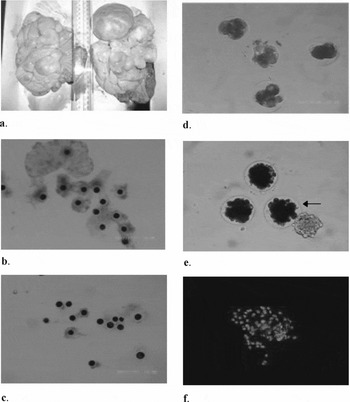
The present study was conducted as a part of the second phase of the Japanese Whale Research Program with Special Permit in the Western North Pacific Ocean (JARPN II) organized by the Institute of Cetacean Research in Tokyo, Japan during the period from 12 May to 18 August 2007. The research area covered from 140 to 170° E and from 35 to 50° N. The grenaded harpoons were used as the primary killing method for all whales that have been recognized by the International Whaling Commission (IWC) as the most humane method for killing whales and are provided by Schedule III (Capture) in the International Convention for the Regulation of Whaling. All samples derived from whales were used in a research-based ship, “Nisshin-maru” belonged to Kyodo Senpaku Kaisha, Ltd., Tokyo, Japan. Sexual maturity of each female was determined by the presence or absence of a corpus luteum (CL) and/or corpus albicans (CA) on either ovary (Fig. 1a). Females without any CL or CA were considered as prepubertal.
Ovaries, COCs and IVF embryos derived from sei whales. (a) Ovaries of a pregnant sei whale (a large corpus luteum on right ovary and many antral follicles on both ovaries). (b) A pubertal non-pregnant sei whale derived Grade A COCs after completion of maturation culture. (c) Same pubertal non-pregnant sei whale derived Grade B COCs after completion of maturation culture. (d) IVF embryos derived from sei whales with different cell numbers. (e) Morula stage sei whale embryos; only the embryo indicated with arrow was confirmed morula after Hoechst stain. (f) Stained nuclei of the same arrow indicated morula (e) with more than 16 nuclei although some nuclei of the attached cumulus cell mass are observed.

In vitro maturation of oocytes
The ovaries were collected within 2 h of death of the whales and kept warm at 20 to 25°C until aspiration. All available superficial antral follicles were aspirated with an 18 gauge needle fitted with a 10 ml disposable plastic syringe. Recovered COCs were selected under a stereomicroscope followed by three times washing in medium 199 (Sigma Aldrich Chemicals, St. Louis, MO, USA) supplemented with 10 mM HEPES (Sigma), 2 mM sodium bicarbonate (Wako Pure Chemical Industries), 75 μg/ml kanamycin (Sigma) and 0.05% (w/v) polyvinyl alcohol (PVA, Sigma). The maturation medium was medium 199 supplemented with 25 mM sodium bicarbonate, 1 mM l-glutamine (Wako), 0.33 mM sodium pyruvate (Wako), 100 μM cysteamine (Sigma), 10 ng/ml epidermal growth factor (EGF, Sigma), 0.02 AU/ml pFSH (Antrin, Denka Pharmaceutical Co.), 1 μg/ml estradiol-17β (Sigma), 75 μg/ml kanamycin and 10% (v/v) whale follicular fluid (wFF). The wFF was collected from a pubertal sei [body length (BL): 14.3 metres, body weight (BW): 26.0 tons] and a pubertal Bryde's (BL: 12.9 metres, BW: 18.1 tons) whale. The wFF was prepared by centrifugation of follicular fluid at 1000 g for 20 min followed by filter of supernatant using 0.45 μm sterile filters and was kept at −20°C until used. After washing once in the maturation medium, 10 to 20 COCs were cultured in 0.5 ml medium covered with mineral oil (Sigma) in 4-well dish (Nunc, Kamstrup, Denmark) at 37°C with 5% CO2 in humidified air for 44 to 48 h. The media used for IVM were adjusted to 390 mOsmol by addition of d-sorbitol (Sigma).
Semen preservation
The semen was recovered from the vas deferens of two sei [BL: 14.3 metres, BW: 22.6 tons, right testis weight (RTW) 3.9 kg, left testis weight (LTW) 4.1 kg and BL: 14.0 metres, BW 23.1 tons, RTW 3.6 kg, LTW 3.5 kg] and one Bryde's (BL: 12.3 metres, BW: 14.6 tons, RTW 2.3 kg, LTW 2.0 kg) whales by squeezing within 2 h of death. After evaluation of semen with respect to volume, motility and concentration, semen samples were diluted five-fold using Embryo Tech medium (Nippon Zenyaku Kogyo Co. Ltd., Fukushima, Japan) and stored in 1 ml aliquots in a microcentrifuge tube at 4°C until used for fertilization.
In vitro fertilization
After IVM culture, COCs were rinsed three times in HEPES-buffered Tyrode's–albumin–lactate–pyruvate (TALP; Fukui, Reference Fukui1990, Fukui et al., Reference Fukui, Mogoe, Ishikawa and Ohsumi1997b) medium supplemented with 0.3% (w/v) BSA (Fatty acid free, Fraction V, Sigma) (washing-TALP) and rinsed once in bicarbonate-buffered TALP supplemented with 0.6% BSA (fertilization-TALP). Three to five oocytes (5 μl) were transferred into a 40 μl droplet of fertilization-TALP under mineral oil in a 96-well plate (Nunc). The IVF was performed by using day 2 to day 8 (day of preservation = day 0) chilled semen derived from the sei and Bryde's whales. The supernatant of liquid stored semen was removed by centrifugation at 600 g for 10 min followed by once washing with calcium-free TALP medium supplemented with 0.6% BSA (capacitation TALP) by centrifugation at 600 g for 10 min. Then the sperm pellet was adjusted to 20 × 106 spermatozoa/ml followed by addition of equal volume of 200 μg/ml heparin (Sigma) solution to give rise the concentration of 10 × 106 spermatozoa/ml and 100 μg/ml heparin concentration. The heparin mixed sperm suspension was incubated for 30 min at 37°C with 5% CO2 in humidified air. A 5 μl aliquot of heparin-treated sperm suspension was added to the 40 μl fertilization-TALP medium containing the oocytes to give rise a final concentration of 1 × 10⁶ spermatozoa/ml. Fertilization was allowed to take place at 37°C with 5% CO2 in humidified air for 20 to 24 h. The TALP media used for IVF were adjusted to 360 mOsmol by addition of d-sorbitol.
In vitro culture
After co-incubation of COCs and spermatozoa for 20 to 24 h, cumulus cells and residual spermatozoa were removed as much as possible from the presumptive zygote by gentle pipetting in HEPES-buffered modified synthetic oviductal fluid (mSOF, Tervit et al., Reference Tervit, Whittingham and Rowson1972; Choi et al., Reference Choi, Lee, Lim, Kang and Hwang2002) medium supplemented with 0.8% BSA (washing-mSOF). After rinsing three times in washing-mSOF, presumptive zygotes were cultured in bicarbonate-buffered mSOF supplemented with 0.34 mM tri-sodium citrate (Merck, Darmstadt, Germany), 2.77 mM myo-inositol (Sigma), 50 μM β-mercaptoethanol (Sigma), 5 ng/ml EGF and either 0.8% BSA or 10% (v/v) FWS. The FWS was collected from the umbilical cord blood of a male sei fetus (fetal length: 157.6 cm, mother BL: 14.6 metres, mother BW: 28.0 tons) and a female Bryde's fetus (fetal length: 235.1 cm, mother BL: 13.0 metres, mother BW: 21.0 tons) followed by clotting by standing at room temperature. The FWS was recovered from the supernatant after centrifugation of clotted blood at 500 g for 10 min followed by inactivation at 59°C for 30 min and stored at −20°C until used. Three to five presumptive zygotes were cultured in 50 μl medium covered with mineral oil in 96-well plate at 37°C with 5% CO2 in humidified air for 10 days. The rates of cleavage and morula/blastocyst formation were evaluated at day 2 and day 10 (day of IVF = day 0), respectively. The media used for IVC were adjusted to 360 mOsmol by addition of d-sorbitol.
Experimental design
The number of COCs recovered with respect to reproductive status of sei and Bryde's whales was determined in Experiment 1. The whales were classified as prepubertal, pubertal non-pregnant and pregnant groups. Twenty six sei whales were used across 21 days (one to three whales per day) and 24 Bryde's whales were used across 12 days (one to four whales per day) in this experiment. All superficial antral follicles of individual sei and Bryde's whales were aspirated and the number of COCs recovered from individual whales was recorded with respect to their reproductive status.
The developmental rates of IVF embryos of sei and Bryde's whales with respect to oocyte grade and protein supplementation in culture medium were determined in Experiment 2. Ten (pubertal 6 and prepubertal 4) sei whales (BL: 13.6 ± 1.2 metres and BW: 20.4 ± 5.0 tons) were used across 8 days and 10 pubertal Bryde's whales (BL: 12.9 ± 0.4 metres and BW: 18.4 ± 2.0 tons) were used across 5 days in this experiment. The COCs were classified into either Grade A (COCs with five or more layers of compact cumulus cells; Fig. 1b) or Grade B (COCs with less than five layers of compact or expanded cumulus cells; Fig. 1c) before being cultured for IVM according to the method described elsewhere (Asada et al., Reference Asada, Tetsuka, Ishikawa, Ohsumi and Fukui2001a). The presumptive zygotes were cultured in medium supplemented with either BSA or FWS. The oocytes without insemination or activation cultured in FWS-supplemented medium were used as control embryos. From cleaved to 8-cell stage, the embryos were evaluated on the basis of morphology. If there was formation of any morula/blastocyst like embryos, they were stained with 5 μg/ml bis-benzimide (Hoechst 33342, Sigma) for 30 min after carrying to the university laboratory in washing-mSOF at 4°C and the blastomere number was determined under fluorescence microscopy. Any morula-like embryo having less than 16 nuclei after staining was not considered as morula.
Statistical analysis
All percentage data were analyzed after arcsine transformation. The data were analyzed by one- or two-way ANOVA. Post hoc analysis was performed by Turkey–Kramer multiple comparison tests to determine significant differences among the groups. Differences were considered significant when the p value was less than 0.05.
Results
Experiment 1
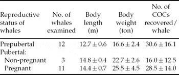
The rates of COCs recovery per sei whale with respect to their reproductive status are shown in Table 1. The number of COCs recovered per sei whale was the highest (30.6 ± 16.1) from prepubertal whales and the lowest (16.0 ± 12.5) from pubertal non-pregnant whales. However, the difference in COCs recovery rate did not vary significantly among whales with different reproductive statuses.
Recovery rates of COCs per sei whale.

Reproductive status of whales was determined by the presence or absence of a fetus in the uterus, CL and/or CA on either ovary. Females without any CL or CA or fetus were considered as prepubertal.
The values on body length, body weight and COCs recovery rates are mean ± SD.
The COCs recovery per whale did not vary significantly (p > 0.05).
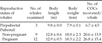
The rates of COCs recovery per Bryde's whale with respect to their reproductive status are shown in Table 2. The number of COCs recovered per Bryde's whale was the highest (26.8 ± 15.4) from pregnant whales and the lowest (6.7 ± 4.5) from prepubertal whales. However, the difference in COCs recovery rate did not vary significantly among whales with different reproductive statuses.
Recovery rates of COCs per Bryde's whale.

Reproductive status of whales was determined by the presence or absence of a fetus in the uterus, CL and/or CA on either ovary. Females without any CL or CA or fetus were considered as prepubertal.
The values on body length, body weight and COCs recovery rate are mean ± SD.
The COCs recovery per whale did not vary significantly (p > 0.05).
Experiment 2
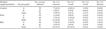
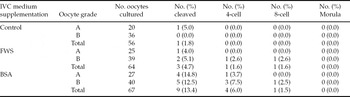
The effects of oocyte grade and protein supplementation in culture medium on development of sei whale IVF embryos are shown in Table 3. The cleavage (embryos with two or more number of blastomeres; Fig. 1d) (12.0 to 19.5%), 4-cell (8.0 to 12.0%) and 8-cell (4.0 to 8.0%) formation rates in sei whales did not vary significantly (p > 0.05) between embryos derived from either grade A or B oocytes and between embryos cultured in either FWS- or BSA-supplemented medium. Only sei whale grade B oocytes cultured in FWS-supplemented medium developed to morula stage (1.1%; Fig. 1e) which was confirmed by observing more than 16 stained nuclei (Fig. 1f). There was no formation of any blastocyst stage embryos. Out of 10 sei whales, cleavage was observed in embryos derived from seven whales after IVF. Parthenogenetic cleavage (8.0 to 21.2%) was observed in embryos derived from three whales.
Effects of oocyte grade and protein supplementation during in vitro culture on development of sei whale embryos following in vitro fertilization.

Control oocytes were cultured in serum supplemented medium without insemination.
COCs with five or more layers of compact cumulus cells were classified as grade A and COCs with less than five layers of compact or expanded cumulus cells were classified as grade B.
Number of whales used = 10.
Number of replicates across the days = 8.
Mean body length = 13.6 ± 1.2 m, Mean body weight = 20.4 ± 5.0 tons.
The values on embryo development within the same column and group did not vary significantly (p > 0.05).
The effects of oocyte grade and protein supplementation in culture medium on development of Bryde's whale IVF embryos are shown in Table 4. The cleavage (embryos with two or more number of blastomeres) (4.0 to 14.8%), 4-cell (0.0 to 7.5%) and 8-cell (0.0 to 2.6%) formation rates in Bryde's whales did not vary significantly (p > 0.05) between embryos derived from either grade A or B oocytes and between embryos cultured in either FWS- or BSA-supplemented medium. There was no formation of any morula or blastocyst stage embryos in Bryde's whales. Out of 10 Bryde's whales, cleavage was observed in embryos derived from six whales after IVF. Parthenogenetic cleavage (5.0%) was observed in embryos derived from one whale.
Effects of oocyte grade and protein supplementation during in vitro culture on development of Bryde's whale embryos following in vitro fertilization.

Control oocytes were cultured in serum supplemented medium without insemination.
COCs with five or more layers of compact cumulus cells were classified as grade A and COCs with less than five layers of compact or expanded cumulus cells were classified as grade B.
Total number of whales used = 10.
Number of replicates across the days = 5.
Mean body length = 12.8 ± 0.4 m, Mean body weight = 18.4 ± 2.0 tons.
The values on embryo development within the same column and group did not vary significantly (p > 0.05).
Discussion
In vitro embryo production using in vivo or IVM oocytes has been demonstrated in almost all domestic animals including humans and several wild animals. However, negligible number of study has been conducted on IVP in cetaceans except some reports on Antarctic and common minke whales (Fukui et al., Reference Fukui, Mogoe, Ishikawa and Ohsumi1997a, Reference Fukui, Mogoe, Ishikawa and Ohsumib, Reference Fukui, Iwayama, Matsuoka, Nagai, Koma, Mogoe, Ishikawa, Fujise, Hirabayashi, Hochi, Kato and Ohsumi2007; Asada et al., Reference Asada, Horii, Mogoe, Fukui, Ishikawa and Ohsumi2000, Reference Asada, Tetsuka, Ishikawa, Ohsumi and Fukui2001a, Reference Asada, Wei, Nagayama, Tetsuka, Ishikawa, Ohsumi and Fukuib; Ikumi et al., Reference Ikumi, Sawai, Takeuchi, Iwayama, Ishikawa, Ohsumi and Fukui2004; Iwayama et al., Reference Iwayama, Hochi, Kato, Hirabayashi, Kuwayama, Ishikawa, Ohsumi and Fukui2004, Reference Iwayama, Ishikawa, Ohsumi and Fukui2005; Fujihira et al., Reference Fujihira, Kobayashi, Hochi, Hirabayashi, Ishikawa, Ohsumi and Fukui2006). The present study is the first report concerning IVM, IVF and embryo development in other whales namely sei and Bryde's whales. The present investigation was conducted to demonstrate the oocyte recovery rate per sei and Bryde's whales (Experiment 1). Moreover, developmental competence of IVF embryos with respect to oocyte grade and IVC medium supplementation was evaluated in sei and Bryde's whales (Experiment 2).
At any IVP programme, the number of oocytes retrieval per ovary or animal is very important to maximize the utilization of oocytes. In the present study, it has been demonstrated that the reproductive status did not influence the rate of oocyte retrieval in sei and Bryde's whales. Similarly, Iwayama et al. (Reference Iwayama, Hochi, Kato, Hirabayashi, Kuwayama, Ishikawa, Ohsumi and Fukui2004) did not observe any difference in rate of oocyte recovery between pubertal and prepubertal Antarctic minke whales although the recovery rate was numerically higher in prepubertal minke whales. Contrasting to the present study, ovaries without corpus luteum yielded a significantly higher number of oocytes than that of ovaries with corpus luteum in sheep (Wani et al., Reference Wani, Wani, Khan and Sidiqi1999). This difference in oocyte recovery between species may be due to variation in reproductive physiology between terrestrial mammals and cetaceans that needs to be investigated. Nevertheless, the difference in reproductive season of the whale species and sampling season may influence the oocytes recovery rates in whales. The Bryde's whale lacks a distinct breeding season in contrast with minke and sei whales. All southern baleen whales except Bryde's whales are thought to undertake seasonal migrations between winter breeding areas in tropical or subtropical waters and summer feeding areas in the Southern Ocean (Mackintosh, Reference Mackintosh and Noris1966). Accordingly, in the present study, the sampling time (May to August) for oocytes might be considered as the breeding season in sei whales.
If IVM oocytes are used, satisfactory rate of oocyte maturation should be achieved in any IVP programme. As the body temperature of cetaceans is lower than other mammals (Folkow & Blix, Reference Folkow and Blix1992), oocytes/embryos of Antarctic and common minke whales used for IVP have been cultured at 37°C (Fukui et al., Reference Fukui, Mogoe, Ishikawa and Ohsumi1997a, Reference Fukui, Mogoe, Ishikawa and Ohsumib, Reference Fukui, Iwayama, Matsuoka, Nagai, Koma, Mogoe, Ishikawa, Fujise, Hirabayashi, Hochi, Kato and Ohsumi2007; Asada et al., Reference Asada, Horii, Mogoe, Fukui, Ishikawa and Ohsumi2000; Reference Asada, Tetsuka, Ishikawa, Ohsumi and Fukui2001a, Reference Asada, Wei, Nagayama, Tetsuka, Ishikawa, Ohsumi and Fukuib; Iwayama et al., Reference Iwayama, Ishikawa, Ohsumi and Fukui2005; Fujihira et al., Reference Fujihira, Kobayashi, Hochi, Hirabayashi, Ishikawa, Ohsumi and Fukui2006). Similarly, in the present study, oocytes of sei and Bryde's whales were incubated at 37°C during culture for IVM, IVF and embryo development, which is lower than that for bovine and porcine (39°C). Fujihira et al. (Reference Fujihira, Kobayashi, Hochi, Hirabayashi, Ishikawa, Ohsumi and Fukui2006) improved the maturation rate of Antarctic minke whale oocytes from 32% (Asada et al., Reference Asada, Tetsuka, Ishikawa, Ohsumi and Fukui2001a) to 61% with shortening of IVM time from 84 to 120 h (Fukui et al., Reference Fukui, Mogoe, Ishikawa and Ohsumi1997a; Asada et al., Reference Asada, Tetsuka, Ishikawa, Ohsumi and Fukui2001a) to 40 to 50 h by changing the osmolarity of the IVM medium to 390 mOsmol and supplementing the medium with wFF instead of FWS. The reason for improvement in maturation rate may be explained by the fact that increased osmolarity of the medium might provided with physiological condition for oocyte maturation as the osmolarity of the minke whale follicular fluid is 388 mOsmol (Iwayama et al., Reference Iwayama, Ishikawa, Ohsumi and Fukui2005) which is higher than that of bovine and porcine (approximately 300 mOsmol). In addition to this, supplementation of wFF in IVM medium may contribute to the improved maturation rate of whale oocytes similar to the positive effect of follicular fluid on oocyte maturation in bovine (Ali et al., Reference Ali, Coenen, Bousequet and Sirard2004) and porcine (Suzuki et al., Reference Suzuki, Misumi, Ozawa, Noguchi, Kaneko, Ohnuma, Fuchimoto, Onishi, Iwamoto, Saito, Nagai and Kikuchi2006) due to containing beneficial hormones and growth factors in it. Significant beneficial effect of FSH and estradiol compared to PMSG and HCG supplementation in IVM medium on oocyte maturation in Antarctic minke whales has also been documented elsewhere (Iwayama et al., Reference Iwayama, Ishikawa, Ohsumi and Fukui2005).
In line with previous studies on minke whale IVM, in the present study, we cultured the immature oocytes for 44 to 48 h in medium with 390 mOsmol osmolarity and, supplemented with wFF, FSH and estradiol. The importance of NaCl concentration and osmolarity of culture media for optimum development of preimplantation embryos has been documented in the bovine (Lim et al., Reference Lim, Kim, Okuda and Niwa1994a; Liu & Foote, Reference Liu and Foote1996), porcine (Beckmann & Day, Reference Beckmann and Day1993), mouse (Lawittis & Biggeres, Reference Lawitts and Biggers1992; Biggers et al., Reference Biggers, Lawitts and Lechene1993), rabbit (Li & Foote, Reference Li and Foote1996) and rat (Miyoshi et al., Reference Miyoshi, Funahashi, Okuda and Niwa1994). Fujihira et al. (Reference Fujihira, Kobayashi, Hochi, Hirabayashi, Ishikawa, Ohsumi and Fukui2006) increased the osmolarity of the medium by addition of NaCl, KCl, MgSO4 and CaCl2 in medium 199 at a constant ratio may be due to higher concentration of sodium in Antarctic minke whale serum (179 mmol) (Fukui et al., Reference Fukui, Mogoe, Terawaki, Ishikawa, Fujise and Ohsumi1995) than those of porcine (147 mmol) (Chang et al., Reference Chang, Jones, Ellefson and Ryan1976) and mouse serum (148 mmol) (Roblero et al., Reference Roblero, Biggers and Lechene1976). Contrasting to the previous study, we increased the osmolarity of medium by addition of sorbitol in the present study because reduced concentration of sodium chloride has been recommended for the development of bovine embryos in vitro (Lim et al., Reference Lim, Kim, Okuda and Niwa1994a; Liu & Foote, Reference Liu and Foote1996).
Numerous attempts have been taken to improve the efficiency of IVM/IVF including the selection of COCs on the basis of morphology. It has been demonstrated that the initial quality of follicular oocytes based on the layers of surrounding cumulus cells is among the important factors affecting rates of IVM, IVF and subsequent development in pigs (Meinecke & Meinecke-Tillmann, Reference Meinecke and Meinecke-Tillmann1993) and cattle (Loos et al., Reference Loos de, van Vliet, van Maurik and Kruip1989). Accordingly, there are reports on influence of oocyte grades with respect to cumulus layers on developmental potential of embryos in buffaloes (Das et al., Reference Das, Chauhan, Palta and Tomer1997), cows, ewes and lambs (Kelly et al., Reference Kelly, Kleemann, Rudiger and Walker2007). Further, Asada et al. (Reference Asada, Tetsuka, Ishikawa, Ohsumi and Fukui2001a) demonstrated variation in embryo development between two grades of COCs derived from Antarctic minke whales. Contrasting to the previous studies, oocytes grade with respect to layers of cumulus cells did not influence the cleavage rates in sei and Bryde's whales in the present study. The difference in embryo development between the two studies may be due to differences in IVM medium supplementation (FWS vs. wFF) and in basic medium used for IVC (TCM vs. mSOF). Moreover, species specific variation may contribute to this difference in embryo development, which needs to be further investigated.
Serum or serum albumin is widely used as protein source for supplementation in embryo culture medium. Previously, when efficacy was tested between supplementation of FWS and BSA in IVF medium, the rates of fertilization in minke whales did not vary between two protein sources (Asada et al., Reference Asada, Tetsuka, Ishikawa, Ohsumi and Fukui2001a). In the present study, when we compared the efficacy between FWS and BSA supplementation in IVC medium, both protein sources were equally effective as evidenced by similar rates of cleavage rates in sei and Bryde's whale embryos. Similarly, Wrenzycki et al. (Reference Wrenzycki, Herrmann, Keskintepe, Martins, Sirisathien, Brackett and Niemann2001) did not observe any difference in cleavage rates of bovine IVF embryos although serum supplementation showed higher morula and blastocyst formation rates compared to BSA counterpart. On the contrary, Bhuiyan et al. (Reference Bhuiyan, Cho, Jang, Park, Kang, Lee and Hwang2004) obtained lower rates of cleavage in bovine IVF embryos cultured in serum-supplemented medium than the BSA-supplemented counterpart although morula and blastocyst formation rates did not differ. This variation between the two studies may be due to variation in sera used (estrus cow serum vs. fetal bovine serum) for supplementation. Moreover, serum supplementation is known to have an inhibitory effect on early stages and a stimulatory effect on later stages of bovine embryo development (Pinyopummintr & Bavister Reference Pinyopummintr and Bavister1991). However, this type of negative effect of FWS (compared to BSA) supplementation on early stage embryo development was not observed in the present study. Similarly, Lim et al. (Reference Lim, Okitsu, Okuda and Niwa1994b) did not observe any negative effect of serum on early stage bovine embryo development.
In the present study, a single criterion as evidence of fertilization based on cleavage to 2 or more cell-stage embryos in whales may not be fully satisfactory even though the cleavage is used as a fertilization parameter in the other domestic species. Moreover, as shown in Fig 1e, some morula-like embryos in morphology did not have enough number of stained nuclei. This finding also emphasizes the need of investigation on other methods as evidence of fertilization. However, due to difficulties in obtaining enough numbers of oocytes available for both species of the whales in our study, investigation on two pronuclei (PN) formation or sperm penetration as evidence of fertilization was not possible to conduct. In the present study, cleavage rate of control unfertilized grade A oocytes in sei whales was comparable with or even better than those of IVF-treated oocytes. These control oocytes were not treated for chemical activation. This means that sei whale oocytes are likely to activate spontaneously in vitro. However, we do not know whether the use of BSA-supplemented medium instead of FWS-supplemented medium suppresses the incidence of spontaneous oocyte activation as we cultured those control oocytes in only FWS-supplemented medium. Artificial induction of sei whale oocyte activation may be interesting to compare with IVF oocytes in future.
To achieve high efficiency in IVF, fresh undiluted and diluted semen rather than frozen-thawed semen are desired. Accordingly, in the present study, we used diluted chilled semen for performing the IVF on sei and Bryde's whales that can be used for 2 weeks for IVF. Storage of Antarctic minke whale diluted semen at chilled condition for similar duration has also been documented for IVF elsewhere (Asada et al., Reference Asada, Tetsuka, Ishikawa, Ohsumi and Fukui2001a). To date, there are no reports on IVF of cetacean oocytes except reports on Antarctic minke whale oocytes using fresh diluted (Asada et al., Reference Asada, Tetsuka, Ishikawa, Ohsumi and Fukui2001a) and frozen-thawed semen (Fukui et al., Reference Fukui, Mogoe, Ishikawa and Ohsumi1997b) with 6 to 15% cleavage rates. Moreover, ICSI has been conducted in Antarctic and common minke whales using MII oocytes (with 1st polar body) with improving the cleavage rates ranging 30 to 46% (Fujihira et al., Reference Fujihira, Kobayashi, Hochi, Hirabayashi, Ishikawa, Ohsumi and Fukui2006; Fukui et al., Reference Fukui, Iwayama, Matsuoka, Nagai, Koma, Mogoe, Ishikawa, Fujise, Hirabayashi, Hochi, Kato and Ohsumi2007). However, no previous studies obtained blastocyst stage embryos although there were reports on production of 3 to 5% morula stage IVF embryos in the Antarctic minke whales (Fukui et al., Reference Fukui, Mogoe, Ishikawa and Ohsumi1997b; Asada et al., Reference Asada, Tetsuka, Ishikawa, Ohsumi and Fukui2001a). This indicates that the conditions of IVM, IVF and IVC for minke whale oocytes are still not optimized and need to be improved. Similarly, in the present study, no IVF embryos in sei whales developed beyond morula stage and no IVF embryos in Bryde's whales developed beyond 8-cell stage although the rate of cleavage was comparable to earlier studies. This emphasizes the importance of further research to optimize the IVP conditions for sei and Bryde's whale oocytes.
In conclusion, the present study indicates that in vitro fertilization of in vitro matured sei whale oocytes is possible to achieve cleaved embryos developing to morula stage. This study is the first in vitro embryo production attempt in sei and Bryde's whales.
Acknowledgements
The authors thank the captain and crew members of the research-based ship Nisshin-maru for their help to obtain ovaries and semen of whales. The authors also acknowledge the Japan Society for the Promotion of Science (JSPS), Tokyo, Japan for financial support for conducting this study.