INTRODUCTION
Acrocnida brachiata (Montagu, Reference Montagu1804) is a common brittle-star, burying in sandy-bottom habitats along the north-western European coastlines. It is widely represented in the subtidal area with an extended distribution in the north-east Atlantic, from the Mediterranean to the shelf seas around the British Isles, but has never been encountered north of 56°N (Koehler, Reference Koehler1921). This species is often observed as densely populated aggregates in subtidal habitats (i.e. 1000 ind. m−2 in Ireland (Keegan & Mercer, Reference Keegan and Mercer1986); 900 ind. m−2 along the coast of Normandy (Gentil & Zakardjian, Reference Gentil and Zakardjian1989)). In contrast, populations are more sparsely distributed within the intertidal habitat (less than 100 ind. m−2 in Brittany (Bourgoin et al., Reference Bourgoin, Guillou and Glémarec1991; Muths, personal observation)), with a presence seemingly restricted to the Irish Sea and the Brittany coastline. Intertidal and subtidal samples of what was regarded as one species Acrocnida brachiata also showed some differences in population dynamics with greater maximum size and life span, earlier breeding and recruitment occurrence in the subtidal area (Bourgoin et al., Reference Bourgoin, Guillou and Glémarec1991). For example, gamete maturation showed a spawning shift, represented by a two-week delay in spawning activity between intertidal and subtidal lineages (Bourgoin et al., Reference Bourgoin, Guillou and Glémarec1991).
These biological characteristics have been confirmed over the same areas, 20 years later (Muths, personal observation). Both size and breeding differences have at first been viewed as the result of differences in the environmental regime affecting individuals within the same habitat; in fact it should correspond to the existence of two distinct lineages. A phylogeographical study of this brittle star (Muths et al., Reference Muths, Davoult, Gentil and Jollivet2006) indeed presented evidence for the existence of two cryptic species, observed on both allozyme data and mtDNA sequences. This divergence coincides with a clear bathymetric segregation at sympatric locations along the western entrance of the English Channel, one lineage being exclusive in the intertidal habitat and the second one mainly in the subtidal habitat. Despite strong differences in habitat preference, it is unlikely that diversifying selective processes were directly implicated in the initial separation of the two lineages. Their emergence is supposedly linked to large scale vicariance which has affected cladogenetic events similarly among a variety of boreal species belonging to muddy-fine sediment communities (Jolly et al., Reference Jolly, Viard, Gentil, Thiebaut and Jollivet2006; Muths et al., Reference Muths, Davoult, Gentil and Jollivet2006). Around 1% of the samples analysed during this molecular study could be suspected to be potential hybrids. Both abrupt bathymetric segregation and spawning asynchrony clearly restrict the level of hybridization.
Finally, if hybridization events are a proof that isolation is not complete, all the previous evidence tends to suggest that the species A. brachiata is in fact composed of two cryptic species. Until now, no morphological differences have been observed within A. brachiata and this species was considered as easily identifiable (Koehler, Reference Koehler1921; Moyse & Tyler, Reference Moyse, Tyler, Hayward and Ryland1995). But, as Knowlton & Weight (Reference Knowlton, Weight, Claridge, Dawah and Wilson1997) underlined in their review on cryptic species, it is often difficult to find qualitative morphological differences between forms that can be unambiguously recognized by other characters. Nevertheless, most of the sibling species exhibit subtle (and sometimes not so subtle) morphological differences that were previously ascribed to intraspecific variation. The aim of the present study was to go further in the morphological description of the A. brachiata lineages and view if morphological differences could support the status of two different species.
Moreover, the genus Acrocnida currently contains two species, the north-east Atlantic type species A. brachiata and the West African A. semisquamata (Koehler, Reference Koehler and Michaelsen1914). A third species, the Mediterranean A. neapolitana (M. Sars, Reference Sars1857), was synonymized with A. brachiata by Madsen (Reference Madsen1970), who also lowered the rank of the taxon to a subgenus of Amphiura. However, in later literature it has been used on generic as well as subgeneric level. Stabilizing the taxonomic position of Acrocnida through further examination is therefore desirable.
The morphology of 15 individuals of each lineage was examined and compared to species of Amphiura, leading to the designation of a neotype for A. brachiata and the description of a new species A. spatulispina.
MATERIALS AND METHODS
Samples were collected in 2006 and 2008 in the Bay of Douarnenez, Brittany, western France, at two stations, each at a different bathymetric level. The first station was situated in the intertidal, at the lowest tidal limit on the shore (zero metre depth) and the second in the subtidal area at a depth of 10 m. Subtidal samples were obtained by diving, sampling the top 10 cm of sediment, whereas intertidal samples were collected by digging at low tide. All these samples have been stored in 95% ethanol.
We selected 15 specimens of each habitat (intertidal and subtidal) and lightly bleached them in diluted household bleach (NaOCl) to remove the upper epidermis without losing papillae and spines. These specimens were then dried and mounted with spray glue (Geiger et al., Reference Geiger, Marshall, Ponder, Sasaki and Warén2007) on aluminium stubs for scanning electron microscopy (SEM). Some arm segments were mounted separately. Skeletal parts (arm vertebrae, lateral, dorsal and ventral plates, oral plates, dental plates, teeth, genital plates and disc scales) of one specimen of 11 mm disc diameter (dd) of each species of Acrocnida, a subtidal specimen of 5.5 mm dd, two intertidal specimens of 5 and 10 mm dd respectively (the latter three after original whole specimen SEM), a Mediterranean specimen of A. brachiata of 4 mm dd, and a 9 mm dd specimen of each of Amphiura filiformis (O.F. Müller, Reference Müller1776) and Amphiura chiajei Forbes, Reference Forbes1843 were prepared by dissolving all soft tissue in concentrated household bleach. The ossicles were then washed in water, mounted wet on the glue and left to dry. All stubs were coated with gold and examined with a Hitachi FE-S4300 scanning electron microscope. Additional images were taken with a digital camera mounted on the ocular tube of a dissecting microscope. A pair of radial shields on one whole specimen of each species was brushed with bleach to remove the overlaying plates and obtain additional data in situ, without sacrificing the whole specimen.
We searched for the type material of A. brachiata by inquiries at The Natural History Museum in London (Andrew Cabrinovic), the regional museums in Plymouth (Jan Freedman) in Devon, in Colchester (Jerry Bowden) and Ipswich (Ann Ainsworth) in Essex, but none of these museums had any records of Montagu's specimens. Ann Ainsworth kindly researched Montagu in the Dictionary of National Biography and found that his material has been purchased by the British Museum, nowadays The Natural History Museum, London, where however A. brachiata could not be found. Gislén (Reference Gislén1926) did not mention any type material of A. brachiata when he described the genus Acrocnida, neither did Madsen (Reference Madsen1970). In all probability the type material has been lost long ago and therefore we describe a neotype to stabilize the species and differentiate it from the closely related Acrocnida spatulispina sp. nov., also described below. In accordance with nomenclatural rules, the neotype of A. brachiata was selected from a sample collected in Salcombe Bay in 1875, housed at the Swedish Museum of Natural History, Stockholm.
Additional abbreviations: SMNH, Swedish Museum of Natural History; spm(s), specimen(s)
SYSTEMATICS
Since only one species of Acrocnida has so far been known from the English Channel and its type material is lost, it is important to confirm the identity of this species, before a sibling species can be distinguished from it. Ideally, material from the type locality should concur with the description. In addition, the current concept and usage of the species needs to be taken into account. Acrocnida brachiata (Montagu, Reference Montagu1804), original genus Asterias, was first described from Salcomb[e] Bay, south coast of Devon, Great Britain, taken from a heap of sand that had been taken for manure. The description mentions oblong, smooth radial shields, small oval disc scales, and arms with 3–400 joints, each bearing 8–9 spines to either side. The ventral disc scales are described as rough, with minute papillae. The dd is given as scarcely half an inch, i.e. about 12.7 mm, and the length of the arms was seven to eight inches, i.e. 178–203 mm. These characters most accurately match the species that is re-described below with neotype designation. They also reflect the concept of this species in the literature up till now.
DIAGNOSIS (AFTER GISLÉN, 1926, MODIFIED)
Disc more or less densely covered by scales. Scales at disc edge and on ventral disc vertically enlarged into tubercles, which may resemble spines. Ventral arm plates with three longitudinal ridges. One to two tentacle scales at least proximally. Arm spines with a tendency towards distal enlargement. Oral plate with lateral rib-like structures.
REMARKS
Gislén (Reference Gislén1926) included the presence of a distinct transverse furrow on the distal margin of the radial shields in his description of the genus Acrocnida. We did not find a furrow, but the end of the radial shield is bent upwards, which perhaps may have given the impression of a furrow. The structure of the oral plate is a new character, found in the present study. Gislén also mistakenly believed that A. semisquamata has a naked ventral disc, which led him to describe the genus as heavily plated at least dorsally. This cannot be maintained, but the scalation of the disc varies in density. We do not regard the distal oral papilla as a generic character at this point and have removed it from the diagnosis.
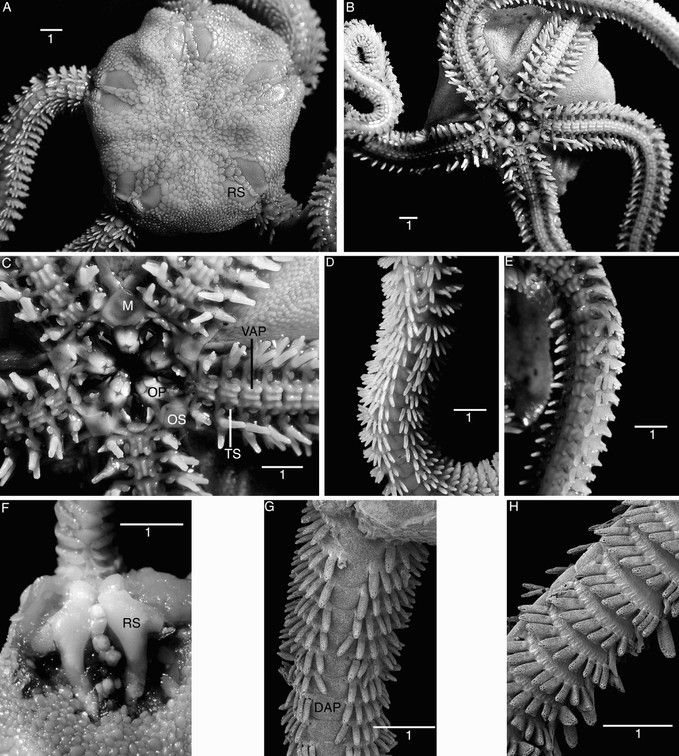
Acrocnida brachiata. (A–E) Neotype; (A) dorsal aspect; (B) ventral aspect; (C) close-up of mouth; (D) arm, ventral aspect; (E) arm, dorsal aspect; (F–H) specimens from Brittany, subtidal; (F) exposed radial shields in situ; (G) proximal arm, dorsal aspect, SEM; (H) arm laterally, SEM, note the enlarged ventral spine. DAP, dorsal arm plate; M, madreporite; OP, oral plate; OS, oral shield; TS, tentacle scale; VAP, ventral arm plate. Scale bars in millimetres.
Asterias brachiata Montagu, Reference Montagu1804: 84
Amphiura (Acrocnida) brachiata: Madsen, Reference Madsen1970: 171
Amphiura neapolitana M. Sars, Reference Sars1857: 94
Acrocnida neapolitana: Gislén, Reference Gislén1926: 16; Mortensen, 1927: 217
TYPE MATERIAL
Neotype: 10 mm dd, in 80% ethanol, SMNH-Type-7775. Salcombe Bay, Devon, south-west England, sand.
COMPARATIVE MATERIAL EXAMINED
Acrocnida brachiata: 2 spms, type locality, same original sample as neotype, (SMNH-105098). 6 spms, (France, Brittany, Douarnenez Bay, 48°68′N 4°20′W, sand, subtidal, 10 m) (SMNH-102859); collected by D. Muths. 55 spms (France, Brittany, Douarnenez Bay, 48°68′N 4°20′W, sand, subtidal, 10 m) (SMNH-102863); collected by C. Boudin, 13 May 2008. Skeletal elements on SEM stubs (France, Brittany, Douarnenez Bay, 48°68′N 4°20′W, sand, subtidal, 10 m) (SMNH-105091–105096). 1 spm, 4 mm dd, skeletal elements on SEM stub and 5 juvenile spms in ethanol (Malta, off Gneja Bay, 40–60 m); collected by C. Mifsud, August 2003 (SMNH-105096, 105097). 1 spm, 2.4 mm dd, SEM stub (France, Port Cros Island, 74 m); collected by M. Bourcier, 28 August 1979 (SMNH-105090) 15 spms and arm pieces, SEM stubs (France, Brittany, Douarnenez Bay, 48°68′N 4°20′W, sand, subtidal, 10 m) (SMNH-102841–102845, 102847–102858). 5 spms, ethanol (Sweden, Bohuslän, Mittskär Island, sand, 30 m); collected 13 August 1924 (SMNH-105107).
Amphiura neapolitana M. Sars, Reference Sars1857, holotype, (Naples, Italy); (Zoological Museum of the University of Oslo, Norway E 735).
DIAGNOSIS
Species of Acrocnida with dense cover of excentrically conical ventral disc scales, up to 11 arm spines, which are usually rod-like, occasionally flattened; three longitudinal ridges on the ventral arm plates, trilobate distal oral papilla; two–three ribs on the abradial side of the jaw (visible only after dissection).
NEOTYPE DESCRIPTION
A specimen of 10 mm dd, arms curled up, longest at least 10 cm. The dd is covered with small, round, overlapping scales, among which six larger scales are conspicuous in the centre of the disc, separated by smaller scales (Figure 1A). Although these are not placed exactly in a circle they are most likely the primary plates. The scales cover the disc completely and no translucent parts are present. The visible part of the radial shields is triangular to half-circular, with straight, upwards bent distal edge, narrow proximal end, convex outer edge and straight inner edge. The radial shields are completely separated by small round scales, distally by an elongated wedge-shaped scale. At the disc edge there are overlapping scales, conically thickened on one side. Distal to the radial shields there are small scales, separating them from the arm and disc edge.
The ventral disc is covered with small, densely overlapping, on one side conically thickened scales (Figure 1B), the cones being higher on the distal part and almost flat on the proximal part of the ventral disc.
At the proximal end of each jaw there is a pair of triangular, massive infradental papillae; further up on the dental plate four teeth are visible, but more may be present higher up (Figure 1C). The lateral oral papillae consist of a low, long, proximally pointed buccal scale high up on the oral plate, and a distinctly trilobed distal papilla on the distalmost part of the oral plate. The latter may be homologous to the adoral shield spine, although it is not inserted on the adoral shield, but instead on the oral plate.
The adoral shields are long, narrow strips, slightly concave proximally, abutting the proximal edge of the oral shield for about half their width. The oral shields are rhombic, slightly wider than long. The madreporite is noticeably larger, also rhombic (Figure 1C).
The dorsal arm plates on the proximalmost joints are slightly wider than long, pentagonal, with strongly convex distal edge, concave proximal edge, and angling lateral edges. On the middle arm joints the dorsal plates are twice as wide as long, with almost straight distal edge and shorter proximal edge. There are 11 spines on proximal joints, the dorsal- and ventralmost as long as an arm joint, other spines shorter, the shortest being in the middle of the row. The spines are rod-like, only slightly tapered, with rounded tip (Figure 1D). On the most proximal joints they form a fan across the arm; distalwards their numbers decrease. The spine articulation ridges on the lateral plates are parallel and of amphiurid type (Martynov, in press).
The ventral arm plates bear three longitudinal parallel ridges (Figure 1E), which are most distinct on the proximal joints and gradually decrease in height along the arm. These plates are rectangular, twice as wide as long, with concave distal edge and lateral edges, contiguous throughout.
At each tentacle pore there are two scales, an oval one along the lateral edge of the ventral plate and a smaller round one on the lateral plate. They do not cover the pore. The ventral scale decreases in size distalwards and vanishes between the 30th and 40th joints, whereas the lateral one is present along most of the arm.
INNER SKELETON
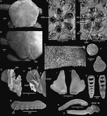
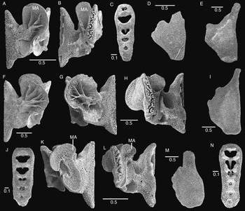
In an 11 mm dd specimen the dental plate is elongated rectangular, somewhat tapering at its ventral end. It is perforated by six foramina, one for each tooth, the largest, heart-shaped, taking almost the entire surface at the dorsal end, the others decreasing in size to a round not completely penetrating hole in the centre of the ventral part of the plate (Figure 2K, L). At the ventralmost end the plate is entire and in the intact animal covered by the infradental papillae. The teeth are rectangular with thickened distal end and acute proximal edge. The dorsalmost tooth is the largest.
Acrocnida brachiata, Douarnenez Bay, Brittany, subtidal. Skeletal parts from 11 mm dd specimen, orientated with dorsal or proximal end up, except N and O. SEM images. (A) Dorsal disc; (B) same animal as A, mouth, note the low, wide ASS; (C) different specimen, mouth, note the trilobate ASS. (D) larger specimen, dorsal disc; (E) ventral interradius with cone-shaped scales; (F) ventral disc scales with eccentric cone; (G) oral plate, adradial aspect, MA slightly damaged; (H) oral plate, abradial aspect, note the rib-like structures; (I) radial shield, underside with ball-like articulation; (J) radial shield, upper side, note the bent distal edge; (K) dental plate, proximal aspect; (L) dental plate, distal aspect; (M) lateral arm plate with spine articulations; (N) abradial genital plate; (O) adradial genital plate. ASS, adoral shield spine; AS, adoral shield; BS, buccal scale; DOP, distal oral plate; MA, muscle attachment flange; OP, oral plate; OS, oral shield; POP, proximal oral plate; R, rib-like structure; S, suture line; SA, spine attachment. Scale bars in millimetres.

The oral plates or half-jaws are high and short, wing-shaped, articulating with each other at a complex, folded adradial edge (Figure 2G, H). Each half-jaw is composed of two ossicles, a proximal and a distal oral plate, joined by an inconspicuous suture line that did not break from bleaching. The abradial side of the distal oral plate is shaped into a large round muscle flange, supported by a large central ridge or rib that branches into three thinner ribs (Figure 2H).
The adradial genital plate (placed alongside the proximal vertebrae) is bar-like with a distal ball-like condyle and pit that articulate with the radial shield above the arm (Figure 2O). The abradial genital plate is thinner, almost as long as the adradial plate, curved, with a distal pit that articulates with the adradial plate from below.
The radial shield is longer than wide with an elongated inner proximal end and a shorter, rounded, outer part. It has a distal round condyle on its underside and the distal edge curves upwards (Figure 2I, J).
The proximal lateral arm plates are short with ten spine articulations, consisting of two parallel ridges and two holes between them for muscle and nerve (Figure 2M).
The vertebrae are short and zygospondylous (condyle-socket articulation), with a dorsal keel directed distalwards (not figured).
The lateral and ventral disc scales extend into a conical tubercle to one side, giving them an asymmetrical appearance (Figure 2E, F).
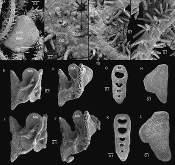
Examination of additional specimens shows that the number of ribs on the jaw varies with size between two and three branches (Figure 3E, I). The number of teeth varies between five and six between specimens of 4 mm dd and 10 mm dd.
Acrocnida brachiata, SEM images. (A–D) 2.4 mm dd juvenile, Mediterranean Sea. (A) Dorsal aspect; (B) section of oral frame, note the spine-like ASS; (C) ventral interradius and arms; (D) arm dorsolaterally; (E–L) size variation in some skeletal elements, orientation with dorsal or distal side up; (E–H) from 4 mm dd specimen; (E) oral plate abradial aspect; (F) oral plate, adradial aspect; (G) dental plate; (H) radial shield, upper side; (I–L) from 5.5 mm dd specimen; (I) oral plate, aboral aspect; (J) oral plate, adoral aspect; (K) dental plate; (L) radial shield. ASS, adoral shield spine; BS, buccal scale; CPP, central primary plate; K, k-plate; MA, muscle attachment flange; OS, oral shield; R, rib-structure; RPP, radial primary plate; RS, radial shield; TS, tentacle scale. Scale bars in millimetres.

MORPHOLOGICAL VARIATION
Of the other two specimens from the same sample as the neotype, one has longer than wide oral shields, a madreporite that is domed in the centre and has a thickened proximal edge, the distal oral papilla is not as clearly trilobate and the primary plates are indistinct. In the other specimen, the distal oral papilla is oval; the madreporite has three round holes in its proximal part, two on one side and a large hole in the centre that is probably caused by some kind of damage. The primary plates are distinct in this specimen.
In the material from Douarnenez Bay sizes range from 5–15 mm dd. Some specimens have widened flat arm spines in the middle of the row of spines at each lateral plate, in addition to rod-like spines. The shape of the radial shield varies, from irregular oval to incised, fork-like with elongated inner edge (Figures 1F, 2A, D, I, J & 3H, L). The arm spines are usually rod-like, but in some specimens a few of the spines in a row may be flattened and distally widened (Figure 1G, H). The shape of the distal oral papilla varies from distinctly trilobed and longer than wide, to low and wide, scale-like (Figure 2B, C). The oral shield varies from rhombic, similar to the holotype, to lozenge-shaped, particularly in smaller specimens. Less variability is present in the cone-like ventral disc scales, which always form a dense cover, and in the shape of the oral plates, which always present two–three ribs on the large flange, independent of size (Figure 3). The disc scalation in A. brachiata is always dense, consisting of small round, overlapping scales (Figure 2A, D). The primary plates are indistinguishable in many large specimens, but sometimes they are larger than the surrounding scales.
A juvenile of 2.4 mm dd from the Mediterranean Sea has a coarse disc scalation with clearly identifiable primary rosette and short oval radial shields that are separated proximally by a wedge-shaped k-plate, but contiguous at their distal ends (Figure 3A). The ridges on the ventral arm plates are not yet present, but the ventral disc scales are conically domed (Figure 3B). There is a large conical spine at the proximal edge of the adoral shield (Figure 3C), which will later form the distal oral papilla (Webb & Tyler, Reference Webb and Tyler1985). The oral shield is wider than long, with obtuse proximal angle and convex distal edge. To either side of the proximal arm joints, four rod-like spines are present, the ventralmost the longest, longer than an arm joint.
REMARKS
The holotype of Amphiura neapolitana of 8 mm dd has up to 11 arm spines, all shorter than an arm joint, forming a continuous row across the proximal dorsal arm joints. On proximal joints, the middle spines are somewhat widened, leaf-like, pointed. The ventral disc is densely covered with small round, conically domed scales. The distal oral papilla is wide and low, trilobate. The oral shield is lozenge-shaped. In all these characters A. neapolitana falls within the variability observed in A. brachiata and we concur with Madsen (Reference Madsen1970) in that it cannot be regarded as a separate species. We re-examined some of the specimens studied by Gislén (Reference Gislén1926) and agree with his assessment that these are A. brachiata, although unusually small, the largest individual being just 6.5 mm dd. Likewise, the species appears only to grow to a small size at Malta (C. Mifsud, personal communication).
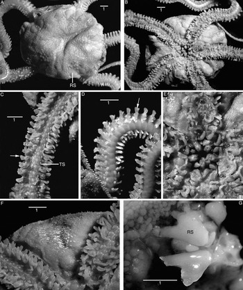
Acrocnida spatulispina. (A–F) Holotype; (A) dorsal aspect; (B) ventral aspect; (C) ventral arm; (D) dorsal arm, arrows point to spatular spines; (E) mouth frame; (F) ventral interradius, note the spine-like scales; (G) paratype, exposed radial shields in situ. ASS, adoral shield spine (distal oral papilla); OP, oral plate, OS, oral shield; RS, radial shield. Scale bars in millimetres.

TYPE MATERIAL
Holotype: 12 mm dd, in ethanol, SMNH-Type-7776. Douarnenez Bay, Brittany, France, intertidal, at low tide.
Paratypes: 24 spms, 5–13 mm dd, in ethanol, SMNH-Type-7777; 7 spms, ethanol, SMNH-Type-7778; 15 spms on SEM stubs, some in parts, SMNH-Types-7779–7796; skeletal elements from a paratype of 11 mm dd on SEM stubs SMNH-Types-7798–7800; all from type locality.
COMPARATIVE MATERIAL EXAMINED
Amphiocnida semisquamata Koehler, Reference Koehler and Michaelsen1914, one syntype, West Africa, no locality (Zoological Institution and Zoological Museum Hamburg, Germany E1678); two syntypes, West Africa, Ghana (Gold Coast), Accra, 5°30′N 0°14′W, 8 m (E 1677). Amphiura filiformis, skeletal elements on SEM stub (Sweden, Halland, Brofjorden, 58°21′N 11°26′E, 20 m, muddy clay bottom); collected by S. Smith, 27 June 1990 (SMNH-105099). Amphiura chiajei, skeletal elements on SEM stub (Sweden, Halland, off Värö, 57°13′N 12°01′E, 39 m); collected by S. Smith, 5 May 1988 (SMNH-105100).
ETYMOLOGY
The specific name refers to the spatula-shaped arm spines.
DIAGNOSIS
Species of Acrocnida with spine-like enlarged, sparsely distributed ventral disc scales, three parallel ridges on the ventral arm plates, two tentacle scales and spatula-shaped arm spines. Maximum size 13 mm dd; more than three ribs on the abradial side of the jaw (visible only after dissection).
HOLOTYPE DESCRIPTION
A specimen of 12 mm dd, with five arms, four of them complete, 8–9 cm long, regenerating distally, fifth arm broken off. Disc soft, wrinkled, more or less excavated in each radius, covered with small, round, overlapping scales; there are larger scales next to the radial shields; the primary plates are indistinct, although there are some larger scales distributed in the centre of the disc which may be the primaries (Figure 4A). At its edge the disc consists of soft, semi-translucent skin, with embedded thin, transparent scales, some of which are thickened at one end into tubercles, and rod-like spinelets.
Larger round scales separate the pairs of radial shields, except at their distal end. The distalmost of these scales is elongated and larger. The visible part of the radial shields is triangular with straight inner edge and narrower proximal end (most of it covered by scales); in one radius partly exposed proximally, showing a fork-like appearance (compare skeletal parts below).
The ventral disc consists of soft, semi-translucent skin, through which the gonads are clearly visible in the proximal part and along the arms. Thin scales with open mesh stereom are embedded in the ventral disc epidermis, sparsely distributed, some thickened into tubercles, interspersed with rod-like spinelets; towards the disc edge the concentration of rods is higher (Figure 4B).
The dorsal arm plates are twice as wide as long, rounded rectangular, contiguous along the entire arm. Each lateral arm plate bears nine arm spines proximally, decreasing distalwards (Figure 4D). The spines are shorter than an arm joint, except the dorsalmost spine on the most proximal joints. On the joints beyond the disc edge, the second ventralmost spine is distally widened, flat, strongly spatula-shaped, even the middle spines are somewhat spatula-shaped, but smaller. The ventralmost spine is flattened, longer than an arm joint. On the distal arm there are only three spines, the ventralmost twice as long as the other two.
The ventral arm plates are rectangular, wider than long, with three longitudinal ridges, the median one most prominent (Figure 4C). The plates are contiguous along the entire arm; the ridges decrease gradually and are hardly visible from the mid-section of the arm. There are two tentacle scales, one wide, low at the lateral edge of the ventral arm plate, another small round at the lateral arm plate. The ventral scale decreases in size along the arm and vanishes beyond joint 33–35, although single scales appear seemingly randomly at some distal joints. The lateral scale is present along the entire arm except the distalmost tip.
The jaws bear two infradental papillae and at least three block-like teeth on the dental plate (Figure 4E). The lateral oral papillae consist of a pointed elongated buccal scale high on the oral plate, and a small round scale (adoral shield spine) on the adoral shield. The adoral shields are narrow bands, curved around the second tentacle pore, bordering the proximal edge of the oral shield, but not reaching around its lateral edges. The oral shields have a wide proximal edge, tapering lateral edges and a narrow distal end. The madreporite is larger, rounded pentagonal and domed.
PARATYPE VARIATIONS
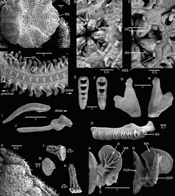
Size ranges from 5–13 mm dd. The shape of the visible part of the radial shield varies, due to variations in the number of overlapping scales, but it is no indication of the true shape of the shield (Figure 5A). The shape and size of the distal oral papilla varies somewhat from low, slightly trilobate to larger, irregularly rounded (Figure 5B, C). In the larger spms at least 10 arm joints are integrated into the disc and lack spatula-shaped spines. These appear on the free arm joints beyond the disc edge, i.e. beyond about the 10th joint, but in some specimens they are present from about the 20th joint (Figure 5D). There are up to 10 arm spines proximally in some specimens. The ventralmost arm spine is longer than the others, sometimes club-shaped, but most often conical. The spatula shape is most strongly expressed in the second ventral arm spine, but the third often also has a weaker spatula shape.
Acrocnida spatulispina. (A–D) Paratype variations; (E–Q) skeletal details, SEM images. Parts orientated with proximal or dorsal end up, except I, J. A, dorsal aspect; (B) oral frame of 5.5 mm dd specimen; (C) oral frame of 10 mm dd specimen; (D) ventral arm, note the spatulate spines; (E, F) dental plate; (E) proximal side; (F) distal side (in situ covering jaws); (G, H) radial shield; (G) upper side; (H) underside with articulation; (I) abradial genital plate; (J) adradial genital plate with articulation; (O) lateral arm plate with spine articulations; (K) ventral interradius, note the spine-like scales; (L–N) ventral disc scales; (P, Q) oral plates; (P) abradial aspect; (Q) adradial aspect. ASS, adoral shield spine; BS, buccal scale; DOP, distal oral plate; MA, muscle attachment flange; OS, oral shield; POP, proximal oral plate; R, rib-structure; RS, radial shield; S, suture line; SA, spine articulation. Scale bars in millimetres.

INNER SKELETON
In an 11 mm dd specimen the dental plate is rectangular, slightly narrower at its ventral end, with five tooth foramina, the dorsalmost of which completely perforate the plate, whereas the ventral ones are not visible from the inner side of the plate (Figure 5E, F).
The radial shields have a fork-like appearance with a longer inner and a shorter outer branch at their proximal end (Figure 5G, H). The distal end is entire, only slightly upwards bent, and bears a ball-like condyle that articulates with the adradial genital plate. The adradial genital plate is strong and bar-like with a ball-like articulation condyle and a pit at its distal end. The abradial genital plate is flat and thin, slightly shorter than the adradial plate.
The oral plate is wing-shaped with a large round distal dorsal muscle attachment flange with dense, smooth stereom and a complex rib-like structure that branches several times (Figure 5P, Q). The suture line between distal and proximal oral plate is present, but indistinct.
Some ventral disc scales are enlarged into spine-like structures with multifid tips, which are longest at the disc edge (Figure 5K–N). Other scales are flat.
The proximal lateral arm plate is short, bearing eight spine articulations, each consisting of two parallel ridges, framing muscle and nerve opening (Figure 5O).
The vertebrae are short and zygospondylous (condyle-socket articulation), with a dorsal keel directed distalwards (not figured).
Examination of additional specimens shows that the multi-branched ribs on the jaw are a stable character, with four branches in the 5.5 mm dd specimen and five main branches, subdividing at their tips, in the 10 mm dd specimen (Figure 6A, F). The number of tooth foramina is five already at the smaller stage (Figure 6C). The shape of the radial shield is variable and likely not size-dependent (Figure 6D, E).
Skeletal elements of Acrocnida spatulispina, Amphiura chiajei and Amphiura filiformis, SEM images, orientation with dorsal/proximal end up, radial shields in upside view. (A–D) A. spatulispina, from 5 mm dd specimen; (A) oral plate, abradial aspect; (B) oral plate, adradial aspect; (C) dental plate; (D) radial shield (distal end broken); (E, F) A. spatulispina, from 10 mm dd specimen; (E) radial shield; (F) oral plate, aboral aspect; (G–J) A. chiajei, from 9 mm dd specimen; (G) oral plate, abradial aspect; (H) oral plate, adradial aspect; (I) radial shield; (J) dental plate; (K–N) A. filiformis, from 9 mm dd specimen; (K) oral plate, abradial aspect; (L) oral plate, adradial aspect; (M) radial shield; (N) dental plate. MA, muscle attachment flange. Scale bars in millimetres.

REMARKS
Acrocnida spatulispina sp. nov. differs from A. brachiata in the sparser disc scalation, in the presence of ventral disc spines instead of tubercles, the spatula-shaped ventral arm spines, fewer arm spines, and a shorter and not as distinctly trilobate adoral shield spine. It may also reach a smaller maximum size and occurs only in the intertidal region, while A. brachiata appears mainly in the subtidal area (only one exception known at that time in the south of Brittany). There are considerable differences between both species also in the shape of the internal skeleton, particularly the oral plates. So far, A. spatulispina is known only from the English Channel.
The syntype E1678 of A. semisquamata has an irregular disc of 5–7 mm dd, the ventral side is scale-clad, not naked as stated by Gislén (Reference Gislén1926). The maximum number of arm spines is eight and they are shorter than an arm joint, conical, and do not meet across the dorsal arm. The distal oral papilla is elongated trilobate. The oral shield is lozenge-shaped, much longer than wide, with a much narrower distal end than A. brachiata and A. spatulispina. The ventral disc is sparsely covered with small, round scales, which are conically thickened only at the disc edge. There is a single tentacle scale on the edge of the ventral arm plate on the five most proximal arm joints, all further joints are scale-less. The syntypes E 1677 are smaller, 3 and 4 mm dd, and have up to seven arm spines. On the proximal joints, the middle spines have a widened base, but a pointed tip. The conical scales are more spine-like at the disc edge in these specimens. Thus A. spatulispina differs from A. semisquamata in an even sparser ventral disc scalation, longer and more spine-like conical scales, two tentacle scales and a different shape of the oral shield and adoral shield spine.
DISCUSSION
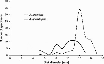
The difference in maximum and mean size between A. brachiata and A. spatulispina in Douarnenez Bay (Figure 7) is small (2 mm), but appears to be consistent over time, since the same difference has been reported for the years 1984–1985 in the same study area (Bourgoin et al., Reference Bourgoin, Guillou and Glémarec1991). Madsen (Reference Madsen1970) gives the maximum size of A. brachiata as 13 mm dd, while Gislén (Reference Gislén1926) stated 17 mm dd. A single one of the 77 specimens of A. brachiata from Brittany measured 15 mm dd, suggesting that larger specimens may be rare.
Size distribution by disc diameter (dd) in populations of Acrocnida brachiata and A. spatulispina in Douarnenez Bay, Brittany, France. The median size of A. brachiata is 12 mm dd and that of A. spatulispina is 10 mm dd, maximum sizes 15 and 13 mm dd, respectively. Sample sizes: N = 77 for A. brachiata and N = 47 for A. spatulispina.

The shape of the dental plate of both A. brachiata and A. spatulispina sp. nov. is typical for the family Amphiuridae (Murakami, Reference Murakami1963) and the jaws with their rib-like structure are similar to that of Amphiura pachybactra (Murakami, Reference Murakami1942) as well as several species of Amphioplus (Murakami, Reference Murakami1963). We found similar jaws also in Amphiura chiajei (Figure 6G), whereas the jaw of Amphiura filiformis lacks the supporting ribs and has a rough, open stereom structure on an ear-shaped abradial muscle attachment area (Figure 6K), in contrast to the smooth, dense surface of A. chiajei and both examined species of Acrocnida.
The shape of the oral plate, distal oral papilla, tentacle scales and oral shield of these species strongly resemble A. chiajei, whereas A. filiformis differs greatly. Acrocnida brachiata (and probably the other two species as well) also shares with A. chiajei the character of a large rod-like adoral shield spine, present in early ontogenetic stages, that is reduced to a scale-like distal oral papilla in the adult, whereas in A. filiformis the spine retains its shape even in mature specimens (Stöhr, Reference Stöhr2005). The ribs on the jaw are the most stable character presented by these highly variable species and may be of high taxonomic value at species level and possibly at genus level. However, the morphology of the ophiuroid jaw and possible phylogenetic implications are still poorly understood. The ribbed jaw structure is common across several amphiurid genera (Stöhr, unpublished observation), whereas the ear-shape is found in Ophiactis and Ophiothrix (Murakami, Reference Murakami1963), but is in amphiurids so far known only in A. filiformis from North Sea populations. These could be convergences caused by functional adaptations to a similar life-style instead of evidence of phylogenetic relationships. Additional data are needed to answer this question.
The combined evidence of population biology and dynamics (Bourgoin et al., Reference Bourgoin, Guillou and Glémarec1991), molecular (Muths et al., Reference Muths, Davoult, Gentil and Jollivet2006) and now morphological differences between intertidal and subtidal populations of Acrocnida strongly support the existence of two distinct species in what was formerly regarded as A. brachiata. However, some of the observed morphological variation and overlap between species with regard to some characters may be explained by an incomplete split of both species and occasional hybridization, as suggested by Muths et al. (Reference Muths, Davoult, Gentil and Jollivet2006).
Our examination of the type material of A. semisquamata confirms its status as a separate species and thus there are currently three possibly closely related species that share the characters of more or less spine-like extended or domed ventral disc scales and three longitudinal ridges on the ventral arm plates. In all three species there is a tendency to form broad, flattened arm spines, but in A. spatulispina sp. nov. the spatulate shape is expressed stronger than in the others and consistently present in all individuals.
Clark (Reference Clark1970) suggested that Acrocnida may be synonymous with Amphiura, but without taking action. Madsen (Reference Madsen1970) regarded Acrocnida as a synonym of Amphiura, because in his view the conically enlarged disc scales were the only distinguishing character. However, with now three species of Acrocnida with different conditions of conical scales, we consider these together with the ribbed oral plate and the three parallel ridges on the ventral arm plate as distinguishing characters. Mortensen (Reference Mortensen1936) remarked on the great resemblance of A. chiajei with Acrocnida semisquamata, but kept the latter as a separate genus due to its conical scales and the ventral arm plate ridges. Indeed, A. chiajei is similar to Acrocnida spp. in the presence of two tentacle scales, a low distal oral papilla that develops from a conical adoral shield spine, a rectangular ventral arm plate and a ribbed oral plate. This may suggest a close relationship to the three Acrocnida species, possibly also to A. pachybactra and other species. Additional information, preferably including molecular data, may support a common ancestor for these species and possibly Amphioplus, if oral plates prove to be of high phylogenetic value.
Synonymizing Acrocnida with Amphiura, even at subgenus level, is at this point unsatisfying, but placing A. chiajei in Acrocnida would have severe taxonomic consequences, because it is the type species of Amphiura. Any future investigation of the relationship between Acrocnida and Amphiura should include other species with tuberculate ventral disc scales such as Amphiura joubini (Koehler, Reference Koehler1922) and additional genera such as Ophiocentrus. Clearly the relationships between the species currently placed in Amphiura are incompletely understood and the information content of internal skeletal parts, particularly the oral plates, has not been utilized since Murakami's (1963) pioneering study. The necessary revision of the large genus Amphiura is, however, beyond the scope of this study.
ACKNOWLEDGEMENTS
We wish to thank Caroline Broudin and the sampling staff of the Station Biologique de Roscoff for collecting specimens in 2008. Many thanks to Andrew Cabrinovic, Jan Freedman, Jerry Bowden and Ann Ainsworth for searching for the lost type of A. brachiata; and to Åse Wilhelmsen and Peter Stiewe for loans of type material. We are grateful to H. Zibrowius and C. Mifsud for donating Mediterranean material. This study was supported by Synthesys grant SE-TAF-4832 to D. Muths. D. Muths is very grateful to D. Davoult and D. Jollivet for supervision during her PhD and encouraging her to construct a Synthesys project. We also thank two anonymous referees for valuable suggestions.