Introduction
Septic cavernous sinus thrombosis is a thrombophlebotic process affecting the cavernous sinus secondary to an infective aetiology. In the antibiotic era, this complication has now become quite rare.Reference Bhatia and Jones1 However, even with modern treatment, the mortality rate of this condition is still high, and is often quoted as 30 per cent.Reference Bhatia and Jones1–Reference Absoud, Hikmet, Dey, Joffe and Thambapillai3
Cavernous sinus thrombosis can result from infection of any of the tissues drained by the cavernous sinus. This includes the mid-face, orbit and sinonasal cavity.Reference Absoud, Hikmet, Dey, Joffe and Thambapillai3 Early reports showed that only 15 per cent of cases of cavernous sinus thrombosis originated from the paranasal sinuses, while most cases were secondary to nasal or mid-facial skin infections.Reference Pascarelli and Lemlich4 However, more recent reports suggest that sinusitis has now become the most prevalent aetiological factor for cavernous sinus thrombosis, possibly as a result of marked reduction in complicated facial skin infections, due to the use of antibiotics.Reference Dale and MacKenzie5–Reference Murthy and Wenstone8
When the paranasal sinuses are the source of cavernous sinus thrombosis, the ethmoid and sphenoid sinuses are often responsible. The mechanism of spread is either through direct extension or by retrograde thrombophlebitis along the ophthalmic veins.Reference Choi, Grunfast, Kennedy, Bolger and Zinreich9
Sphenoid sinusitis occurs in a significant proportion of cases of septic cavernous sinus thrombosis, either on its own or in conjunction with other sinus involvement.Reference Bhatia and Jones1 Although Deveze et al. described a case of bilateral cavernous sinus thrombosis with a right ophthalmic vein thrombosis secondary to left sphenoid sinus noninvasive aspergillosis,Reference Deveze, Facon, Latil, Moulin, Payan-Cassin and Dessi10 unilateral cavernous sinus thrombosis secondary to contralateral sphenoid sinusitis has not previously been described in the English language literature, to the best of our knowledge.
Case report
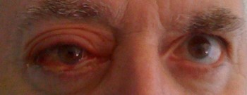
A 62-year-old, Caucasian man with no previous history of sinusitis presented to his general practitioner with a severe, right-sided, temporal headache. The general practitioner made a diagnosis of temporal arteritis and commenced the patient on systemic steroids. Five days later, the patient's headache was unimproved, and he had started to show signs of proptosis, ptosis and ophthalmoplegia in the right eye (Figure 1). He was referred urgently to the neurology department.
Clinical photograph showing right eye proptosis, ophthalmoplegia and ptosis.

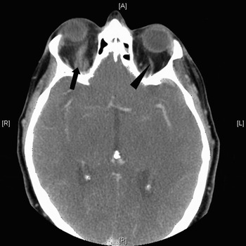
An urgent computed tomography (CT) head scan and a contrast-enhanced CT angiogram were performed, which showed right proptosis with marked congestion and dilation of the right superior ophthalmic vein (Figure 2). This was initially suspected to be due to a carotid cavernous fistula. However, a cerebral angiogram excluded this.
Axial, contrast-enhanced, computed tomography scan showing marked congestion and dilation of the right superior ophthalmic vein (arrow), in comparison with a normal left superior ophthalmic vein (arrowhead). A = anterior; P = posterior; R = right; L = left

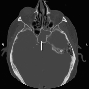
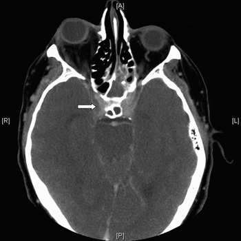
Review of the original CT angiogram identified asymmetrical opacification of the cavernous sinuses consistent with a right cavernous sinus thrombosis. The scans also showed complete opacification of the left sphenoid sinus (Figures 3 and 4). The patient was thus referred to the otolaryngology team with a diagnosis of right cavernous sinus thrombosis, most probably secondary to left sphenoid sinusitis.
Axial, computed tomography scan showing total opacity of the left sphenoid sinus (arrow). The left sphenoid sinus can be seen extending posterior to the right sinus and lying in contact with both cavernous sinuses. A = anterior; P = posterior; R = right; L = left

Axial, contrast-enhanced computed tomography scan showing filling defect of the right cavernous sinus due to the thrombosis (arrow). A = anterior; P = posterior; R = right; L = left
The patient was commenced on intravenous amoxicillin-clavulanate and a therapeutic dose of the low molecular weight heparin enoxaparin (80 mg daily).
Two days later, the patient was taken to the operating theatre for endoscopic sphenoidotomies (after ceasing enoxaparin the night before). Intra-operatively, the right sphenoid sinus was found to be clear, whereas the left sphenoid sinus contained pus, polypoid mucosa and a whitish material morphologically resembling a fungal infection.
Post-operatively, the antifungal voriconazole was added to the patient's treatment, and enoxaparin was resumed the following day.
All microbiological cultures of the samples obtained from the sphenoid sinus failed to show any bacterial or fungal growth. This may have been because the patient had already received a few days' treatment with a broad-spectrum antibiotic. Histological analysis of samples showed inflamed sinus mucosa but failed to detect the presence of any fungal hyphae. However, due to the seriousness of the condition, the microbiologists recommended 14 days' treatment with amoxicillin-clavulanate and 28 days' treatment with antifungals, replacing voriconazole with itraconazole on the fifth day of treatment.
Following endoscopic drainage of the sphenoid sinus, the patient's symptoms and signs improved dramatically. The enoxaparin was continued for five days post-operatively (at 80 mg daily), being then replaced by warfarin for three months, aiming for a target international normalised ratio (INR) of 2–3. The patient was discharged from hospital on the fifth post-operative day and was seen regularly for follow up in the out-patients department.
After four months of follow up, the patient was discharged to the care of his general practitioner, with completely normal ophthalmic and neurological status.
Discussion
Sphenoid sinusitis can frequently be misdiagnosed on presentation and referred initially to clinicians other than otolaryngologists.Reference Bhatia and Jones1 This is due to the central location of the sphenoid sinus within the skull and the vague symptomatology associated with its infections.Reference Sofferman and Burlington11 This potential for misinterpretation of the clinical picture is clearly demonstrated in our patient's case. The fact that he developed an intracranial complication on the contralateral side to his sinusitis made the diagnosis even more obscure. The sphenoid sinus has important anatomical relationships. As a midline structure, it can present with bilateral or contralateral intracranial complications. This fact must be taken into consideration when dealing with sphenoid sinus pathology. In our patient's case, CT scanning showed that the left sphenoid sinus was the dominant one, extending posterior to the right sphenoid sinus and lying in contact with both cavernous sinuses (Figure 3). This could have been the cause of the spread of infection to the right cavernous sinus.
The consensus on treatment of septic cavernous sinus thrombosis is that it should include intravenous antibiotics and surgical drainage of the source of infection in the paranasal sinuses.Reference Bhatia and Jones1–Reference Absoud, Hikmet, Dey, Joffe and Thambapillai3, Reference Southwick, Richardson and Swartz6, Reference Choi, Grunfast, Kennedy, Bolger and Zinreich9 Where fungi are involved as a causative organism, antifungals have occasionally been used.Reference Cheung, Seurry, Isaacson and McGinn12
The use of anticoagulants in cavernous sinus thrombosis remains controversial. The theoretical basis for their use is that they will prevent thrombus propagation. The biggest concern, on the other hand, is that they can increase the risk of intracranial or intraorbital bleeding, particularly in patients with concurrent complications such as cortical venous infarcts or intracavernous carotid artery necrosis.Reference Bhatia and Jones1
• Cavernous sinus thrombosis is a life-threatening condition which can be very difficult to diagnose
• The sphenoid sinus is anatomically a midline structure, and may thus lead to bilateral or contralateral intracranial complications
• Treatment for septic cavernous sinus thrombosis should include intravenous antibiotics and surgical drainage to remove the source of infection
• Anticoagulation therapy has been shown to be effective in some cases of cavernous sinus thrombosis; however, its role remains controversial due to the risk of complications
The benefits of anticoagulation in cases of cavernous sinus thrombosis have however been shown in a number of reports. Southwick et al., in a retrospective literature review, showed a statistically significant reduction in mortality with the use of anticoagulation in cavernous sinus thrombosis cases.Reference Southwick, Richardson and Swartz6 However, they did not recommend anticoagulation for other types of dural sinus thrombosis. Levine et al. also conducted a retrospective review, and found no significant change in mortality associated with the addition of anticoagulation to intravenous antibiotics when treating cavernous sinus thrombosis.Reference Levine, Twyman and Gilman13 Nevertheless, they did find a significant reduction in morbidity when anticoagulants were added early to the treatment.
Despite the above encouraging reports, there have been two reported cases (in the English language literature) of intracranial haemorrhages attributed to anticoagulation for cavernous sinus thrombosis. One of these resulted in a fatal haemorrhagic cerebral infarction, while the other patient experienced a subarachnoid haemorrhage but made a full recovery after the heparin was reversed with protamine.Reference Bhatia and Jones1
There is not enough published evidence on safe and effective levels of anticoagulation in cavernous sinus thrombosis. Bhatia and Jones recommended keeping the activated partial thromboplastin time ratio between 1.5 and 2.5, and the INR between 2 and 3.Reference Bhatia and Jones1 They also recommended anticoagulation for three months. These figures correlate well with our experience in the reported case.
Conclusions
Cavernous sinus thrombosis can be the initial presentation of sinusitis. Sphenoid sinusitis can lead to contralateral cavernous sinus thrombosis. Intravenous antibiotics and urgent surgical drainage may constitute a very successful treatment for this life-threatening condition. The use of anticoagulation remains controversial.