Introduction
The recruitment of eosinophils into gastrointestinal (GI) tissue is a complex process induced by systemic diseases or primary GI disorders. Eosinophils tend to be located in the tissue rather than the peripheral blood. In fact, eosinophils persist in the circulation for less than 1 h in dogs and 6–8 h in humans and then migrate quickly into tissues (Tizard, Reference Tizard and Tizerd2009; Young and Meadows, Reference Young, Meadows, Weiss and Wardrop2010). The GI tract has been described as the major site of eosinophilic migration (Lamouse-Smith and Furuta, Reference Lamouse-Smith and Furuta2006; Tizard, Reference Tizard and Tizerd2009). Human GI eosinophilia falls into three categories: primary eosinophilic GI disorders (EGIDs); hypereosinophilic syndrome (HES), which can induce GI eosinophilia; and GI eosinophilia due to known causes (Zuo and Rothenberg, Reference Zuo and Rothenberg2007). In human cases, the term inflammatory bowel disease (IBD) refers to Crohn's disease and ulcerative colitis (Xavier and Podolsky, Reference Xavier and Podolsky2007). Consequently, human eosinophilic gastroenteritis is not classified as IBD, even though inflammation of the bowel is present and corticosteroids are routinely utilized for the treatment. Eosinophilic gastroenteritis is marked by the presence of GI symptoms and eosinophilic infiltration from the esophagus to the colon with no evidence of parasitic or extraintestinal disease (Blackshaw and Levison, Reference Blackshaw and Levison1986; Talley et al., Reference Talley, Shorter, Phillips and Zinsmeister1990).
Unlike the human disease, canine eosinophilic gastroenteritis is currently classified as a form of idiopathic IBD (Hall and German, Reference Hall, German and Steiner2008; Jergens, Reference Jergens, Washabau and Day2013). IBD is a chronic idiopathic GI disorder and is diagnosed based on: (1) chronic GI signs (usually >3 weeks); (2) histopathologic evidence of mucosal inflammation; (3) lack of other identifiable causes of GI inflammation; (4) inadequate response to dietary, antibiotic, or anthelmintic treatment; and (5) clinical response to anti-inflammatory or immunosuppressive agents (Washabau et al., Reference Washabau, Day, Willard, Hall, Jergens, Mansell, Minami and Bilzer2010). The histopathological findings in dogs with IBD vary among dogs and the infiltrating inflammatory cell type can either be based on a single cell type or a mixed cell population. Lymphocytic–plasmacytic enteritis (LPE) is the most common form of canine IBD (German, Reference German, Washabau and Day2013). Eosinophilic gastroenteritis is less common than LPE, granulomatous enteritis and histiocytic ulcerative colitis (HUC) are considered rare (Washabau, Reference Washabau, Washabau and Day2013). Canine eosinophilic gastroenteritis can be seen in dogs of all ages and breeds, but it is most commonly found in Boxers, Doberman pinschers, German shepherds, Rottweilers, and Shar-Peis (Dossin, Reference Dossin and Steiner2008; Hall and German, Reference Hall, German and Steiner2008). Eosinophilic gastroenteritis and other forms of GI eosinophilia in dogs are still poorly understood.
In general, an immune response to parasites or to diets is considered to be the main causes of eosinophilic infiltration of the GI tract in dogs (Kleinschmidt et al., Reference Kleinschmidt, Meneses, Nolte and Hewicker-Trautwein2007). Eosinophils have long been recognized as inflammatory leukocytes that are specifically involved in a response to parasites. However, recent studies have shown infiltration of the GI tract with eosinophils unrelated to parasitic infestation (McTavish, Reference Mctavish2002; Mazzei et al., Reference Mazzei, Bissett, Murphy, Hunter and Neel2009). The present review primarily focuses on the GI eosinophilic infiltration in dogs and summarizes the clinical signs, diagnosis, and treatment of different eosinophilic disorders. Because relatively little is known about GI eosinophilia in dogs, this review also provides comparisons to the human form, which is better understood.
Eosinophils: morphology, activation, degranulation, and mediators
Morphology of eosinophils
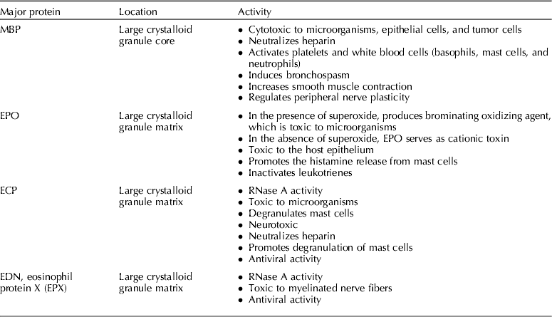
Eosinophils are polymorphonuclear white blood cells that play a role in innate, acquired, and adaptive immunity, as well as in tissue remodeling. Eosinophils are characterized by the fact that their cytoplasm can be stained intensively with anionic dyes, such as eosin (Khan, Reference Khan2005; Young and Meadows, Reference Young, Meadows, Weiss and Wardrop2010). Eosinophils contain small primary granules that contain acrylsulfatase, peroxidase, and acid phosphatase and large crystalloid granules that contain major basic proteins (MBPs), eosinophil cationic protein (ECP), eosinophil peroxidase (EPO), and eosinophil-derived neurotoxin (EDN) (Tizard, Reference Tizard and Tizerd2009) (Table 1, Fig. 1). MBPs, which are located in the core of large granules, have activities that affect smooth muscle contraction and peripheral nerve plasticity (Rothenberg and Hogan, Reference Rothenberg and Hogan2006). ECP, EPO, and EDN are found in the cellular matrix of the large granules. ECP is a ribonuclease A (RNase A) that has both cytotoxic and noncytotoxic activities. ECP has anti-viral activity and can also suppress the proliferation of T cells and immunoglobulin synthesis by B cells (Rothenberg and Hogan, Reference Rothenberg and Hogan2006). EPO is more potent than myeloperoxidase (MPO), which is secreted by neutrophils to kill infectious organisms. EPO, in contrast to MPO, forms a highly reactive oxygen species (hypobromous acid (HOBr)) in the presence of superoxide (Weiss et al., Reference Weiss, Test, Eckmann, Roos and Regiani1986). EDN, which is an RNase A and a cytotoxic agent, is associated with host defense against viruses and can be secreted by other inflammatory cells, such as mononuclear cells and neutrophils (Rothenberg and Hogan, Reference Rothenberg and Hogan2006; Young and Meadows, Reference Young, Meadows, Weiss and Wardrop2010).
The intracellular structure of an eosinophil contains small primary granules and large crystalloid granules.

The location and activity of the major proteins in large crystalloid granules of eosinophils
Eosinophils are considered late-phase cells of the host immune response (Rothenberg and Hogan, Reference Rothenberg and Hogan2006) . They originate and develop from pluripotent stem cells in the bone marrow. The regulation of eosinophil expansion is controlled by eosinophilopoietins, interleukin-1 (IL-1), IL-3, IL-5, and granulocyte-macrophage colony-stimulating factor (GM-CSF) (Yang et al., Reference Yang, Sellins, Weber and Mccall2001; Rothenberg and Hogan, Reference Rothenberg and Hogan2006). Primitive stem cells become eosinophilic precursor myeloblasts in the presence of IL-1, IL-3, and GM-CSF (Yan and Shaffer, Reference Yan and Shaffer2009). IL-1, IL-3, and GM-CSF play a major role in eosinophil development, whereas IL-5, which is produced by mast cells, is the major cytokine that regulates the migration of eosinophils from bone marrow to blood circulation (Latimer and Prasse, Reference Latimer, Prasse, Latimer, Mahaffey and Prasse2003). Eosinophils cross the endothelium into target tissues by the regulation of chemokine eotaxin-1, eosinophil adhesion molecules, and adhesion receptors on the endothelium (Fig. 2). Because eosinophils are predominantly tissue-dwelling cells, only a small number of eosinophils can be found in the circulation. The major target organ of eosinophils in dogs is the GI tract (Khan, Reference Khan2005; Rothenberg and Hogan, Reference Rothenberg and Hogan2006; Tizard, Reference Tizard and Tizerd2009; Young and Meadows, Reference Young, Meadows, Weiss and Wardrop2010).
The development, migration, and activation of eosinophils. IL-1, IL-3, and GM-CSF stimulate the generation of eosinophil colony-stimulating units from stem cells in the bone marrow. Antigen-presenting cells present the antigen to Th2 cells. Th2 cells release IL-5, which stimulates eosinophil differentiation and proliferation. IL-4 from Th2 cells promotes the accumulation of eosinophils and IgE production from B cells. Th2 cells and activated mast cells produce IL-13 and TNF to promote local inflammation.
Activation of eosinophils
Various changes in cell morphology, cell surface characteristics, and functional activities occur during eosinophil activation. Eosinophilic mobilization is orchestrated by T helper 2 (Th2) cells, mast cells, and eotaxins. Once antigen-presenting cells (APCs) present the antigen to Th2 cells, the activated Th2 cells produce IL-4, IL-5, and tumor necrosis factor (TNF) (Yan and Shaffer, Reference Yan and Shaffer2009). IL-4 stimulates eosinophilic accumulation and an immunoglobulin E (IgE) response from B cells. IL-5 is important for the termination of differentiation and proliferation of eosinophils (Yan and Shaffer, Reference Yan and Shaffer2009). In addition, mast cells release IL-13 and TNF, which play a role in promoting the local inflammation (Yan and Shaffer, Reference Yan and Shaffer2009) (Fig. 2).
Eotaxins are chemokines that act as eosinophilic chemoattractants to the mast cell degranulation site. CCR3 is a receptor on eosinophils for eotaxins. Gurish et al. (Reference Gurish, Humbles, Tao, Finkelstein, Boyce, Gerard, Friend and Austen2002) found that a low abundance of CCR3 was associated with a decreased response of eosinophil recruitment in mice during parasitic infestation. In addition, activated eosinophils can express MHC class II and immunosuppressive enzymes. The mobilization of eosinophils to the site of mast cell degranulation and the activation of eosinophils increase their ability to kill and respond to the inflammation of other eosinophils (Young and Meadows, Reference Young, Meadows, Weiss and Wardrop2010).
Eosinophil degranulation and mediators
Eosinophils can destroy small particles through exocytosis and large particles through extracellular destruction. Exocytosis is regulated by the formation of a docking complex that consists of the soluble N-ethylmaleimide-sensitive factor attachment protein and its receptor (SNARE) on the vesicle and the target membrane (Blackshaw and Levison, Reference Blackshaw and Levison1986). Eosinophil sombrero vesicle (EoSV), a unique vesicular compartment of eosinophils, migrates to the docking site on the plasma membrane to release protein mediators during eosinophil activation. This piecemeal degranulation plays a role in the elimination of large parasitic infections. Eosinophils degranulate in response to IgE, chemokines, C5a, and platelet-activating factor (PAF) (Rothenberg and Hogan, Reference Rothenberg and Hogan2006; Young and Meadows, Reference Young, Meadows, Weiss and Wardrop2010). Lipid mediators, such as prostaglandins, leukotrienes, and thromboxane A2, are produced and released from eosinophils to play a role in the host defense mechanism; however, prostaglandins may also down regulate eosinophil functions.
Eosinophils also release inflammatory and toxic mediators, such as ECP, MBP, and EOP. Interestingly, EOP selectively generates reactive brominating reagents (OBr−) when chloride concentrations are higher than bromide concentrations (100 mM Cl−, 20–150 μM Br−, and 0.1–0.6 μM I−) (Shen et al., Reference Shen, Mitra, Wu, Chen, Yang, Qin and Hazen2001). Superoxide and hydrogen peroxide that are generated during the respiratory burst of eosinophils react with EOP to produce HOBr through an EPO–H2O2–halide system (Fig. 3) (Weiss et al., Reference Weiss, Test, Eckmann, Roos and Regiani1986; Senthilmohan and Kettle, Reference Senthilmohan and Kettle2006). HOBr is a highly potent non-stable oxidizing agent that is generated by eosinophils (Mayeno et al., Reference Mayeno, Curran, Roberts and Foote1989; van Dalen and Kettle, Reference van Dalen and Kettle2001). It has activities against various microorganisms, such as viruses, bacteria, fungi, and parasites (Mayeno et al., Reference Mayeno, Curran, Roberts and Foote1989), and will react with tyrosine in physiological conditions (Weiss et al., Reference Weiss, Test, Eckmann, Roos and Regiani1986; van Dalen and Kettle, Reference van Dalen and Kettle2001). The reaction of HOBr and tyrosine generates 3-bromotyrosine, which is a stable cytotoxic product that marks the activation of eosinophils (Weiss et al., Reference Weiss, Test, Eckmann, Roos and Regiani1986; van Dalen and Kettle, Reference van Dalen and Kettle2001). Eosinophils also release an array of cytokines: IL-1, IL-2, TNFα, TNFβ, IL-4, IL-6, IL-8, IL-10, IL-12, IL-13, IL-16, IL-18, TGFα/β, and CCL5 (Blackshaw and Levison, Reference Blackshaw and Levison1986).
The generation of an EPO–H2O2–halide system during an eosinophil respiratory burst. Superoxide is formed during the respiratory burst of eosinophils and generates hydrogen peroxide in the presence of superoxide dismutase. Hydrogen peroxide then reacts with EPO to produce HOBr through the EPO–H2O2–halide system. HOBr is toxic to microorganisms, which will be destroyed.
The release of granule proteins, lipid mediators, and cytokines during eosinophil degranulation not only can eliminate bacterial and parasitic infections, but can also harm the cell or surrounding tissues. Mast cells and eosinophils coordinate with each other during allergic reactions. In contrast, the uncontrolled or excessive release of inflammatory mediators from mast cells and eosinophils leads to type I hypersensitivity (Abbas et al., Reference Abbas, Lichtman, Pillai, Abbas, Lichtman and Pillai2007; Tizard, Reference Tizard and Tizerd2009).
Eosinophilic disorders
Eosinophilic esophagitis
Eosinophilic esophagitis, also called idiopathic eosinophilic esophagitis, is the most common eosinophil-associated GI tract disorder in human patients (Blanchard and Rothenberg, Reference Blanchard and Rothenberg2008). Eosinophilic esophagitis manifests itself as a chronic immune-mediated disease characterized by esophageal dysfunction with a predominantly eosinophilic inflammation (Liacouras et al., Reference Liacouras, Furuta, Hirano, Atkins, Attwood, Bonis, Burks, Chehade, Collins, Dellon, Dohil, Falk, Gonsalves, Gupta, Katzka, Lucendo, Markowitz, Noel, Odze, Putnam, Richter, Romero, Ruchelli, Sampson, Schoepfer, Shaheen, Sicherer, Spechler, Spergel, Straumann, Wershil, Rothenberg and Aceves2011). Young male adults appear predisposed to eosinophilic esophagitis, which is also commonly associated with allergic diseases (Liacouras et al., Reference Liacouras, Furuta, Hirano, Atkins, Attwood, Bonis, Burks, Chehade, Collins, Dellon, Dohil, Falk, Gonsalves, Gupta, Katzka, Lucendo, Markowitz, Noel, Odze, Putnam, Richter, Romero, Ruchelli, Sampson, Schoepfer, Shaheen, Sicherer, Spechler, Spergel, Straumann, Wershil, Rothenberg and Aceves2011). Eosinophilic esophagitis is characterized by an infiltration of eosinophils into the esophagus. Normally, eosinophils are not present in the mucosa of the esophagus. Therefore, the presence of eosinophils in the esophageal mucosa is diagnostic for eosinophilic esophagitis (Rothenberg et al., Reference Rothenberg, Mishra, Collins and Putnam2001). In people, eosinophilic esophagitis can be diagnosed by the exclusion of other causes of esophageal disease, such as parasites or neoplasia, and by the presence of ⩾15 eosinophils per high power field (hpf) in mucosal biopsy specimens (Hogan, Reference Hogan2009; Liacouras et al., Reference Liacouras, Furuta, Hirano, Atkins, Attwood, Bonis, Burks, Chehade, Collins, Dellon, Dohil, Falk, Gonsalves, Gupta, Katzka, Lucendo, Markowitz, Noel, Odze, Putnam, Richter, Romero, Ruchelli, Sampson, Schoepfer, Shaheen, Sicherer, Spechler, Spergel, Straumann, Wershil, Rothenberg and Aceves2011).
Canine eosinophilic esophagitis was apparently first reportedly in 2009 (Mazzei et al., Reference Mazzei, Bissett, Murphy, Hunter and Neel2009). The dog reported had a history of allergic skin disease. The underlying cause of eosinophilic esophagitis in this instance was believed to be food allergy (Mazzei et al., Reference Mazzei, Bissett, Murphy, Hunter and Neel2009). The dog had clinical signs of esophageal disease, such as regurgitation, coughing, and dysphagia. The dog was unresponsive to anti-reflux therapy, but responded well to corticosteroid therapy and an elimination diet. In this case, the diagnosis of eosinophilic esophagitis was based on clinical signs, endoscopic and histopathologic findings, failure of prokinetic and gastric protectant drug therapy, and response to corticosteroid administration and allergen restriction (Mazzei et al., Reference Mazzei, Bissett, Murphy, Hunter and Neel2009). Other diseases associated with eosinophilic infiltration, such as spirocercosis, must also be ruled out before eosinophilic esophagitis can be diagnosed (Van der Merwe et al., Reference Van Der Merwe, Kirberger, Clift, Williams, Keller and Naidoo2008).
Eosinophilic gastritis
Gastritis and duodenitis often occur concurrently, which may be related to the close anatomical and physiological relationship between the stomach and small intestine (Lidbury et al., Reference Lidbury, Suchodolski and Steiner2009). Lymphocytic–plasmacytic gastritis is the most common form of canine gastritis (Lidbury et al., Reference Lidbury, Suchodolski and Steiner2009; Simpson, Reference Simpson, Washabau and Day2013). Eosinophilic gastritis has also been reported, but occurs less frequently (Lidbury et al., Reference Lidbury, Suchodolski and Steiner2009). The eosinophilic infiltration is mainly limited to the gastric mucosa and rarely extends into the muscularis or serosa of the stomach wall. The infiltration can cause hypertrophy of the rugal folds as well as an ulcerated mucosa (Neiger, Reference Neiger and Steiner2008). In severe cases, eosinophilia may also be present in the blood. Eosinophilic gastritis is more common in dogs under 5 years of age (van der Gaag, Reference Van Der Gaag1988b). Eosinophilic gastritis can be associated with urticaria and allergic skin lesions (Neiger, Reference Neiger and Steiner2008). The cause of eosinophilic gastritis remains unknown; however, several factors, such as a genetic predisposition and diet, are likely involved in its pathogenesis.
In contrast to the esophagus, eosinophils can normally be found in the stomach and intestines. Therefore, the diagnosis of eosinophilic gastritis is more complicated than that of eosinophilic esophagitis. In human medicine, there is no ‘gold standard’ for diagnosis of eosinophilic gastritis, but combinations of clinical and histopathological findings are usually used to diagnose the disease. An increase in eosinophils found in gastric biopsies that are characterized by the infiltration of eosinophils into the gastric glands combined with the exclusion of other causes of eosinophilia, such as infection, support a diagnosis of eosinophilic gastroenteritis (Rothenberg, Reference Rothenberg2004).
In dogs, a diagnosis of eosinophilic gastritis requires the exclusion of other causes of eosinophilic infiltration into the gastric mucosa. These other causes include parasitic infections of the stomach that are caused by Physaloptera spp., Ollulanustricuspis, Gnathostoma spp., and Spirocerca spp., can also cause an infiltration of a dog's stomach wall by eosinophils (Neiger, Reference Neiger and Steiner2008; Simpson, Reference Simpson, Ettinger and Feldman2010). The major clinical sign of eosinophilic gastritis is chronic, persistent, or intermittent vomiting. Delayed gastric emptying, anorexia, and weight loss can also be found in dogs with chronic gastritis (Neiger, Reference Neiger and Steiner2008). Because the clinical signs of eosinophilic gastritis cannot be distinguished from other forms of gastritis, a gastric biopsy is the only diagnostic tool to identify eosinophilic gastritis. The International GI Standardization Group of the World Small Animal Veterinary Association (WSAVA) has provided guidelines for the normal histology of the stomach (Washabau et al., Reference Washabau, Day, Willard, Hall, Jergens, Mansell, Minami and Bilzer2010). The mucosa of the normal gastric body and gastric antrum may have 0–2 (mean: 0.5) and 0–6 (mean: 2.7) eosinophils per 10,000 μm2, respectively (Fig. 4A). Eosinophil counts above these levels would suggest eosinophilic gastritis.
Histopathological appearance of the canine gastrointestinal tract infiltrated with eosinophils (arrows). (A) Stomach: mild eosinophilic infiltration of the gastric antrum. (B) Duodenum: moderate eosinophilic duodenitis. (C) Ileum: moderate eosinophilic ileitis. (D) Ileum: severe eosinophilic enteritis. (E) Colon: severe eosinophilic colitis. (F) Colon: moderate eosinophilic colitis (stain: H&E).
Eosinophilic enteritis
Eosinophilic enteritis includes eosinophilic duodenitis, eosinophilic jejunitis, and eosinophilic ileitis. In human patients, the causes of IBD and eosinophilic enteritis remain unknown. However, it is widely hypothesized that these disorders are due to a loss of one's tolerance to luminal antigens. Disruption of the mucosal barrier, dysregulation of the immune system, disturbances in the intestinal microbiota, or a combination of these factors is thought to cause the loss of tolerance seen in this condition (Elson et al., Reference Elson, Cong, Brandwein, Weaver, Mccabe, Mähler, Sundberg and Leiter1998). Eosinophilic enteritis is uncommon and found more frequently in men than in women (1.4:1) (Edelman, Reference Edelman1998). Eosinophilic enteritis is considered an idiopathic disease. Histopathological findings associated with eosinophilic enteritis include increased numbers of eosinophils, hyperplastic crypts, villous atrophy, and epithelial cell necrosis (Collins, Reference Collins2009). Peripheral eosinophilia is reported in 75% of patients with eosinophilic enteritis; thus, peripheral eosinophilia is not a reliable indicator of the disease (Talley et al., Reference Talley, Shorter, Phillips and Zinsmeister1990).
Ancylostoma caninum, a canine hookworm, can cause eosinophilic enteritis and peripheral eosinophilia in dogs and, on rare occasions, in people (Walker et al., Reference Walker, Croese, Clouston, Parry, Loukas and Prociv1995). Primary eosinophilic enteritis has been reported to cause small intestinal obstruction in humans without peripheral eosinophilia (Uenishi et al., Reference Uenishi, Sakata, Tanaka, Yamamoto, Shuto, Hirohashi, Kubo and Kinoshita2003; Yun et al., Reference Yun, Cho, Park, Choi, Kim, Shin and Kim2007). Eosinophils infiltrate the muscularis layer of the small intestinal mucosa. This infiltration leads to thickening of the intestinal wall and obstruction of the intestinal lumen (Yun et al., Reference Yun, Cho, Park, Choi, Kim, Shin and Kim2007).
In dogs, eosinophilic enteritis is the second most commonly diagnosed form of IBD, while LPE is most common (Hall and German, Reference Hall, German and Steiner2008). Parasitic infection and food allergy must be excluded before making a diagnosis of eosinophilic IBD. Eosinophilic enteritis may be associated with HES. Diarrhea, weight loss, and abdominal pain are the most common clinical signs of eosinophilic enteritis. Peripheral eosinophilia can also be found in patients with eosinophilic enteritis (Quigley and Henry, Reference Quigley and Henry1981). Chronic and often bloody diarrhea are commonly found in dogs with eosinophilic enteritis (O'Brien, Reference O'brien1989), and mucosal erosion and ulceration can also be found in this disorder (van der Gaag et al., Reference Van Der Gaag, Happe and Wolvekamp1983). Hypoalbuminemia may also be associated with chronic eosinophilic enteritis due to protein-losing enteropathy.
Eosinophilia is not pathognomonic of eosinophilic enteritis, so intestinal biopsies are needed for diagnosis (Yang et al., Reference Yang, Sellins, Weber and Mccall2001; Yan and Shaffer, Reference Yan and Shaffer2009). Biopsies of both the duodenum and ileum should be evaluated for a diagnosis of small intestinal inflammation (Dossin et al., Reference Dossin, Tesseydre, Concordet and Raymond2007). The intestinal mucosa may contain a small number of eosinophils physiologically; thus it is important to perform the intestinal histopathological interpretation based on the WSAVA standardization guidelines (Washabau et al., Reference Washabau, Day, Willard, Hall, Jergens, Mansell, Minami and Bilzer2010). In an adult dog, the normal eosinophilic count in the cryptal lamina propria, villous lamina propria, and tip of the villi are 9.8±7.5, 3.7±3.5, and 3.8±6.1 per 10,000 μm2, respectively (Washabau et al., Reference Washabau, Day, Willard, Hall, Jergens, Mansell, Minami and Bilzer2010) (Fig. 4B–D).
Eosinophilic colitis
Eosinophilic colitis is the least common of human eosinophilic GI diseases (Alfadda et al., Reference Alfadda, Storr and Shaffer2011; Yen and Pardi, Reference Yen and Pardi2012). The cause of primary eosinophilic colitis, which mainly affects infants and young children, is unknown (Yen and Pardi, Reference Yen and Pardi2012). Secondary eosinophilic colitis may result from an IgE-mediated food allergy, drug-induced eosinophilic colitis, or IBD (Tortora et al., Reference Tortora, Purchiaroni, Scarpellini, Ojetti, Gabrielli, Vitale, Giovanni and Gasbarrini2012; Yen and Pardi, Reference Yen and Pardi2012). Non-IgE-associated eosinophilic colitis is mainly found in adults (Yen and Pardi, Reference Yen and Pardi2012). The clinical signs of eosinophilic colitis include abdominal pain, bloody or non-bloody diarrhea, and weight loss. The diagnosis of eosinophilic colitis in human patients is based on increased numbers of mucosal eosinophils and other abnormalities, such as architectural changes in crypts, Paneth cell metaplasia in the distal colon, basal lymphoid aggregates, diffuse plasmacytosis, and eosinophils in the muscularis mucosa (Collins, Reference Collins2009).
Eosinophilic colitis (eosinophilic ulcerative colitis) is considered to be a form of IBD in dogs that is characterized by eosinophilic infiltration of the colon (van der Gaag et al., Reference Van Der Gaag, Van Der Linde-Sipman, Van Sluys and Wolvekamp1990; Leib, Reference Leib and Steiner2008). Eosinophilic colitis is rare in dogs, while atrophic colitis, diffuse colitis, and canine HUC are more common (van der Gaag, Reference Van Der Gaag1988a). The cause of eosinophilic colitis is still unclear, but the average age of dogs diagnosed was 3.9 years (van der Gaag and van der Linde-Sipman, Reference Van Der Gaag and Van Der Linde-Sipman1987). van der Gaag and van der Linde-Sipman (Reference Van Der Gaag and Van Der Linde-Sipman1987) reported a case of eosinophilic ulcerative colitis in a 3-year-old dog. The dog presented with hemorrhagic diarrhea, anorexia, weight loss, hypoalbuminemia, and anemia. However, eosinophilia was not present. Before diagnosing eosinophilic colitis, one should eliminate other causes of eosinophilia in the colon, such as endoparasites, autoimmune disease, and hypersensitivity. A colonoscopy and biopsy should be performed to diagnose eosinophilic colitis (van der Gaag et al., Reference Van Der Gaag, Van Der Linde-Sipman, Van Sluys and Wolvekamp1990). The WSAVA guidelines suggest that the physiologic count of eosinophils in the lamina propria between the basal crypts of the colon is 3.8±3.7 cells per 10,000 μm2 (Washabau et al., Reference Washabau, Day, Willard, Hall, Jergens, Mansell, Minami and Bilzer2010) (Fig. 4E, F).
Eosinophilic gastroenteritis and eosinophilic gastroenterocolitis
Eosinophilia in more than one segment of the GI tract of dogs has been reported in several publications (Van Der Gaag et al., Reference Van Der Gaag, Happe and Wolvekamp1983; Rodriguez et al., Reference Rodriguez, Rodriguez, Pena, Flores, Gonzalez and Castano1995; McTavish, Reference Mctavish2002; Brellou et al., Reference Brellou, Kleinschmidt, Meneses, Nolte and Hewicker-Trautwein2006; Fonseca-Alves et al., Reference Fonseca-Alves, Correa and Elias2012). Eosinophilic gastroenteritis has been reported in a German shepherd, a Basset hound, a Siberian husky, and a mixed-breed dog (Van Der Gaag et al., Reference Van Der Gaag, Happe and Wolvekamp1983; Rodriguez et al., Reference Rodriguez, Rodriguez, Pena, Flores, Gonzalez and Castano1995; McTavish, Reference Mctavish2002; Brellou et al., Reference Brellou, Kleinschmidt, Meneses, Nolte and Hewicker-Trautwein2006; Fonseca-Alves et al., Reference Fonseca-Alves, Correa and Elias2012). Several studies have attempted to identify the cause of eosinophilic gastroenteritis/colitis in dogs. Kleinschmidt et al. (Reference Kleinschmidt, Meneses, Nolte and Hewicker-Trautwein2007) found an increased abundance of mast cells in the area of the eosinophilic gastroenterocolitis, and concluded that a type I hypersensitivity reaction was involved in eosinophilic gastroenterocolitis. Mast cell tumors and lymphomas also release cytokines that secrete eosinophil polymorphonuclear leukocyte chemotaxis factors (Marchetti et al., Reference Marchetti, Benetti, Citi and Taccini2005; Ozaki et al., Reference Ozaki, Yamagami, Nomura and Narama2006; Tomiyasu et al., Reference Tomiyasu, Fujino, Ugai, Goto-Koshino, Ide, Takahashi, Ohno, Uchida, Nakayam and Tsujimoto2010). The factors responsible for this stimulation include IL-5, IL-3, GM-CSF, and eotaxin. These tumors result in paraneoplastic eosinophilia and eosinophilic infiltrates in GI tract.
The clinical signs of eosinophilic gastroenterocolitis are chronic GI signs, such as vomiting, hematemesis, inappetence, weight loss, abdominal pain, melena, and bloody diarrhea (van der Gaag et al., Reference Van Der Gaag, Happe and Wolvekamp1983; Rodriguez et al., Reference Rodriguez, Rodriguez, Pena, Flores, Gonzalez and Castano1995; McTavish, Reference Mctavish2002; Brellou et al., Reference Brellou, Kleinschmidt, Meneses, Nolte and Hewicker-Trautwein2006; Mazzei et al., Reference Mazzei, Bissett, Murphy, Hunter and Neel2009; Fonseca-Alves et al., Reference Fonseca-Alves, Correa and Elias2012). Hematemesis, hematochezia, melena, weight loss, and regenerative anemia are thought to be due to GI ulceration (McTavish, Reference Mctavish2002). However, peripheral eosinophilia is not always present in these cases (Fonseca-Alves et al., Reference Fonseca-Alves, Correa and Elias2012). Thickening of the gastric and intestinal wall can also be found in eosinophilic gastroenterocolitis (van der Gaag et al., Reference Van Der Gaag, Happe and Wolvekamp1983; Fonseca-Alves et al., Reference Fonseca-Alves, Correa and Elias2012). The diagnosis of eosinophilic gastroenteritis and gastroentrocolitis in dogs is based on clinical signs along with histopathological findings in the GI tract (McTavish, Reference Mctavish2002).
Currently, there is no non-invasive marker for eosinophil activity available. However, the development of such a non-invasive biomarker for the diagnosis of GI eosinophilia would significantly advance the diagnostic capabilities for eosinophilic gastroenteritis as well as other eosinophilic diseases.
Treatments
Management of EGIDs in dogs involves the use of immunosuppressive drugs and allergen restriction along with symptomatic therapy, including anti-emetics, antacids, anthelminthics, and antibiotics (Mazzei et al., Reference Mazzei, Bissett, Murphy, Hunter and Neel2009). A combination of immunosuppressive drugs, such as glucocorticoids with symptomatic therapy, is crucial to improve clinical signs. Food elimination trials may also aid in treating these conditions (Talley et al., Reference Talley, Shorter, Phillips and Zinsmeister1990).
The treatment of eosinophilic esophagitis requires a combination of allergen restriction and corticosteroid administration. Relief of clinical signs comes mainly from the combination of corticosteroids and a food-elimination trial (Sellon and Willard, Reference Sellon and Willard2003). Sellon and Willard (Reference Sellon and Willard2003) reported on a case in which a small oral dose of prednisone and intra-lesion triamcinolone given to a dog with eosinophilic esophagitis led to clinical improvement. The minimization of exposure of the esophageal mucosa to gastric acid also helps alleviate esophagitis (Sellon and Willard, Reference Sellon and Willard2003). H2-receptor antagonists, such as famotidine, but more importantly proton pump inhibitors (PPIs), such as omeprazole, are very helpful in the reduction of gastric acid secretion (Sellon and Willard, Reference Sellon and Willard2003). However, therapy with H2-receptor antagonists and/or PPIs alone is not effective in dogs with eosinophilic esophagitis. Not surprisingly, the exclusive use of prokinetic medications (such as metoclopramide) and anti-emetic medication (such as ondansetron or maropitant) did not able resolve the clinical signs in one report (Mazzei et al., Reference Mazzei, Bissett, Murphy, Hunter and Neel2009). The use of immunosuppressive agents, such as anti-IL-5, anti-IL-3, and anti-eotaxin, has been recommended for people with eosinophilic esophagitis, but the beneficial effects of these treatments are still under scrutiny (Liacouras et al., Reference Liacouras, Furuta, Hirano, Atkins, Attwood, Bonis, Burks, Chehade, Collins, Dellon, Dohil, Falk, Gonsalves, Gupta, Katzka, Lucendo, Markowitz, Noel, Odze, Putnam, Richter, Romero, Ruchelli, Sampson, Schoepfer, Shaheen, Sicherer, Spechler, Spergel, Straumann, Wershil, Rothenberg and Aceves2011).
The treatment of chronic gastritis in dogs should begin by treatment of the underlying causes. However, because the underlying cause of eosinophilic gastritis is often not identified, the treatment of eosinophilic gastritis is complicated. In human patients, identification of food allergies is part of the initial approach. If a specific type of food cannot be identified or restricted, immunosuppressive drugs are the choice of treatment (Rothenberg, Reference Rothenberg2004). Monteleukast, a leukotriene receptor blocker, has a similar successful treatment outcome as corticosteroids in human patients (Jawairia et al., Reference Jawairia, Shahzad and Mustacchia2012). Recommended treatments of this disorder in dogs include dietary management, immunosuppressive therapy, and inhibition of gastric acid secretion (Neiger, Reference Neiger and Steiner2008; Simpson, Reference Simpson, Ettinger and Feldman2010; Simpson, Reference Simpson, Washabau and Day2013). The neutralization of gastric acid by H2-receptor antagonists or, more importantly, PPIs, is also effective in alleviating the clinical signs of gastritis, and promoting the healing of the gastric mucosa (Rothenberg, Reference Rothenberg2004; Simpson, Reference Simpson, Ettinger and Feldman2010). The aim of dietary management is to avoid allergens that can activate the body's immune response. A single novel protein and single-source carbohydrate diet or a hydrolyzed protein diet are ideal for a feeding trial in such patients (Guilford et al., Reference Gurish, Humbles, Tao, Finkelstein, Boyce, Gerard, Friend and Austen2001; Neiger, Reference Neiger and Steiner2008). Most of the animals show improvement 2 weeks after the treatment is started (Guilford et al., Reference Gurish, Humbles, Tao, Finkelstein, Boyce, Gerard, Friend and Austen2001; Neiger, Reference Neiger and Steiner2008). Immunosuppressive drugs, such as corticosteroids, azathioprine, or cyclophosphamide, are also recommended for the treatment of canine eosinophilic gastritis (Simpson, Reference Simpson, Washabau and Day2013).
The treatment of eosinophilic enteritis in dogs centers first on eliminating known causes of eosinophilic infiltration of the intestines (Neiger, Reference Neiger and Steiner2008). For example, anthelmintic and antiprotozoal drugs should be given to eliminate possible infections. Antigen-restricted or protein hydrolysate-based diets should be given if there is no response to anthelmintic and antiprotozoal trials. The use of immunosuppressive drugs is considered the last choice of treatment for eosinophilic enteritis (Simpson and Jergens, Reference Simpson and Jergens2011). Hypoalbuminemia due to excessive intestinal protein loss should be monitored to prevent unexpected complications. Eosinophilic enteritis in dogs commonly recurs (Neiger, Reference Neiger and Steiner2008). In human medicine, the treatment of eosinophilic enteritis usually relies on corticosteroid therapy, which results in a 90% successful response rate within 2 weeks (Rothenberg, Reference Rothenberg2004; Tortora et al., Reference Tortora, Purchiaroni, Scarpellini, Ojetti, Gabrielli, Vitale, Giovanni and Gasbarrini2012). However, for human patients, surgery may be needed in the case of obstruction or perforation of the small intestine (Uenishi et al., Reference Uenishi, Sakata, Tanaka, Yamamoto, Shuto, Hirohashi, Kubo and Kinoshita2003; Yun et al., Reference Yun, Cho, Park, Choi, Kim, Shin and Kim2007).
In dogs, the treatment of eosinophilic colitis resembles that of other eosinophilic conditions, with an elimination diet being an important component of the treatment (Hall and German, Reference Hall, German and Steiner2008). Patients tend to respond well to corticosteroid therapy but may relapse when the medication is discontinued or tapered. In humans, eosinophilic colitis is a non-IgE-related disease (Rothenberg, Reference Rothenberg2004). Therefore, the use of IgE-modulating drugs, such as cromoglycate and histamine receptor antagonists, is not effective in eosinophilic colitis (Zuo and Rothenberg, Reference Zuo and Rothenberg2007). Immunosuppressive therapy using corticosteroids is the main treatment option for human patients with eosinophilic colitis and usually leads to a successful response (Rothenberg, Reference Rothenberg2004; Tortora et al., Reference Tortora, Purchiaroni, Scarpellini, Ojetti, Gabrielli, Vitale, Giovanni and Gasbarrini2012; Yen and Pardi, Reference Yen and Pardi2012).
Future development of immunomodulator regimens aiming to inhibit chemokine eotaxins might help reduce the number of eosinophils infiltrating the affected tissue. Furthermore, the development of novel therapeutic agents that can inhibit activation or degranulation of eosinophils may transform the treatment of eosinophilic gastroenteritis.
Significance for comparative research
Rodent models of induced eosinophilic gastroenteritis are widely used to study human eosinophilic gastroenteritis (Mishra et al., Reference Mishra, Hogan, Lee, Foster and Rothenberg1999; Hogan et al., Reference Hogan, Mishra, Brandt, Foster and Rothenberg2000; Hogan et al., Reference Hogan, Mishra, Brandt, Royalty, Pope, Zimmermann, Foster and Rothenberg2001; Mishra et al., Reference Mishra, Hogan, Brandt and Rothenberg2001; Wan et al., Reference Wan, Zhang, Liu and Li2013). These studies have several limitations as these induced models of disease do not adequately mimic naturally occurring disease in human patients (Mishra et al., Reference Mishra, Hogan, Brandt and Rothenberg2001; Kweon and Kiyono, Reference Kweon and Kiyono2003). For example, Ascaris pseudocoelomic fluid failed to induce eosinophilic gastroesophageal reflux in mice. Thus, it was concluded that the pathogenesis of human eosinophilic esophagitis is different than that of induced eosinophilic esophagitis in mice. Consequently, there is a need for non-rodent models and models where EGE develops spontaneously. As discussed above, human and canine eosinophilic gastroenteritis have many similarities in terms of clinical features and histopathological changes. Moreover, the similarities in living environment and the diet of humans and dogs gives rise to the possibility that canine EGE may be a good model for the disease in human patients. The study of disease pathogenesis, identification of novel non-invasive diagnostic markers, and the development of novel therapeutic agents in either human or canine patients could benefit the other species. For example, leukotriene blockers used to treat human patients may be useful as a novel therapeutic agent for canine eosinophilic gastroenteritis.
Conclusion
Currently, the diagnosis of canine EGIDs requires the collection and histopathological evaluation of a GI biopsy and the exclusion of an identifiable underlying disease, such as parasitic infestation or neoplasia (Dossin et al., Reference Dossin, Tesseydre, Concordet and Raymond2007). Combination therapy involving the use of immunosuppressive drugs together with other symptomatic treatments, such as anti-emetic, antacid, or antibiotic therapy, is pivotal for treatment success. Identification of more specific biomarkers for eosinophil activation, such as 3-bromotyrosine, ECP, or EDN, may improve the diagnostic capabilities for eosinophilic GI diseases (Peterson et al., Reference Peterson, Eklund, Taha, Raab and Carlson2002; Mita et al., Reference Mita, Higashi, Taniguchi, Higashi, Kawagishi and Akiyama2004; Wagner et al., Reference Wagner, Peterson, Stolt, Sangfelt, Agnarsdottir, Lampinen and Carlson2011). Researchers and practitioners await the development of specific therapeutic strategies that target mucosal eosinophilia. Such a breakthrough would significantly improve the prognosis and quality of life of canine patients with EGIDs.
Acknowledgments
We would like to thank Dr. Jonathan Lidbury, Dr. Micah Bishop, and Mary Beth Schaefer for editorial suggestions. We also thank Dr. Kathrin Burke for the histomicrographs and Phuong Huynh for the photoshop work for the generation of Figs. 1–3.