Introduction
Loop mediated isothermal amplification (LAMP) is a simple, yet a powerful state-of-the-art gene amplification technique (Notomi et al., Reference Notomi, Okayama, Masubuchi, Yonekawa, Watanabe, Amino and Hase2000); used for the rapid diagnosis and detection of microbial diseases (Mori and Notomi, Reference Mori and Notomi2009). It amplifies nucleic acid under constant optimal temperature conditions of approximately 65°C in three stages; an initial step, a cyclic reaction, and an elongation step. The technique employs a DNA polymerase with strand-displacement activity, along with two forward and backward inner primers (FIP and BIP) and other outer primers (F3, B3), to recognize six separate regions within a target DNA (Fig. 1). The loops serve as a hybridization site for the inner primers initiating a new cycling step followed by elongation, and again by the next cycling step. The reaction produces a mixture of stem-loop and multi-loop cauliflower-like structures that are all constructed from multiple repeats of only the targeted genomic sequence (Fig. 1). Combination with reverse transcription, LAMP can amplify RNA sequences with high efficiency (Notomi et al., Reference Notomi, Okayama, Masubuchi, Yonekawa, Watanabe, Amino and Hase2000).
LAMP assay (a) primer design of the LAMP reaction. For ease of explanation, six distinct regions are designated on the target DNA, labeled F3, F2, F1, B1c, B2c, and B3 from the 5′ end. As c represents a complementary sequence, the F1c sequence is complementary to the F1 sequence. Two inner primers (FIP and BIP) and outer primers (F3 and B3) are used in the LAMP method. FIP (BIP) is a hybrid primer consisting of the F1c (B1c) sequence and the F2 (B2) sequence. (b) Starting structure producing step. DNA synthesis initiated from FIP proceeds as follows. The F2 region anneals to the F2c region on the target DNA and initiates the elongation. DNA amplification proceeds with BIP in a similar manner. The F3 primer anneals to the F3c region on the target DNA, and strand displacement DNA synthesis takes place. The DNA strand elongated from FIP is replaced and released. The released single strand forms a loop structure at its 3′ end (structure 3). DNA synthesis proceeds with the single-strand DNA as the template, and BIP and B3 primer, in the same manner as described earlier, to generate structure 5, which possesses the loop structure at both ends (dumbbell-like structure). (c) Cycling amplification step. Using self-structure as the template, self-primed DNA synthesis is initiated from the 3′ end F1 region, and the elongation starts from FIP annealing to the single strand of the F2c region in the loop structure. Passing through several steps, structure 7 is generated, which is complementary to structure 5, and structure 5 is produced from structure 8 in a reaction similar to that which led from structures 5–7. Structures 9 and 10 are produced from structures 6 and 8, respectively, and more elongated structures (11, 12) are also produced. (Tomita et al., Reference Tomita, Mori, Kanda and Notomi2008).

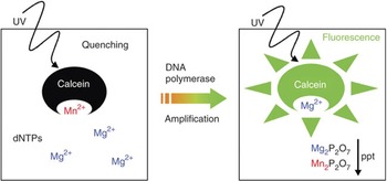
During the LAMP reaction, magnesium pyrophosphate is produced in proportional amounts to the amplified product generating turbidity. A pulse centrifuge to the reaction tubes deposit the magnesium pyrophosphate in the bottom of the tubes allowing the detection of amplification by naked eye (Fig. 2). Moreover, real-time monitoring of LAMP amplification can be achieved through spectrophotometric analysis with turbidimeter allowing the quantification of the pathogens. Visual detection can be achieved by adding intercalating dye, ethidium bromide, SYBR Green I, or calcein acetoxymethyl (AM) ester (fluorexon) to the final reaction mixture. An advantage of SYBR Green I is the ability to obtain results immediately, as the original orange color of the dye will change into green when examined under ultra-violet (UV) light (302 nm). Calcein AM on the other hand is an indirect method as the calcein AM combines with manganese and then quenches the fluorescence. A second reaction is required where the pyrophosphate ions will remove manganese ions from calcein AM to produce fluorescence (Tomita et al., Reference Tomita, Mori, Kanda and Notomi2008) (Fig. 3). The LAMP-amplified products are separated by molecular-grade agarose gel electrophoresis in Tris-borate EDTA buffer stained with ethidium bromide and visualized using an UV transilluminator. The amplicons reveal a ladder-like pattern. Additionally, restriction analysis alone or followed by sequencing reactions can be performed for verification of new protocols that are tested for the first time (Notomi et al., Reference Notomi, Okayama, Masubuchi, Yonekawa, Watanabe, Amino and Hase2000).
The calcein dye detection method of the LAMP product. The dye, calcein, binds manganese ions that quench fluorescence. DNA polymerase LAMP results in the production of pyrophosphate that binds with the calcein bound manganese ions as well as magnesium ions resulting in two detection methods that indicate that the LAMP reaction was successful: fluorescent emissions from calcein and/or the production of manganese phosphate/ that forms a precipitate that is visually detected. The presence of magnesium ions in the buffer system of the LAMP system will enhance calcein fluorescence. (Tomita et al., Reference Tomita, Mori, Kanda and Notomi2008).

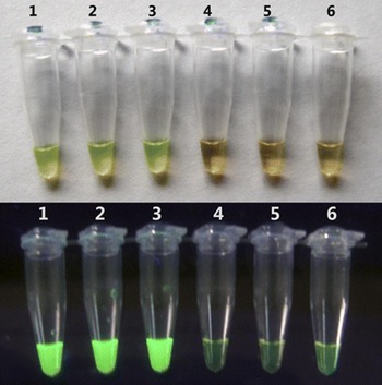
Visual detection RT-LAMP with a fluorescent detection reagent. Ten-fold serial dilutions of viral RNA (lanes 1–6, 1–0.00001 PFU per reaction mixture, respectively) were detected. (a) Evaluation under normal light. The color of positive samples changed from orange to green, whereas the color of negative samples and the negative control remained orange. (b) Evaluation under UV light. Positive samples were bright green under UV light, while negative samples or the negative control remained weakly green. (Jiang et al., Reference Jiang, Liu, Deng, Xu, Li, Han, Cao, Qin and Qin2011)

The novelty of the LAMP is its ability to amplify nucleic acid under isothermal condition; it allows the use of simple and cost-effective reaction equipment (thermostat or water bath) to produce a tremendous amount of a targeted sequence (109–1010 copies) within 15–60 min (Notomi et al., Reference Notomi, Kanda, Taguch, Minekaw, Itamura, Odagiri and Tashiro2004). The assay is highly specific as the amplification reaction occurs only when all six regions within a target DNA are correctly recognized by the primers (Fig. 1). It is comparable to PCR in terms of sensitivity, but is less affected by presence of non-targeted DNA and inhibitory molecules (Kaneko et al., Reference Kaneko, Kawana, Fukushima and Suzutani2007). LAMP can allow amplification of specific product without prior extraction procedure, by directly adding the reaction mixture to swab specimens or sera (Yamada et al., Reference Yamada, Itoh and Yoshida2006; Ihira et al., Reference Ihira, Akimoto, Miyake, Fujita, Sugata, Suga, Ohashi, Nishimura, Ozaki, Asano and Yoshikawa2007). A simple extraction method or exclusion of an extraction step reduces the processing time and costs, thus further simplifying the procedure for use under field conditions. Since the reverse transcription LAMP (RT-LAMP) is a one-step assay, the cDNA synthesis step is eliminated, decreasing the risk of contamination. The RT-LAMP assay coupled with use of Flinders Technology Associated filter paper (FTA® cards) for the preservation of RNA samples has proved to be a practical approach for the collection and identification of RNA viruses in the field and in the laboratory (Madhanmohan et al., Reference Madhanmohan, Nagendrakumar, Manikumar, Yuvaraj, Parida and Srinivasan2013).
This technology has several intrinsic disadvantages including restricted availability of reagents and instruments, limited multiplex capability and limitations related to primer design. LAMP does not allow for the inclusion of an internal PCR inhibition control (IC). As each initial target generates one unique PCR product in the form of precise copies of the same template, LAMP is less versatile than PCR. Primer design requirements and constraints on target sites selection can limit the procedures resolution and/or specificity. It requires specific software for analysis (Parida et al., Reference Parida, Sannarangaiah, Dash, Rao and Morita2008). Because the final product is a mixture of large fragments, LAMP is not useful for cloning or other molecular biology applications enabled by PCR.
Because of its simplicity, rapid amplification and easy detection, LAMP has been used for diagnosis of several important emerging and re-emerging diseases, detection of pathogenic microbes in food, quantification of template DNA, single nucleotide polymorphism typing (Iwasaki et al., Reference Iwasaki, Yonekawa, Otsuka, Suzuki, Nagamine, Hase, Tatsumi, Horigome, Notomi and Kanda2003; Nakamura et al., Reference Nakamura, Ito, Takahashi, Hashimoto, Kawamoto, Yamanaka, Taniguchi, Kamatani and Gemma2007) and tumor detection (Horibe et al., Reference Horibe, Ochiai, Shimada, Tomonaga, Nomura, Gun, Tanizawa and Hayashi2006). This review will discuss the scope and capability of LAMP for the early detection and identification of World Organization for Animal Health (OIE) notifiable viral pathogens of ruminants, swine, and avian species.
LAMP assays for notifiable OIE livestock diseases
Ruminants
Foot and mouth disease virus
Foot-and-mouth disease virus (FMDV), a member of the genus Aphthovirus, family Picornaviridae, exists as seven antigenically distinct serotypes (O, A, C, Asia 1, SAT 1, SAT 2, and SAT 3), plus numerous constantly evolving subtypes (Carrillo et al., Reference Carrillo, Tulman, Delhon, Lu, Carreno, Vagnozzi, Kutish and Rock2005). The virus is responsible for a highly contagious and economically devastating disease in cloven-hoofed livestock. Its transboundary nature can result in devastating losses to the agricultural industry, national and international trade of livestock and animal products (King et al., Reference King, Ferris, Shaw, Reid, Hutchings, Giuffre, Robida, Callahan, Nelson and Beckham2006).
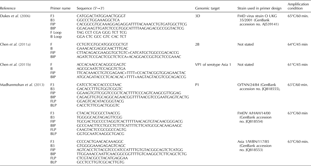
Rapid and accurate diagnosis of suspected FMDV outbreaks is important for controlling this extremely contagious virus. Several FMDV LAMP assays have been developed. A first generation one-step RT-LAMP assay (Table 1) for rapid detection of all seven serotypes of the virus was developed, amplifying 174-bp target sequence of the FMDV 3D RNA polymerase gene with an equal sensitivity to the real-time RT-PCR (Dukes et al., Reference Dukes, King and Alexandersen2006). This RT-LAMP assay was able to detect 10 copies of FMDV transcript in 22 min, rather than the 55 min required for the real time RT-PCR. Another FMDV RT-LAMP assay was developed for the rapid detection of FMDV within 1 h at 65°C, using six primers for FMDV polyprotein gene (Li et al., Reference Li, Chen, Xiong and Fang2009a). The detection limit of this method was reported to be 10−5 dilution of RNA sample, which was 100-fold higher than that of conventional PCR and 10-fold higher than real-time PCR. In 2011, an even faster FMDV RT-LAMP assay was developed using a set of four primers (Table 1) targeting FMDV 2B (Chen et al., Reference Chen, Zhang, Liu and Liu2011a) in a reaction that takes 45 min. Another RT-LAMP assay for typing FMDV serotype Asia 1 using four primers (Table 1) was developed by this same group that targets the conserved regions of VP1 sequence from FMDV serotype Asia1 (Chen et al., Reference Chen, Zhang, Liu and Liu2011b). Recently, a LAMP assay for serotyping of FMDV O, A and Asia 1 using three primer sets (Table 1) was developed (Madhanmohan et al., Reference Madhanmohan, Nagendrakumar, Manikumar, Yuvaraj, Parida and Srinivasan2013). A FMDV serotype C specific assay with no cross-reactivity with serotype A, Asia 1, O FMDV, and SVDV has also been developed (Ding et al., Reference Ding, Zhou, Ma, Qi, Wei and Zhang2014). The high sensitivity and specificity of the FMDV RT-LAMP assays make them great tools for use as a point-of-care (POC) test.
Published RT-LAMP assays for detection of FMDV.

Bovine viral diarrhea virus
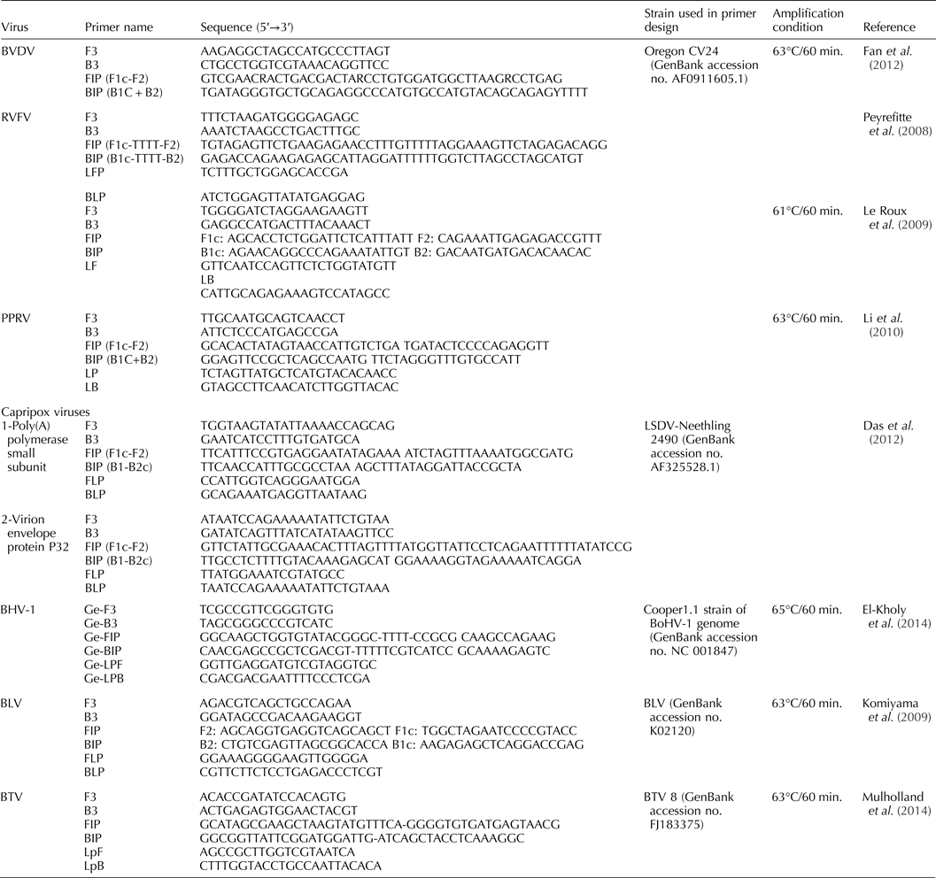
Bovine viral diarrhea virus (BVDV), a positive-sense single strand RNA virus, is a member of the genus Pestivirus in the family Flaviviridae (Wengler, Reference Wengler, Francki, Fauquet, Knudson and Brown1991). There are two major genotypes of BVDV (BVDV1 and BVDV2), which are further classified into two biotypes, cytopathic and noncytopathic, based on the presence or absence of the cytopathic effects in cell cultures (Ridpath et al., Reference Ridpath, Bolin and Dubovi1994). Non-cytopathic viruses of both species can establish persistent infections in utero that may spontaneously mutate to the cytopathogenic biotype, resulting in the onset of fatal mucosal disease (Brownlie et al., Reference Brownlie, Clarke and Howard1984). BVDV is a contaminant in biological products derived from bovine serum such as fetal calf serum, a common growth supplement for cell cultures intended for virus isolation, biological studies, and vaccine production (Liu et al., Reference Liu, Xia, Belák and Baule2008). Identification of persistently infected reservoir animals is crucial to a successful control of BVDV (Paton et al., Reference Paton, Sharp and Ibata1999). A highly sensitive RT-LAMP assay to detect BVDV-RNA using six primer sets (Table 2), capable of amplifying six target sequences at the 5′UTR region of BVDV Oregon CV24 was developed and optimized (Fan et al., Reference Fan, Xie, Xie, Liu, Pang, Deng, Xie, Peng and Wang2012).
Published RT-LAMP assays for detection of BVDV, RVFV, PPRV, Capripox, BHV-1, BLV, and BTV.

Rift Valley fever virus
Rift Valley fever virus (RVFV) is a zoonotic mosquito-borne virus of the genus Phlebovirus in the family Bunyaviridae. Its tripartite negative-strand RNA genome is composed of a large segment (L), a medium segment (M), and a small segment (S) (Schmaljohn, Reference Schmaljohn, Fields, Knipe and Howley1996). The virus causes explosive outbreaks with high mortality in newly born ruminants and abortion storms in pregnant ewes, nannies and cows at any stage of gestation (Pepin et al., Reference Pepin, Bouloy, Bird, Kemp and Paweska2010). The first RT-LAMP assay for the detection of RVFV amplified 282-nucleotide segment from the L RNA segment of RVFV using a set of six primers (Table 2) at 63°C (Peyrefitte et al., Reference Peyrefitte, Boubis, Coudrier, Bouloy, Grandadam, Tolou and Plumet2008). The assay surpassed conventional RT-PCR with high specificity and positive results obtained within 30 min. Another real-time RT-LAMP assay targeting the genomic L RNA segment of RVFV was developed using a six primer set (Table 2) with no cross-reactivity with other arboviruses in less than 30 min (Le Roux et al., Reference Le Roux, Kubo, Grobbelaar, van Vuren, Weyer, Nel, Swanepoel, Morita and Paweska2009). Despite these advantages, nucleic acid-based assays like RT-LAMP should be run in parallel with additional tests because viremia in RVFV-infected individuals is of short duration (~30 days), increasing the likelihood of false negative animals. Detection of RVFV immunoglobulin M and/or immunoglobulin G antibodies would be useful to avoid misdiagnosis (Paweska et al., Reference Paweska, Burt, Anthony, Smith, Grobbelaar, Croft, Ksiazek and Swanepoel2003).
Peste des petits ruminants virus
Peste des petits ruminants virus (PPRV), a member of genus Morbillivirus in the family Paramyxoviridae is the causative agent of peste des petits ruminants (PPR), an acute, highly contagious, and economically important viral disease of small ruminants. The morbidity and mortality rates of PPR can reach as high as 100 and 90%, respectively (Abu Elzein et al., Reference Abu Elzein, Hassanien, Al-Afaleq, Abd Elhadi and Housawi1990). A one-step, single-tube, RT-LAMP assay for detection of PPRV with a set of six LAMP primers (Table 2) within 60 min at 63◦C has been developed (Li et al., Reference Li, Bao, Wu, Wang, Wang, Gong, Liu and Li2010). No cross-reactivity with other related viruses’, including rinderpest virus, canine distemper virus, and measles virus, was detected. The sensitivity of the assay was similar to that of real-time RT-PCR and 10-fold higher than that of conventional RT-PCR in the tested clinical samples.
Lumpy skin disease virus
Lumpy skin disease virus (LSDV), a member of genus Capripoxvirus (CaPV) of the family Poxviridae, causes an occasionally fatal disease of cattle. The major impact of LSD is associated with production losses, including abortion, sterility, decreased weight gain, reduced milk production, and damage to hides (Babiuk et al., Reference Babiuk, Bowden, Parkyn, Dalman, Manning, Neufeld, Embury-Hyatt, Copps and Boyle2008a). The disease is endemic in sub-Saharan Africa and Egypt but has the potential to spread to other regions of Africa and the Middle East (Babiuk et al., Reference Babiuk, Bowden, Boyle, Wallace and Kitching2008b). A highly specific CaPV LAMP assay using primers (Table 2) designed to target a conserved gene encoding the CaPV poly(A) polymerase small subunit (VP39) has been developed (Das et al., Reference Das, Babiuk and McIntosh2012). Hydroxynaphthol blue (HNB) was incorporated to monitor assay progress by color change from violet when negative to sky blue when positive, and results were verified by agarose gel electrophoresis. In comparison studies, the LAMP assay was 10–100-fold higher in sensitivity than virus isolation, with 90–95% specificity and 89–100% sensitivity when compared to real time PCR (Das et al., Reference Das, Babiuk and McIntosh2012).
Bovine herpesvirus 1
Bovine herpesvirus-1 (BHV-1), a member of family Herpesviridae, subfamily Alphaherpesvirinae, genus Varicellovirus, is a serious viral pathogen of domestic and wild cattle (Roizman, Reference Roizman1992). The virus has been associated with respiratory infection, genital disease, and abortions (Kahrs, Reference Kahrs and Kahrs2001). Infected animals may run an undetectable subclinical course or may become latently infected surviving and shedding the virus for their lifetime. Furthermore, cattle vaccinated with conventional vaccines cannot be distinguished from infected cattle in the field by serological tests. A rapid (less than 1 h) LAMP assay was developed for detection of BoHV-1 using primers (Table 2) targeting the glycoprotein E gene (El-Kholy et al., Reference El-Kholy, Abdelrahman and Soliman2014).
Bovine leukemia virus
Bovine leukemia virus (BLV), an oncogenic retrovirus, belongs to the genus Deltaretrovirus of the Retroviridae family (Kettmann et al., Reference Kettmann, Burny, Callebaut, Droogmans, Mammerickx, Willems, Portetelle and Levy1994). It is associated with B-cell lymphocytosis, leukemia, and lymphosarcoma in the ovine and bovine species (Tajima et al., Reference Tajima, Ikawa and Aida1998). BLV infection is characterized by a long period of viral latency and by the absence of viremia. A rapid, sensitive RT-LAMP assay was established for diagnosis of BLV infection (Table 2) (Komiyama et al., Reference Komiyama, Suzuki, Miura and Sentsui2009).
Bluetongue virus
Bluetongue virus (BTV) is an arthropod-borne virus (arbovirus) that belongs to the family Reoviridae, genus Orbivirus (Murphy et al., Reference Murphy, Borden, Shope and Harrison1971) and infects both domestic and wild ruminants. Early detection and identification of BTV is essential for disease control. A one-step accelerated RT-LAMP assay was designed to target a specific region of VP2 gene of BTV 8 (Mulholland et al., Reference Mulholland, Hoffmann, McMenamy, Korthase, Earley, Markey, Cassidy, McKillen, Allan and Welsh2014). Because the VP2 gene is serotype specific, LAMP assays for other BTV serotypes could easily be developed.
Swine
Classical swine fever virus
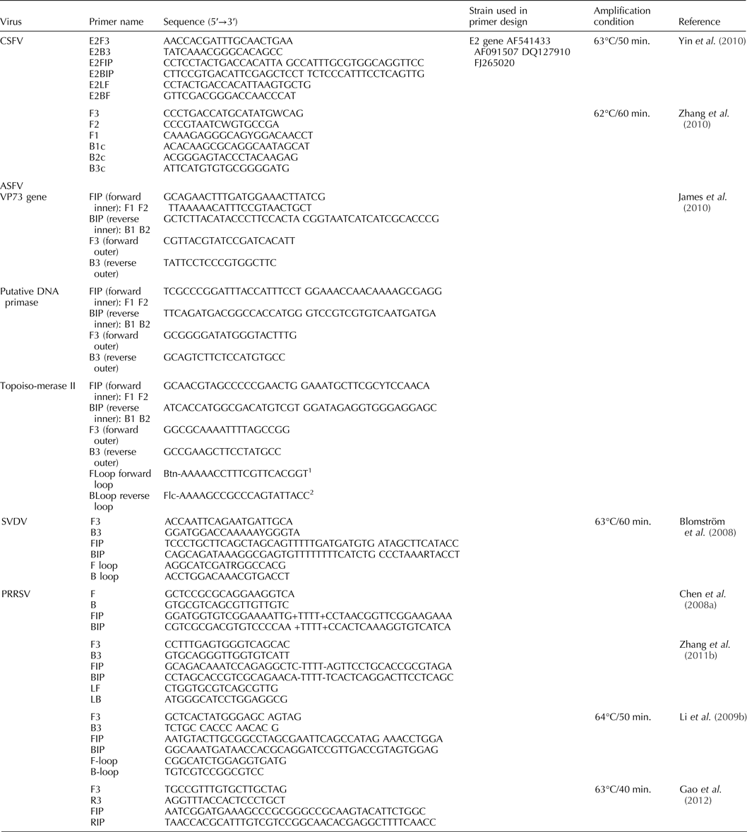
Classical swine fever virus (CSFV) is a member of the genus Pestivirus in the family Flaviviridae (Thiel et al., Reference Thiel, Collett, Gould, Heinz, Houghton, Meyers, Purcell, Rice, Fauquet, Mayo, Maniloff, Desselberger and Ball2005). CSFV causes a highly contagious and often fatal disease of swine with severe economic losses (Edwards et al., Reference Edwards, Fukusho, Lefevre, Lipowski, Pejsak, Roehe and Westergaard2000). Clinical signs of CSF can remain undetected, particularly during infections with low virulence CSFV strains. Rapid and precise detection of CSFV is critical for disease containment. RT-LAMP for rapid pre-clinical detection of CSFV infection using a set of four primers targeting the 5′ untranslated region of CSFV and with no cross-reactivity with other porcine viruses has been developed (Chen et al., Reference Chen, Zhang, Ma, Ma, Ding, Liu, Chen, Ma, Zhang and Liu2009). A one-step RT-LAMP for the detection of CSFV using a set of primers designed based on the E2 gene (Table 3) within 50 min at 63°C has been developed (Yin et al., Reference Yin, Shang, Zhou, Tian, Liu, Cai and Liu2010). Another similar RT-LAMP assay for detection of CSFV at 63°C within 1 h was also developed (Chen et al., Reference Chen, Fan, Wang, Xu, Zhao, Zhou, Liu, Tang and Zou2010a).
Published RT-LAMP assays for detection of swine viral infections.

1 Oligo labeled with biotin for lateral flow device studies.
2 Oligo labeled with fluorescein (FITC) for lateral flow device studies.
These two CSFV RT-LAMP assays target both wild-type CSFV and the heat-treated cell lysate vaccine (HCLV) strain, making the interpretation of field vs. vaccine exposure complicated in terms of eradication and control when vaccination is used (Chen et al., Reference Chen, Zhang, Ma, Ma, Ding, Liu, Chen, Ma, Zhang and Liu2009; Yin et al., Reference Yin, Shang, Zhou, Tian, Liu, Cai and Liu2010). A specific LAMP assay was developed and evaluated for identification of pigs infected with wild-type CSFV in herds vaccinated with a rabbit-passaged attenuated live vaccine (the most frequently used vaccine) using a set of four primers (Table 3) targeting six positions in the NS5B gene region of the viral genome (Zhang et al., Reference Zhang, Sun, Liu, Belák and Qiu2010). Another RT-LAMP assay for detection of the CSF HCLV strain has been developed by the same laboratory (Zhang et al., Reference Zhang, Han, Sun, Belák, Liu and Qiu2011a). Another rapid RT-LAMP assay targeting the 5′UTR gene for the detection of CSFV at 65°C employing a set of six primers was developed (Wongsawat et al., Reference Wongsawat, Dharakul, Narat and Rabablert2011).
African swine fever virus
African swine fever virus, a member of genus Asfivirus, family Asfarviridae, is a large enveloped virus with a double-stranded DNA genome (Murphy et al., Reference Murphy, Gibbs, Horzinek, Studdert, Murphy, Gibbs, Horzinek and Studdert1999). The virus is responsible for severe economic losses in domesticated pigs and wild boar, causing acute hemorrhagic fever with high morbidity and mortality. The disease severity ranges from lethal to moderately virulent or even non-virulent depending on the host species and strain of the virus involved (Tulman et al., Reference Tulman, Delhon, Ku and Rock2009).
The rapid, sensitive and specific detection of ASFV is essential, not only for the implementation of control measures to prevent the spread of ASF, but also in the differential diagnosis of other pig diseases with similar clinical findings. The LAMP assay for the detection of ASFV topoisomerase II gene (Table 3) has been developed (James et al., Reference James, Ebert, McGonigle, Reid, Boonham, Tomlinson, Hutchings, Denyer, Oura, Dukes and King2010). The assay specificity was confirmed by restriction enzyme digestion of the reaction products. This study also demonstrated that the LAMP amplicons can be detected reliably using lateral flow devices (LFDs).
Swine vesicular disease virus
Swine vesicular disease virus (SVDV), a positive single stranded RNA virus belonging to the genus Enterovirus in the Picornaviridae family (Lin and Kitching, Reference Lin and Kitching2000), is the causative agent of swine vesicular disease (SVD). A rapid diagnostic tool that can distinguish SVDV from FMDV and other viruses causing similar vesicular diseases is essential. A one-step RT-LAMP assay that can detect SVDV within 30–60 min (Table 3) has been developed that targets the highly conserved SVDV 3D polymerase gene (Blomström et al., Reference Blomström, Hakhverdyan, Reid, Dukes, King, Belák and Berg2008).
Porcine reproductive and respiratory syndrome virus
Porcine reproductive and respiratory syndrome virus (PRRSV), a member of the genus Arterivirus in the family Arteriviridae, is a small, enveloped, positive-strand RNA virus (Conzelmann et al., Reference Conzelmann, Vissner, van Woensel and Thiel1993). It is associated with reproductive failure in sows and respiratory disease in growing pigs and has a great economic impact on swine production worldwide (Zimmerman et al., Reference Zimmerman, Benfield, Murtaugh, Osorio, Stevenson, Torremorell, Straw, Zimmerman, D'Allaire and Taylor2006). A variant of PRRSV, atypical PRRSV, has symptoms including shivering, high fever and erythematous blanching rash, that resemble CSF; a rapid and accurate identification tool is necessary (Tian et al., Reference Tian, Yu, Zhao, Feng, Cao, Wang, Hu, Chen, Hu, Tian, Liu, Zhang, Deng, Ding, Yang, Zhang, Xiao, Qiao, Wang, Hou, Wang, Yang, Kang, Sun, Jin, Wang, Kitamura, Yan and Gao2007).
A PRRSV RT-LAMP assay targeting the open reading frames 1a (nucleotides 2710–2946) of highly pathogenic PRRSV genome (Table 3) has been developed for the detection of PRRSV from blood, semen, and lung samples (Chen et al., Reference Chen, Zhang, Sun, Ma, Liu, Quan and Liu2008a). Rovira et al. (Reference Rovira, Abrahante, Murtaugh and Claudia2009) successfully detected PRRSV from serum samples of acutely infected animals by RT-LAMP. Another RT-LAMP assay was developed (Table 3) targeting the M protein (Li et al., Reference Li, Zhou, Xue, Ma, Zhu and Cao2009b). A RT-LAMP to detect PRRSV using set a of Nsp2 primers (Table 3) was established (Zhang et al., Reference Zhang, Fu, Li, Jiang, Li and Wu2011b, Reference Zhang, Liu, Chen, Wang and Ningc). Another RT-LAMP assay (Table 3) targeting the M gene of type II PRRSV has been developed (Gao et al., Reference Gao, Cui, Ren, Suo, Li, Sun, Su, Opriessnig and Ren2012).
Avian
Avian influenza virus
Influenza A viruses belong to the family Orthomyxoviridae, having an eight-segmented, single-stranded, negative sense RNA genome. Influenza A viruses are classified into 17 haemagglutinin (HA) subtypes and 10 neuraminidase subtypes (Tong et al., Reference Tong, Li, Rivailler, Conrardy, Castillo, Chen, Recuenco, Ellison, Davis, York, Turmelle, Moran, Rogers, Shi, Tao, Weil, Tang, Rowe, Sammons, Xu, Frace, Lindblade, Cox, Anderson, Rupprecht and Donis2012) infecting animals and humans. The LAMP assay is the most convenient diagnostic tool for rapid and accurate detection of highly pathogenic avian influenza (HPAI) for pandemic preparedness. Several RT-LAMP assays have been developed for the detection of H5N1 viruses. The first assay to detect H5N1 virus using RT-LAMP technology detected virus in 35 min (Imai et al., Reference Imai, Ninomiya, Minekawa, Notomi, Ishizaki, Tashiro and Odagiri2006). Next, a H5 subtype RT-LAMP system was developed (Table 4) (Imai et al., Reference Imai, Ninomiya, Minekawa, Notomi, Ishizaki, Tu, Tien, Tashiro and Odagiri2007). This system detected H5-HA genes in throat swabs collected from human beings as well as from wild birds. A one-step RT-LAMP assay for detection of HPAI subtype H5N1 viruses has also been described (Jayawardena et al., Reference Jayawardena, Cheung, Barr, Chan, Chen, Guan, Peiris and Poon2007). The assay produced negative results for all non-H5N1 subtypes. Another study evaluated the diagnostic performance of two commercial real-time RT-LAMP kits, avian Flu H5 and avian Flu H7, in detecting AIV infections (Postel et al., Reference Postel, Letzel, Frischmann, Grund, Beer and Harder2010). Updating the primer sequences is necessary to maintain the high sensitivity of the H5 RT-LAMP assay. New sets of primers (Table 4) for RT-LAMP to specifically amplify the HA gene of avian influenza viruses subtype H5N1 have been developed (Dinh et al., Reference Dinh, Le, Vuong, Hasebe and Morita2011).
Published RT-LAMP assays for detection of avian viral infections.

1 The restriction enzyme site sequences of EcoR I and EcoR V were added in FIP and BIP primer, respectively.
2 Tsai et al. (Reference Tsai, Liu, Shien, Lee, Chang and Wang2012) have used IBDV-P: Probe GGGACCAGATGTCATGG; modified with 5′-terminal FITC.
3 Primer labeled with biotin.
The LAMP technique has also been implemented for the detection of other HA subtypes; H9 AIV subtype (Chen et al., Reference Chen, Zhang, Sun, Ma, Liu, Cai and Liu2008b), H3 subtype AIV (Peng et al., Reference Peng, Xie, Liu, Pang, Deng, Xie, Xie, Fan, Feng and Khan2011), H6 subtype AIV (Chen-Yu et al., Reference Chen-Yu, Zhi-Xun, Yang, Yi, An-Li, Jia-Bo, Yao-Shan, Xian-Wen, Zhi-Qin and Li-Ji2011), and H7 (Bao et al., Reference Bao, Wang, Zhao, Sun, Li, Xiong and Chen2012) have all been detected using HA-specific primers (Table 4). RT-LAMP method may provide a new avenue for the recognition of HPAIV, and may be employed to screen for potential carriers in wild and domestic birds. A RT-LAMP for detection of avian influenza viruses in field specimens has been developed (Table 4). This assay targets the most conserved region of the matrix gene (Shivakoti et al., Reference Shivakoti, Ito, Murase, Ono, Takakuwa, Yamashiro, Otsuki and Ito2010).
Infectious bronchitis virus
Infectious bronchitis virus (IBV), a member of family Coronaviridae, genus Coronavirus, is the causative agent of an acute, highly contagious avian disease affecting the respiratory and urogential tracts (Cavanagh and Naqi, Reference Cavanagh, Naqi, Calnek, Barnes, Beard, McDougald and Saif1997). There are considerable economic losses in the poultry industry attributed to the diminished performance of both meat-type and egg-laying birds (Cavanagh and Gelb, Reference Cavanagh, Gelb and Saif2008). Antigenic variants of IBV complicate the diagnosis process; hence differentiation is required for proper control and using appropriate preventive measures. An IBV RT-LAMP assay targeting the nucleocapsid phosphoprotein gene of infectious bronchitis virus (Table 4) has been reported (Chen et al., Reference Chen, Zhang, Ma, Ma, Ding, Liu, Cai, Ma, Zhang and Liu2010b). The sensitivity of RT-LAMP and RT-PCR assays for detecting IBV RNA in clinical specimens was 99.5 and 98.4%, respectively.
Infectious bursal disease virus
Infectious bursal disease virus (IBDV), a member of family Birnaviridae, genus Avibirnavirus, is the etiologic agent of IBD, an acute and highly contagious immunosuppressive disease in young chickens (Becht and Müller, Reference Becht and Müller1991). IBDV causes a huge economic loss to the poultry industry worldwide. The virus causes destruction of B-lymphocytes in the bursa of Fabricius leading to deficiencies in humoral immunity that leaves birds vulnerable to other pathogens (Saif, Reference Saif1991). A RT-LAMP method for rapid detection of IBDV using four primers (Table 4) specific to six regions of the VP3 gene has been developed (Xu et al., Reference Xu, Zhang, Yin, Cui, Xu, Guo, Li, Wang, Liu and Han2009). Subsequently a RT-LAMP assay for rapid identification of IBDV using a set of four primers (Table 4) to amplify the VP2 gene has been developed (Xue et al., Reference Xue, Zhang, Zhou, Xu, Li and Cao2009; Lee et al., Reference Lee, Lin, Lai, Lai, Chen and Wang2011). The assay detected 30 different IBDV isolates and had no cross-reaction with three other avian viruses (IBV, NDV and AIV). A VP5-based RT-LAMP method for the detection and discrimination of IBDV, using a set of two inner, two outer, and two loop primers (Table 4) has been developed (Wang et al., Reference Wang, Kang, Gao, Gao, Qin, Lin, Yu, Qi and Wang2011). In combination with Tfi I restriction analysis, virulent IBDV strain could be distinguished from current vaccine strains. A combination assay using a RT-LAMP combined with a chromatographic lateral flow dipstick (LFD) for the detection of IBDV using biotin-labeled primers (Table 4), fluorescein isothiocyanate FITC-labeled DNA probe, and the LFD has been reported (Tsai et al., Reference Tsai, Liu, Shien, Lee, Chang and Wang2012). False positive results were not detected during the testing of other unrelated viruses and cells. In addition, the amplification efficiency of RT-LAMP was enhanced when a loop primer was used accelerating the RT-LAMP-LFD product detection time from 70 min (without loop primer) to 40 min. This RT-LAMP-LFD assay stands out as a promising alternative for the diagnosis of IBDV under field conditions, in rural areas and developing countries where there is a lack of complex laboratory services.
Newcastle disease virus
Newcastle disease virus (NDV), a member of family Paramyxoviridae, is the causative agent of a highly contagious widespread disease of poultry worldwide (Alexander, Reference Alexander, Calnek, Barnes, Beard, McDougald and Saif1997). Despite the significant reduction in reported outbreaks after vaccination campaigns and restricted importation quarantine measurements, the disease remains a potential threat to both commercial and backyard poultry producers with outbreaks being reported in many countries with strict eradication measures (Kho et al., Reference Kho, Mohd-Azmi, Arshad and Yusoff2000). A LAMP-based NDV diagnostic system has been evaluated for rapid, simple, and sensitive detection of NDV from culture isolates as well as clinical samples using one set of specific primers (Table 4) that target the fusion protein gene (Pham et al., Reference Pham, Nakajima, Ohashi and Onuma2005). NDV LAMP was as sensitive and specific as nested PCR. Another one-step RT-LAMP assay with degenerate primers for detection of NDV in clinical field samples (Table 4) has been developed with improved sensitivity and specificity (Li et al., Reference Li, Xue, Qin, Zhou, Chen, Bi and Cao2009c). Recently, a RT-LAMP assay was developed that could detect NDV in clinical cases using cloacal or oropharyngeal swabs in less than 60 min. Analysis was done using specific RT-LAMP primers (Table 4) targeting the fusion protein (Kirunda et al., Reference Kirunda, Thekisoe, Kasaija, Kerfua, Nasinyama, Opuda-Asibo and Inoue2012).
Duck hepatitis virus
Duck hepatitis virus type 1 (DHV-1), a member of family Picornaviridae and genus Avihepatovirus, is a single-stranded RNA virus responsible for an acute and highly contagious disease affecting ducklings under 3 weeks of age with a high mortality rate of up to 95% worldwide (Tseng et al., Reference Tseng, Knowles and Tsai2007). A RT-LAMP assay was designed for detection of DHV-1 using primers (Table 4) specific to the highly conserved 3D gene (Song et al., Reference Song, Wan, Yu, Han, Qiu, Hu, Tan and Ding2012) or 2C gene (Yang et al., Reference Yang, Li, Bi, Xu and Liu2012).The RT-LAMP assay was highly specific; no cross-reactivity was observed from the samples of other related pathogens.
West Nile virus
West Nile virus (WNV), an arbovirus, belongs to the family Flaviviridae, genus Flavivirus (Gubler et al., Reference Gubler, Kuno, Markoff, Knipe and Howley2007) and exists in a transmission cycle between birds and mosquitoes, where human beings and other mammals are incidental hosts. Rapid and early detection of virus activity among mosquitoes, migratory birds, domestic poultry and equids is necessary for the prediction and prevention of large-scale epidemics. A one-step, single tube, real-time accelerated RT-LAMP assay was developed for detecting the envelope gene of WNV (Parida et al., Reference Parida, Posadas, Inoue, Hasebe and Morita2004) (Table 4).
Conclusions
This review details the use of LAMP techniques to improve diagnosis of 18 viral diseases of international veterinary importance. The technique is highly sensitive, highly specific, quantitative, and yet simple and requiring no sophisticated equipment. The reduced risk of cross-contamination is an additional important feature of this technology. The assay can be incorporated with automated platforms to provide diagnostic laboratories with the ability to test large numbers of samples that might be received during outbreaks of livestock disease.
The simplicity and ease of implementation of the LAMP assay makes it the diagnostic test of choice in ‘low-resource laboratories’ and under field conditions. The assay can be read directly by visual examination. In contrast to PCR, highly skilled personnel are not required, as persons with basic skills in molecular biology can perform the assay. The technique would be of great value for veterinary authorities, pathologists and field veterinarians to facilitate rapid and accurate diagnosis of diseases, which is the necessary first step toward disease control, so the proper vaccination or eradication programs can be implemented for the specific disease.
In spite of the obvious advantages of LAMP diagnostics for veterinary application, field implementation has not occurred. Although OIE recognizes the importance of the LAMP technology in their guideline ‘Development and Optimisation of Nucleic Acid Detection Assays’ (OIE 2014a), there is no mention of any of the specific LAMP tests described in this review in the ‘Diagnostic Techniques’ section for any of the 18 selected agents in the ‘OIE Manual of Diagnostic Tests and Vaccines for Terrestrial Animals 2014’ (OIE, 2014b). In contrast, conventional and/or real time reverse transcription PCR (RT-PCR) is described in each agent's ‘Diagnostic Techniques’ section. Without specific inclusion of LAMP technology in the OIE Manual of Diagnostic Tests, its use will be limited. Another limitation is the lack of standardized reagent assay kits with thermally stable reagents for veterinary application. In human medicine, there have been two major LAMP field successes, tuberculosis and malaria (WHO, 2013; Patel et al., Reference Patel, Lucchi, Srivastava, Lin, Sug-aram, Aruncharus, Bharti, Shukla, Congpuong, Satimai, Singh, Udhayakumar and Meshnick2014). In both cases standardized reagent assay kits with thermally stable reagents have been used patient side in developing countries. These LAMP assays have been demonstrated to be sensitive, specific, less costly, and faster. In the malaria and TB studies, one of the major needs was a sensitive point-of-care field molecular test to replace blood smears. With malaria, LAMP test results were available 30–75 min after drawing the sample compared to an average of 8 days to receive PCR diagnostic results from a central reference laboratory in Thailand (Patel et al., Reference Patel, Lucchi, Srivastava, Lin, Sug-aram, Aruncharus, Bharti, Shukla, Congpuong, Satimai, Singh, Udhayakumar and Meshnick2014). In both cases, the World Health Organization has worked extensively for the implementation of LAMP technology in the field.
In summary, LAMP veterinary diagnostic testing holds great promise for field and low-resource laboratory use. However, for its widespread application in veterinary diagnostics, there has to be a recognition and promotion of the LAMP technology by OIE and other animal health regulatory agencies. The adoption of LAMP would likely lead to the production of standardized reagent assay kits with thermally stable reagents that could be used worldwide.
Acknowledgments
The authors wish to acknowledge funding received from the South Dakota State University Agricultural Experiment Station and Animal Disease Research and Diagnostic Laboratory.