Introduction
Soil-transmitted helminths (STH) are a major public health concern in developing countries (Hotez et al., Reference Hotez, Bundy, Beegle, Brooker, Drake, de Silva, Montresor, Engels, Jukes, Chitsulo, Chow, Laxminarayan, Michaud, Bethony, Correa-Oliveira, Shuhua, Fenwick, Savioli, Jamison, Breman, Measham, Alleyne, Claeson, Evans, Jha, Mills and Musgrove2006, Reference Hotez, Brindley, Bethony, King, Pearce and Jacobson2008), affecting more than 1.4 billion people (Pullan et al., Reference Pullan, Smith, Jasrasaria and Brooker2014; Dunn et al., Reference Dunn, Turner, Tun and Anderson2016). Diagnosis of helminth infections and evaluation of parasite load have usually been carried out using the Kato–Katz (KK) thick smear method (Katz et al., Reference Katz, Chaves and Pellegrino1972). Although this method has been tested extensively (Stoltzfus et al., Reference Stoltzfus, Chwaya, Tielsch, Schulze, Albonico and Savioli1997; Goodman et al., Reference Goodman, Haji, Bickle, Stoltzfus, Tielsch, Ramsan, Savioli and Albonico2007; Knopp et al., Reference Knopp, Mgeni, Khamis, Steinmann, Stothard, Rollinson, Marti and Utzinger2008, Reference Knopp, Speich, Hattendorf, Rinaldi, Mohammed, Khamis, Mohammed, Albonico, Rollinson, Marti, Cringoli and Utzinger2011; Glinz et al., Reference Glinz, Silue, Knopp, Lohourignon, Yao, Steinmann, Rinaldi, Cringoli, N'Goran and Utzinger2010; Tarafder et al., Reference Tarafder, Carabin, Joseph, Balolong, Olveda and McGarvey2010; Levecke et al., Reference Levecke, Behnke, Ajjampur, Albonico, Ame, Charlier, Geiger, Hoa, Kamwa Ngassam, Kotze, McCarthy, Montresor, Periago, Roy, Tchuem Tchuente, Thach and Vercruysse2011; Barda et al., Reference Barda, Rinaldi, Ianniello, Zepherine, Salvo, Sadutshang, Cringoli, Clementi and Albonico2013, Reference Barda, Cajal, Villagran, Cimino, Juarez, Krolewiecki and Rinaldi2014; Assefa et al., Reference Assefa, Crellen, Kepha, Kihara, Njenga, Pullan and Brooker2014; Periago et al., Reference Periago, Diniz, Pinto, Yakovleva, Correa-Oliveira, Diemert and Bethony2015) and recommended by the World Health Organization (WHO, 1991), it has some recognized drawbacks. The KK method is used mainly for detecting helminth ova since it is not suitable for the diagnosis of nematode larvae and protozoans. Moreover, it is a tedious and time-consuming method. Relatively newer methods, such as the McMaster (Levecque et al., Reference Levecke, Behnke, Ajjampur, Albonico, Ame, Charlier, Geiger, Hoa, Kamwa Ngassam, Kotze, McCarthy, Montresor, Periago, Roy, Tchuem Tchuente, Thach and Vercruysse2011) and FLOTAC techniques (Cringoli et al., Reference Cringoli, Rinaldi, Maurelli and Utzinger2010; Barda et al., Reference Barda, Rinaldi, Ianniello, Zepherine, Salvo, Sadutshang, Cringoli, Clementi and Albonico2013) are reported to have better, albeit variable, performance for diagnosis and monitoring of treatment programmes for STH. However, some field studies indicate a similar sensitivity to KK, although FLOTAC seems to have a higher cost-effectiveness (Assefa et al., Reference Assefa, Crellen, Kepha, Kihara, Njenga, Pullan and Brooker2014; Nikolay et al., Reference Nikolay, Brooker and Pullan2014). In contrast to KK, fixed suspension samples have the advantage of drastically reducing the unpleasantness of working with fresh stool samples, eliminating the need for prompt examination to prevent destruction of non-resistant stages of parasites, which is a great advantage for field workers, and they are suitable for the diagnosis of all intestinal parasites (Mank et al., Reference Mank, Zaat, Blotkamp and Polderman1995; Pietrzak-Johnston et al., Reference Pietrzak-Johnston, Bishop, Wahlquist, Moura, Da Silva, Da Silva and Nguyen-Dinh2000; Vandenberg et al., Reference Vandenberg, Van Laethem, Souayah, Kutane, van Gool and Dediste2006; Dacombe et al., Reference Dacombe, Crampin, Floyd, Randall, Ndhlovu, Bickle and Fine2007). Additionally, like the KK, it is possible to quantify the intensity of infection using these methods. The only disadvantage is that diluting the sample may reduce the sensitivity of detection of ova, particularly in low-intensity infections, and cannot be used in assays such as the Baermann method for Strongyloides stercoralis, with which live larvae are detected. Recently, it has been found that eggs in fixed faeces can be floated and diagnosed using the mini-FLOTAC technique (Barda et al., Reference Barda, Cajal, Villagran, Cimino, Juarez, Krolewiecki, Rinaldi, Cringoli, Burioni and Albonico2015).
The use of several fixatives has been reported and the two most popular are MIF (merthiolate, iodine, formaldehyde) (Sapero & Lawless, Reference Sapero and Lawless1953) and SAF (sodium acetate, acetic acid, formaldehyde) (Vandenberg et al., Reference Vandenberg, Van Laethem, Souayah, Kutane, van Gool and Dediste2006). In this study, we assessed the performance of MIF vs. KK methods in diagnosing helminth infections and in determining the intensity of such infections in stool samples from people living in a Venezuelan rural community with moderate to high prevalence of intestinal helminth infections (Homan, Reference Homan2009).
Materials and methods
Ethical considerations
This study was part of a larger project intending to elucidate and evaluate the risk factors of helminth infections in the rural community ‘Caserio El 25, Carabobo State, Venezuela’. The objectives of the project were explained to the members of each household in the community to obtain a written informed consent from adults and parents or legal representatives of children. The director and teachers of the local school were also informed about the study purposes, and procedures involving the attending children. At the end of the study, all participants were offered (free of charge) a single dose of Pirantel© as deworming agent.
Sampling
The rural community ‘Caserio El 25’ was selected because both Ascaris lumbricoides and Trichuris trichiura infections in this community have been found to exceed by 30% their average prevalence in cross-sectional studies (Homan, Reference Homan2009). Data collection took place in June 2009. Since the community is relatively small, we attempted to enrol all inhabitants (n = 470), but eventually the sample size was based on those household members who were directly approachable and provided the informed consent, including students attending the local primary school who were present during a 3-day visit to the community. In total, we obtained an individual faecal sample from 227 participants.
Coprological examination
Faecal samples were provided in standard faeces containers and were processed within 32 h from collection, meaning that they were homogenized in the container and fixed in MIF the same day of collection and also prepared for KK (Katz et al., Reference Katz, Chaves and Pellegrino1972) the day after. One faecal sample was obtained from each inhabitant. A commercial KK kit (HELM-Tec, Brazil) was used according to the manufacturer's recommendations, based on the template equivalent to 41.7 mg of faeces. Faecal smears on glass slides were prepared and stored in the refrigerator in humid chambers and then clarified by light exposure at room temperature for 20 min in batches to be observed within 1 h. The slides were kept covered to reduce over-clarification. Two KK slides were prepared from each sample and observed by two experienced laboratory technicians.
For the MIF technique (Sapero & Lawless, Reference Sapero and Lawless1953), 7-ml red-plug Vacutainer/Venoject type tubes with 4 ml merthiolate/iodine/formaldehyde solution were pre-weighed. Faecal samples of approximately 1 cc (until a mark for 5 ml in the tube) were added to the fixation tubes with the help of two wooden rods, fragmented in the fixative, plugged with rubber stopper and mixed with a Vortex to ensure fixation. The tubes were post-weighed and the difference of pre- and post-weight was the amount of faeces added to the tube. After vortexing, duplicate 100 μl samples were taken from each tube and placed in microscope slides, covered with 24 × 50 mm cover slips, sealed with melted paraffin wax and examined thoroughly at × 100 total magnification (objective lens 10×, ocular lens 10×) for counting eggs of parasites. The number of eggs per gram (EPG) of faeces for a total volume of faecal suspension of 5 ml was calculated as follows: 5000 μl × n eggs counted per slide (in 100 μl)/100 μl sample × weight of faeces in the tube, or as n eggs per slide × 50/weight of faeces in the tube.
Data analysis
The analysis of the outcomes from each diagnostic method and from both methods combined (which is used here as the ‘gold standard’) was performed using SPSS statistical software (SPSS Inc., Chicago, Illinois, USA). For both diagnostic methods, we calculated the prevalence (proportion of people testing positive to a given parasite under study), sensitivity (proportion of positives that are correctly identified as such), negative predictive value (NPV, proportion of people whose test results are negative and who are truly parasite-free), and their corresponding 95% confidence intervals (95% CIs). Spearman's rank correlation coefficient (rho) was also calculated to assess the correlation between the total EPGs of each helminth species between the two diagnostic methods. Comparison of prevalences, sensitivity and NPV between methods was performed using the Z-test. The Mann–Whitney U-test was used to test differences in median EPGs of the intensity of infection for each parasite between the two diagnostic methods, according to the thresholds proposed by the WHO (Montresor et al., Reference Montresor, Cromptom, Hall, Bundy and Savioli1998; WHO, 2002). For A. lumbricoides, the thresholds were: low, 1–4999; moderate, 5000–49,999; and high, >49,999; for T. trichiura, the thresholds were: low, 1–999; moderate, 1000–9999; and high, >9999; for hookworms, the thresholds were: low, 1–1999; moderate, 2000–3999; and high, >3999; and for Schistosoma mansoni, the thresholds were: low, 1–99; moderate, 100–299; and high, >299. For A. lumbricoides, the EPGs were further classified into fertile and infertile. The agreement of MIF or KK with the gold standard regarding the presence/absence of each helminth species was assessed using Cohen's kappa statistic (k). The values of this statistic may indicate a slight (k < 0.2), fair (0.2 ≤ k < 0.4), moderate (0.4 ≤ k < 0.6), substantial (0.6 ≤ k < 0.8) and an almost perfect agreement (k ≥ 0.8) (Levecke et al., Reference Levecke, Behnke, Ajjampur, Albonico, Ame, Charlier, Geiger, Hoa, Kamwa Ngassam, Kotze, McCarthy, Montresor, Periago, Roy, Tchuem Tchuente, Thach and Vercruysse2011).
Results
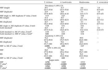
The prevalence of T. trichiura diagnosed by MIF (33.9%) was significantly lower (P = 0.01) than that derived from both KK and MIF combined, i.e. the ‘gold standard’ (44.5%) (table 1). The prevalence of A. lumbricoides and that of hookworms, as measured by MIF (34.8% and 7.5%) and KK (38.3% and 5.3%), were not significantly different from the gold standard (41.0% and 8.8%). Although examining duplicate slides for each method increased the number of detections, this did not lead to additional significant differences.
Prevalence, sensitivity, negative predictive values and Kappa index of agreement for the techniques MIF and Kato–Katz for the detection of intestinal helminths in first and duplicate examined slides (n = 227). Significant differences are shown in bold.

MIF, merthiolate, iodine, formaldehyde; KK, Kato–Katz; NPV, negative predictive value; 95% CI, 95% confidence interval.
aGold standard: outcomes of MIF + KK (duplicate slides). For sensitivity and NPV, the gold standard was set at 100%.
bComparing results of duplicate slides of any method vs. the gold standard. For the Kappa index the P value corresponds to statistical significance of the concordance of results.
Strongyloides stercoralis (prevalence = 2.6%, 95% CI = 1.0–5.7) and Schistosoma mansoni (prevalence = 0.4%, 95% CI = 0.01–2.4) were only observed with MIF.
Statistics for E. vermicularis, S. stercoralis and S. mansoni were not performed because of their very low prevalence, or because one or both of the methods (KK and MIF) were not specific for these parasites.
Regarding sensitivity, KK performed better than MIF for T. trichiura (P < 0.001) and A. lumbricoides (P = 0.02), but not for hookworms (P = 0.07) (table 1). The NPV for both methods was high (>80%) (Levecke et al., Reference Levecke, Behnke, Ajjampur, Albonico, Ame, Charlier, Geiger, Hoa, Kamwa Ngassam, Kotze, McCarthy, Montresor, Periago, Roy, Tchuem Tchuente, Thach and Vercruysse2011). However, KK performed significantly better than MIF for T. trichiura and A. lumbricoides (table 1).
The Kappa index showed substantial to almost perfect agreement of both KK and MIF with the gold standard for T. trichiura, A. lumbricoides and hookworms. It is worth mentioning that four and five cases of Enterobius vermicularis were detected with MIF and KK, respectively (both methods are not specific for this parasite), and that six cases of S. stercoralis and one case of S. mansoni (parasite load moderate, 185 EPG) were detected with MIF alone.
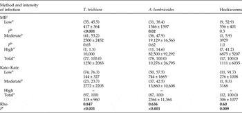
The intensity of infection showed important differences between KK and MIF methods (table 2). For most parasites, MIF detected higher EPGs than KK, and no high parasite loads were detected with KK. The rho values for the total EPGs for each parasite showed significant correlations between the two methods, with MIF performing better than KK (table 2).
Intensity of infection (EPG) (n, %) of intestinal helminth infections using MIF and Kato–Katz methods. Significant differences are shown in bold.

Rho, Spearman's rank correlation coefficient.
aMedian ± interquartile range of eggs per gram (EPG) of faeces.
b P value (Mann–Whitney U-test) for the comparison of EPGs between MIF and KK methods for each group (low, moderate and high) and for each parasite.
c P value for the correlation of total EPGs between MIF and KK methods for each parasite.
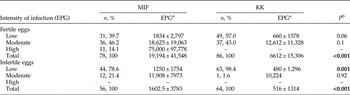
The parasite load of A. lumbricoides was divided into fertile and infertile eggs to check whether these influenced the outcome of the analysis. Results were similar to those obtained for the total EPGs mentioned above. However, KK showed a higher prevalence of both fertile and infertile eggs in low and total parasitic loads, but the median EPG of infertile eggs was significantly higher in MIF for the categories of low and total parasitic loads. Additionally, in all categories, most eggs were fertile. The proportion of infertile eggs was close to 40% in low parasite loads (MIF and KK) and close to 30% in moderate (MIF) parasite loads (percentages calculated from table 3), some participants had only infertile eggs. No infertile eggs were detected in the group of high parasitic loads (table 3).
Ascaris lumbricoides fertile and infertile eggs detected by MIF and KK methods. Significant differences are shown in bold.

aMedian ± interquartile range of the eggs per gram (EPG).
b P value for the comparison in EPGs between MIF and KK methods for each group (low, moderate and high) using the Mann–Whitney U-test.
Discussion
The unpleasantness of working with fresh faecal material hampers the clinical diagnosis of intestinal parasites, particularly in resource-limited settings. Therefore, the time dedicated to slide examination tends to be minimal, and the applied techniques are usually restricted to thin wet mounts with isotonic saline and iodine stain under a 22 × 22 mm cover slip. In such conditions, only heavy, and sometimes moderate, helminth infections can be diagnosed. In clinical practice in resource-limited settings, the Kato technique is used sporadically, and the KK and MIF methods are hardly used, if ever. Additionally, some intestinal parasites (e.g. trophozoites of protozoans and larvae of S. stercoralis) are fragile and require a short time interval between faecal collection and examination, which is often difficult to accomplish. KK is the most frequently applied technique for field studies of soil-transmitted intestinal helminths, despite the limitations occasionally posed by the time span elapsed between collection and examination when the examined communities are far from laboratories. Our results suggest that a simple fixation method like MIF can overcome not only the practical drawbacks of direct examination of faeces, but can also provide highly competitive performances regarding the diagnosis and assessment of intensity of infection for clinical and field work.
Due to the lack of a gold standard for soil-transmitted helminths, we compared the MIF and KK results with one another, as well as with those of an artificial gold standard represented by the results of both MIF and KK combined, as other authors have done previously (Goodman et al., Reference Goodman, Haji, Bickle, Stoltzfus, Tielsch, Ramsan, Savioli and Albonico2007; Knopp et al., Reference Knopp, Mgeni, Khamis, Steinmann, Stothard, Rollinson, Marti and Utzinger2008, Reference Knopp, Speich, Hattendorf, Rinaldi, Mohammed, Khamis, Mohammed, Albonico, Rollinson, Marti, Cringoli and Utzinger2011; Steinmann et al., Reference Steinmann, Du, Wang, Wang, Jiang, Li, Marti, Zhou and Utzinger2008; Utzinger et al., Reference Utzinger, Rinaldi, Lohourignon, Rohner, Zimmermann, Tschannen, N'Goran and Cringoli2008; Periago et al., Reference Periago, Diniz, Pinto, Yakovleva, Correa-Oliveira, Diemert and Bethony2015). With regard to the three most commonly detected helminth species for which we had enough data to perform the statistical analysis (i.e. T. trichiura, A. lumbricoides and hookworms), both MIF and KK methods performed similarly, except for cases with low parasite loads of T. trichiura and A. lumbricoides. In general, it is conceivable that methods based on dilution, such as MIF, will miss some cases with low parasite loads, and the same applies to sensitivity and NPV, for which KK showed a better performance over MIF in detecting T. trichiura and A. lumbricoides. Hookworm infections, for which KK is known to have a limited performance (Dacombe et al., Reference Dacombe, Crampin, Floyd, Randall, Ndhlovu, Bickle and Fine2007; Glinz et al., Reference Glinz, Silue, Knopp, Lohourignon, Yao, Steinmann, Rinaldi, Cringoli, N'Goran and Utzinger2010; Tarafder et al., Reference Tarafder, Carabin, Joseph, Balolong, Olveda and McGarvey2010), showed a lower (albeit not statistically significant) prevalence and sensitivity compared to MIF, possibly due to the low prevalence found in the community under study. A better gold standard for the detection of hookworms would have been a flotation method, such as FLOTAC or mini-FLOTAC (Utzinger et al., Reference Utzinger, Rinaldi, Lohourignon, Rohner, Zimmermann, Tschannen, N'Goran and Cringoli2008; Glinz et al., Reference Glinz, Silue, Knopp, Lohourignon, Yao, Steinmann, Rinaldi, Cringoli, N'Goran and Utzinger2010; Barda et al., Reference Barda, Rinaldi, Ianniello, Zepherine, Salvo, Sadutshang, Cringoli, Clementi and Albonico2013; Assefa et al., Reference Assefa, Crellen, Kepha, Kihara, Njenga, Pullan and Brooker2014), which proved useful in the detection and quantitation of helminth infections, although it may fail with relatively heavy eggs, such as those of S. mansoni, depending on the flotation solution used (Barda et al., Reference Barda, Rinaldi, Ianniello, Zepherine, Salvo, Sadutshang, Cringoli, Clementi and Albonico2013). Indeed, any flotation method will have drawbacks due to differential buoyancies of helminth eggs (Levecke et al., Reference Levecke, Behnke, Ajjampur, Albonico, Ame, Charlier, Geiger, Hoa, Kamwa Ngassam, Kotze, McCarthy, Montresor, Periago, Roy, Tchuem Tchuente, Thach and Vercruysse2011). The kappa index of concordance of KK and MIF methods with the gold standard showed an almost perfect agreement for A. lumbricoides, T. trichiura and hookworms, another indication of good performance of the MIF method. Additionally, six cases of S. stercoralis (for which KK is not a specific method) could be detected with MIF and one case of S. mansoni (for which KK is specific). Very low prevalences and low parasite loads of S. mansoni are difficult to detect at all with any method, and high prevalence of other intestinal helminths may shadow the diagnosis with the KK technique. Both methods were able to detect a similar low prevalence of pinworms, for which neither of them is specific. Altogether, these findings support the application of MIF.
MIF was able to detect significantly higher EPGs, either at total or low parasite loads, and was the only method able to detect high loads for A. lumbricoides, T. trichiura and hookworms, while at moderate loads both methods performed equally. This is intriguing and, to the best of our knowledge, has never been reported before. Counting high EPGs in a KK slide is an inefficient and tiring task when the number of eggs in the slide is high, and yet when multiplying the egg numbers counted by the fixed KK factor of 24 (according to the manufacturer), high parasitic loads were never achieved with any of the three helminths we evaluated. This has led others (Stoltzfus et al., Reference Stoltzfus, Chwaya, Tielsch, Schulze, Albonico and Savioli1997) to propose a second dilution method for counts above 10,000 EPG, i.e. about 417 eggs per slide. It can be argued that the fixed multiplication factor of the KK method (×24) does not take into consideration the actual mass of the faeces placed into each template, which may be responsible for the high level of variation in egg counts, since the mass of faeces under examination also varies considerably (Levecke et al., Reference Levecke, Behnke, Ajjampur, Albonico, Ame, Charlier, Geiger, Hoa, Kamwa Ngassam, Kotze, McCarthy, Montresor, Periago, Roy, Tchuem Tchuente, Thach and Vercruysse2011). However, these authors found that correcting the egg counts for the actual mass of faeces placed in the template reduces the number of EPGs. In our case it was the opposite as, in the MIF method where the mass of faeces is known, the egg counts were higher. We speculated that there might be other (hitherto unknown) variables, such as dietary factors in the community, which may alter the weight of the faeces, or the ease with which laboratory technicians use MIF in ‘clean’ microscopic fields, with eggs being fairly dispersed and therefore more reasonably counted. Additionally, KK seems to be able to better detect cases characterized by lower parasite loads than MIF. This may explain why the median EPG from KK at low parasitic loads was lower than that of MIF.
We further explored the intensity of infection in A. lumbricoides, differentiating fertile from infertile eggs with MIF and KK. Interestingly, the count of infertile eggs, although always lower than that of the fertile ones (total infertile eggs always close to 10%), was high in low (close to 40%) and moderate (close to 30%) parasite loads, and absent at higher loads using the MIF method, suggesting that the male/female ratio of adult worms becomes more homogeneous as parasite load increases. The presence of a substantial number of infertile eggs at low and moderate parasite loads is a fact to be taken into account when using flotation methods for diagnosis, as these eggs are heavy (Levecke et al., Reference Levecke, Behnke, Ajjampur, Albonico, Ame, Charlier, Geiger, Hoa, Kamwa Ngassam, Kotze, McCarthy, Montresor, Periago, Roy, Tchuem Tchuente, Thach and Vercruysse2011) and may alter the sensitivity in conditions of reduced intensity of infection and during evaluation of reduced parasite loads after deworming. Another obvious, but not usually explored, advantage of fixation methods is that samples can be re-examined for confirmation a long time after collection. We have re-examined our samples for verification of qualitative diagnoses of helminths 3 years after collection, and all three helminths (A. lumbricoides, T. trichiura and hookworms) could still be observed perfectly in similar proportions, with the addition of some more cases of S. stercoralis being detected; the protozoan parasites diagnosed earlier were also present 3 years after.
Another remark can be made about the amount of faeces examined. KK examines a mean of 41.7 mg of faeces, according to the commercial kit used. In our conditions, the mean weight of the sample contained in the 100 μl of MIF examined per slide was approximately 3 mg (mean over the 227 samples), which is approximately 14 times lower than the amount of faeces examined with the KK method. Yet, the differences in the results from both methods were not so pronounced, either qualitatively or quantitatively. Furthermore, with so few faeces and without previous sieving, which would have meant adding a further undesirable step, the MIF was able to detect more hookworm infections, further stressing the advantage of the fixation method.
Other fixatives are available, with variable performance, such as formalin, Schaudinn, polyvinyl alcohol and sodium acetate–acetic acid–formaldehyde (SAF) (Ash & Orihel, Reference Ash and Orihel1997). Some contain very toxic components, such as mercury (Schaudinn). Others, produced by commercial companies and advertised as being environmentally friendly, such as ECOFIX, Proto-fix Parasafe and Streck tissue fixative (STF), still need to be evaluated in the field (Pietrzak-Johnston et al., Reference Pietrzak-Johnston, Bishop, Wahlquist, Moura, Da Silva, Da Silva and Nguyen-Dinh2000). At present, SAF is widely used in clinical and field work (Mank et al., Reference Mank, Zaat, Blotkamp and Polderman1995; Glinz et al., Reference Glinz, Silue, Knopp, Lohourignon, Yao, Steinmann, Rinaldi, Cringoli, N'Goran and Utzinger2010). Polymerase chain reaction (PCR) techniques are highly sensitive and have been used for some protozoan and helminth infections (Basuni et al., Reference Basuni, Muhi, Othman, Verweij, Ahmad, Miswan, Rahumatullah, Aziz, Zainudin and Noordin2011; Verweij & van Lieshout, Reference Verweij and van Lieshout2011; Becker et al., Reference Becker, Chatigre, Gohou, Coulibaly, Leuppi, Polman, Chappuis, Mertens, Herrmann, N'Goran, Utzinger and von Muller2015). They have the drawback that to set up proper PCR laboratories in underdeveloped countries is very expensive and that not all parasites have sequences worked out for PCR diagnosis. Consequently, fixing samples is very advantageous for field work, with the addition that both patent infections and their intensities can be evaluated. It would also be a desirable goal to complement diagnosis by microscopic morphology and PCR.
In conclusion, the use of a fixation method such as MIF is highly desirable to overcome the issues of examining fresh samples of faeces, and this proved to be a highly competitive method for field studies.
Acknowledgements
We acknowledge technical help in the field and in the laboratory by the following persons: technicians Esmirna Colmenares, Milagro Armas, Gleisa Medina, Maria Simons and Magdoris Romero, and medical student Maria Alejandra Marquez (Department of Parasitology, Faculty of Health Sciences, Universidad de Carabobo).
Financial support
This work received financial support from Ley Organica de Ciencia, Tecnologia e Innovacion (LOCTI)-Universidad de Carabobo, project numbers 1.21 and 1.235, and Fondo Nacional de Investigaciones Cientificas y Tecnologicas (FONACIT) 2012 000996.
Conflict of interest
None.
Ethical standards
Ethical approval was obtained from the Ethical Committee of the Carabobo State Health Authority (INSALUD).