Implications
L-type amino acid transporter-1 (LAT1) is very important for animal and human nutrition and health. Our finding in this study is that the expression of LAT1 was increased by arginine supplementation. The findings provide a potential animal model that could be used to further study dietary treatments using essential amino acids for the elucidation of effective diets for human nutrition.
Introduction
Amino acid transport across the cellular membrane is mediated through amino acid transporters located in the membrane. The supply of amino acids in the brain is regulated by the activity of the large neutral amino acid transporters of the brain capillary endothelial cells that form the blood–brain barrier (BBB) (Boado et al., Reference Boado, Li, Nagaya, Zhang and Pardridge1999). The L-type amino acid transporter-1 (LAT1) is regarded as the predominant amino acid transporter at the BBB (Mann et al., Reference Mann, Yudilevich and Sobrevia2003). LAT1 transports large, branched-chain, aromatic and neutral amino acids (Kanai et al., Reference Kanai, Segawa, Miyamoto, Uchino, Takeda and Endou1998). LAT1 is highly expressed in rapidly dividing cells such as cultured cells and malignant tumors, and is important for providing nutrients to support continuous cell growth and proliferation (Campbell and Thompson, Reference Campbell and Thompson2001; Yanagida et al., Reference Yanagida, Kanai, Chairoungdua, Kim, Segawa, Nii, Cha, Matsuo, Fukushima, Fukasawa, Tani, Taketani, Uchino, Kim, Inatomi, Okayasu, Miyamoto, Takeda, Goya and Endou2001). Nutrition is one of the critical factors ensuring adequate growth and development in all species. In particular, brain development is sensitive to specific nutrient intake including protein and energy (Walker, Reference Walker2005). The objective of this study was to investigate how LAT1 expression changes with different dietary levels of CP and energy, as well as arginine (Arg).
Material and methods
Animal, diets and treatment
About 64 Duroc × Landrace × Yorkshire crossbreed pigs with an average body weight (BW) of 55.6 ± 7.0 kg were housed individually in an environment-controlled (24°C to 28°C) nursery facility with hard plastic completely slotted flooring. Pigs were fed one of the eight experimental diets for 60 days: low, control and high levels of CP (13.62%, 16.74% and 19.77%) or energy (13.50, 14.60 and 16.47 MJ/kg), and control CP or energy plus 1% Arg. The dietary contents of Lys, Met, minerals and vitamins were formulated to meet the levels recommended by NRC (1998). Pigs were fed equal amounts of diet three times daily at: 0730, 1130 and 1530 h. All the pigs had free access to both their diets and drinking water.
At 3.5 h post final feeding, 10 ml blood samples were collected into heparinized tubes, followed immediately by centrifugation at 3000 × g for 10 min at 4°C. The supernatant fluid (serum) were collected and immediately frozen at −20°C for amino acids and biochemical analyses. And then four pigs that were identified as being closest in BW to the average within each pen were given a lethal intracardial injection of pentobarbital sodium (50 mg/kg of BW) and bled to exsanguination. Brain samples were harvested rapidly after sawing the skull in the identical location for each pig. The white matter structures that were sampled were: the corpus callosum without the induseum griseum, the subcortical part of the pyramidal tracts before their entry into the internal capsule, the subcortical white matter underlying the occipital cortex and the fimbria. All experiments were conducted in accordance with the Chinese guidelines for animal welfare and experimental protocol.
Determination of serum biochemical analyses and amino acids
An automated biochemistry analyzer (Synchron CX Pro, Beckman Coulter, Fullerton, CA, USA) was used to determine the concentrations of serum glucose (GLU), urea and ammonia. All the kits were purchased from Beijing Chemlin Biotech Co., Ltd (Beijing, China). The HPLC method used for the quantitative analysis of serum amino acids was adapted from a procedure using ortho-phthaldialdehyde as a derivatisation reagent and l-norvaline as an internal standard (both from Sigma Aldrich, New York, USA).
Western blot and quantification analysis
Samples were homogenized in six volumes of buffer A (20 mM HEPES, pH 7.4, 100 mM KCl, 0.2 mM EDTA, 2 mM EGTA, 1 mM DTT, 50 mM NaF, 50 mM β-glycerolphosphate, 0.1 mM PMSF, 1 mM benzamidine, 0.5 mM sodium vanadate and 1 μM microcystin LR) and centrifuged at 10000 g for 10 min at 4°C. The pellet was discarded and the supernatant was aliquoted into microcentrifuge tubes. The proteins were separated by electrophoresis on a 4% to 10% gradient sodium dodecyl sulfate polyacrylamide gel and transferred to polyvinylidene difluoride membranes (Immobilon-P, Millipore, Bedford, MA) for 1 h at room temperature (100 mA), then incubated with a blocking solution (0.05% Tween 20, 50 mM Tris, pH 8.0, 150 mM NaCl and 5% powdered non-fat milk) for 2 h. The membranes were then incubated with anti-LAT1 antibody (1 : 400 dilution) at 4°C overnight. After three washes with Tris Buffered Saline with Tween 20 (TBST), secondary goat anti-rabbit antibody (horseradish peroxidase-conjugated, Zhongsan gold bridge, China) were applied at a 1 : 5000 dilution and the membranes were incubated at room temperature for 3 h. The affinity purified rabbit anti-LAT1 was purchased from Santa Cruz Biotechnology Inc. (CA, USA).
Immunohistochemical staining was performed on the Alpha Innotech (San Leandro, CA) 8800 image station with FLUORCHEM software using enhanced chemiluminescence as the chromagen.
Statistical analysis
The relative intensities of the Western blot membranes were compared using Alpha Ease software and the resulting values were compared using one-way ANOVA using SPSS 11.0. Differences were considered significant when P < 0.05.
Results
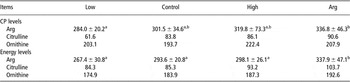
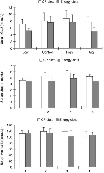
Compared with the energy control and low CP groups, dietary supplementation with 1% Arg increased serum Arg (P < 0.05). There were no differences (P > 0.05) in ornithine or citrulline between the groups (Table 1). Citrulline, produced from Arg as a by-product of the reaction catalyzed by members of the nitric oxide synthases family, was also increased by 21.6% in the Arg group compared with the energy control group (Table 1). Serum GLU had a high trend with the dietary CP or energy higher. Arg significantly decreased serum GLU concentration (P < 0.05) (Figure 1a). Serum urea N and ammonia level of piglets fed Arg was low comparing to piglets in control group (P > 0.05) (Figure 1b and c).
Serum amino acids of the pigs fed dietaries with different levels of CP and energy with Arg (μmol/l)

Arg = arginine.
Values are expressed as mean ± s.e.m., n = 8 pigs per treatment.
a,bMeans within a row with different superscript letters differ (P < 0.05).
Serum biochemical analyses of the pigs offered diets with different CP and energy levels, and the control diet plus 1% arginine. Values are expressed as mean ± s.d.; mean values with different letters are significantly different from each other (P < 0.05); n = 8.

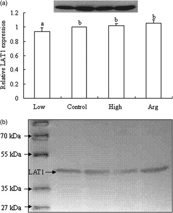
LAT1 staining was present in the brain tissues as a single band with a molecular mass of 45 kDa (Figure 2). Pigs fed the low-CP diet had a lower level of LAT1 presented in the brain tissues of pigs fed the low protein level diet (P < 0.05), much higher level were found in pigs fed the higher CP level diet, with moderate to intense staining seen in pigs fed the diet supplemented with 1% Arg (Figure 2).
(a) Western blots of membrane fractions from the brain tissues of the pigs fed dietaries with low, control and high CP levels (13.62%, 16.74% and 19.77% dietary CP) and 16.74% with arginine detected with the anti-l-type amino acid transporter-1 (LAT1). Values are expressed as mean ± s.d.; mean values with different letters are significantly different from each other (P < 0.05); n = 4. (b) Western blots of membrane fractions from the tissues with 3-3′ Diaminobenzidine detection. LAT1 was observed as a band with a molecular mass of 45 kDa.

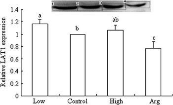
In contrast, tissue collected from pigs fed the control-energy diet showed a lower LAT1 expression than that the low-energy diet. However, pigs fed the diet supplemented with 1% Arg showed lowest LAT1 expression (P < 0.05) (Figure 3).
Western blots of membrane fractions from the brain tissues of the pigs fed dietaries with low, control and high energy levels (13.50, 14.60 and 16.47 MJ/kg energy and 14.60 MJ/kg with 1% arginine). Values are expressed as mean ± s.d.; mean values with different letters are significantly different from each other (P < 0.05); n = 4.

Discussion
Arg plays important roles in regulating both growth and metabolic function in pigs. Dietary supplementation with 1% Arg increased serum Arg. Differences in the expression of LAT1 under different levels of exogenous protein have been reported in normal and various tumor cells (Campbell et al., Reference Campbell, Sah, Medina, Albina, Coleman and Thompson2000; Campbell and Thompson, Reference Campbell and Thompson2001). The rate of amino acid efflux from individual cells must be capable of adapting to meet the cellular demands for growth, and plays a central role in the control of extracellular amino acid homeostasis. We hypothesized that the cells in the brain tissues expressing LAT1 would be sensitive to low protein. In this study, the Western blot analysis allowed for this sensitivity to be evaluated, and reduced levels of LAT1 were observed. The lower LAT1 expression was due to the decreased level of protein uptake, while the level of energy in the provided diets affected on LAT1 expression significantly. Moreover, the expression of LAT1 was found to be regulated by amino acid availability. Brust et al. (Reference Brust, Vorwieger, Walter, Fuchtner, Stark, Kuwabara, Herzau, Opfermann, Steinbach, Ganapathy and Bauer2004) indicated that LAT1 underlies developmental changes after birth causing a decrease in the permeability of the BBB permeability for those amino acids required during brain development, however, there is no strong correlation between the amino acid supply to the brain and the demands of protein synthesis in the brain tissue (Brust et al., Reference Brust, Vorwieger, Walter, Fuchtner, Stark, Kuwabara, Herzau, Opfermann, Steinbach, Ganapathy and Bauer2004). Thus, whether LAT1 expression affects protein synthesis requires further study.
Surprisingly, Arg, which is not a substrate of LAT1, has the most profound effect on transcription, and the depletion of l-Arg lead to an increase of LAT1 expression in non-tumourigenic hepatic cell lines (Yanagida et al., Reference Yanagida, Kanai, Chairoungdua, Kim, Segawa, Nii, Cha, Matsuo, Fukushima, Fukasawa, Tani, Taketani, Uchino, Kim, Inatomi, Okayasu, Miyamoto, Takeda, Goya and Endou2001; Tomi et al., Reference Tomi, Mori, Tachikawa, Katayama, Terasaki and Hosoya2005). In our study, there was no difference in the expression of LAT1 between samples with or without Arg. Interestingly, pigs fed the diet containing control-CP supplemented with Arg demonstrated high LAT1 expression while pigs fed the control-energy diet supplemented with Arg had lower expression. More research is needed to understand the role of Arg and its transporters in the forebrain white matter.
In conclusion, the results of the current study suggest that the expression of LAT1 adapts to nutritional changes, and that treatment with essential amino acids such as Arg may be beneficial to increase the expression of LAT1. The findings provide a potential animal model that could be used to further study dietary treatments using essential amino acids for the elucidation of effective diets for human nutrition.
Acknowledgements
This work was supported in part by the Chinese Academy of Science Knowledge Innovation Project (no. KSCX2-YW-N-051 and no. KSCX2-SW-323), NSFC (no. 30771558 and no. 30528006), the National 863 Program of China (no. 2008AA10Z316).