Introduction
The vomeronasal organ, or Jacobson's organ, is an accessory olfactory organ found in all vertebrates, except fish, that specialises in the chemoreception of pheromones.Reference Keverne1 Its evolutionary path starts in amphibians as a thickening of the nasal mucosa on each antero-inferior side of the nasal septum. In higher orders of vertebrates, the organ is a fully developed and independent epithelial invagination of the mucosa, with specialised olfactory cells covering the canal on each side of the nasal septum and unmyelinated nerve fibres found upon histological examination.Reference Døving and Trotier2 The sensory innervation of the organ comes from fibres deviated from the accessory olfactory bulb. The primary function of the organ is chemoreception of pheromones, and it has been shown that the organ plays a vital role in the formation of social and sexual behaviour in animals.Reference Kudjakova, Sarycheva and Kamensky3, Reference Eisthen, Wysocki and Beauchamp4
The vomeronasal organ was first described in humans by Dutch botanist and anatomist Frederik Ruysch in 1703 when describing and sketching the nasal septum of a young male cadaver, and was later rediscovered in 1813 by the Danish surgeon Ludwig Jacobson, after whom the organ is trivially named.Reference Knecht, Witt, Abolmaali, Huttenbrink and Hummel5, Reference Bhatnagar and Smith6 Nevertheless, the presence of the organ and its functionality in primates has been widely disputed. There is general agreement that the organ is present in the embryos of primates, but it is supposed that around birth the organ completely disappears.Reference Bhatnagar and Smith7, Reference Smith and Bhatnagar8 Furthermore, an accessory olfactory bulb, as a structure, is not present in primates; therefore, it is believed that even if the organ is present, it has no special sensory innervation, although there are arguments that contradict this.Reference Lehman, Newman and Silverman9
This study aimed to retrospectively evaluate the incidence of the vomeronasal organ in adults from the north-eastern Bulgarian population and compare these results to other studies of the organ carried out in different populations.
Materials and methods
All procedures contributing to this work complied with the ethical standards of the Helsinki Declaration of 1975, as revised in 2008, and with the ethical standards of the Bulgarian Ministry of Healthcare.
A total of 966 consecutive video recordings of nasopharyngolaryngoscopies were retrieved from the digital patient record system of a single institution, St Marina University Hospital, in Varna, Bulgaria, and retrospectively reviewed. All examinations were carried out for other ENT diagnostic indications, by one of the senior authors. The routine examinations were carried out under local anaesthesia with lidocaine, apart from two patients with allergy.
Two types of endoscopes were used to obtain the video recordings: a flexible nasopharyngolaryngoscope (model 11001RD1; Karl Storz, Tuttlingen, Germany) and a Hopkins rigid 4 mm 0° endoscope (Richard Wolf, Knittlingen, Germany). These were used together with a video-recording endoscopy camera (Telecam 20210030 PAL; Karl Storz) and a video documentation system (AIDA; Karl Storz). The indexing of the video files allowed for exact identification of the patients' sex and age at the time of examination, without disclosing any further personal information.
A structure was identified as a vomeronasal organ if it was a pit- or duct-shaped diverticulum on the antero-inferior side of the nasal septum, and was no more than 1.5–2 cm away from the nares. The selected criteria were nearly identical to those of other study groups researching the presence of the organ, underlining the similarity in the observed structures and allowing for the opportunity to compare the organ's incidence between different studies.Reference Won, Mair, Bolger and Conran10–Reference Smith, Siegel, Burrows, Mooney, Burdi and Fabrizio14
Results
Of the reviewed 966 video recordings, only 205 (21.22 per cent of the total recordings) were considered suitable for evaluation. Most video recordings were excluded because they began in the epipharynx. Some were considered not suitable because of a blurred lens. Others were excluded because of the presence of septum pathologies, which varied from severe inflammation to complete absence of a septum.
Of these 205 recordings, 85 were bilateral and therefore the septum could be examined on both sides. Forty-nine recordings were evaluable only on the left side and 71 were evaluable only on the right side of the septum. Therefore, the total number of nasal halves examined was 290; the total number of nasal halves evaluable on the left side was 134 and the total number of nasal halves evaluable on the right side was 156.
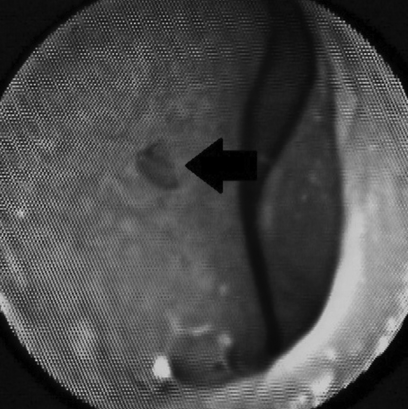
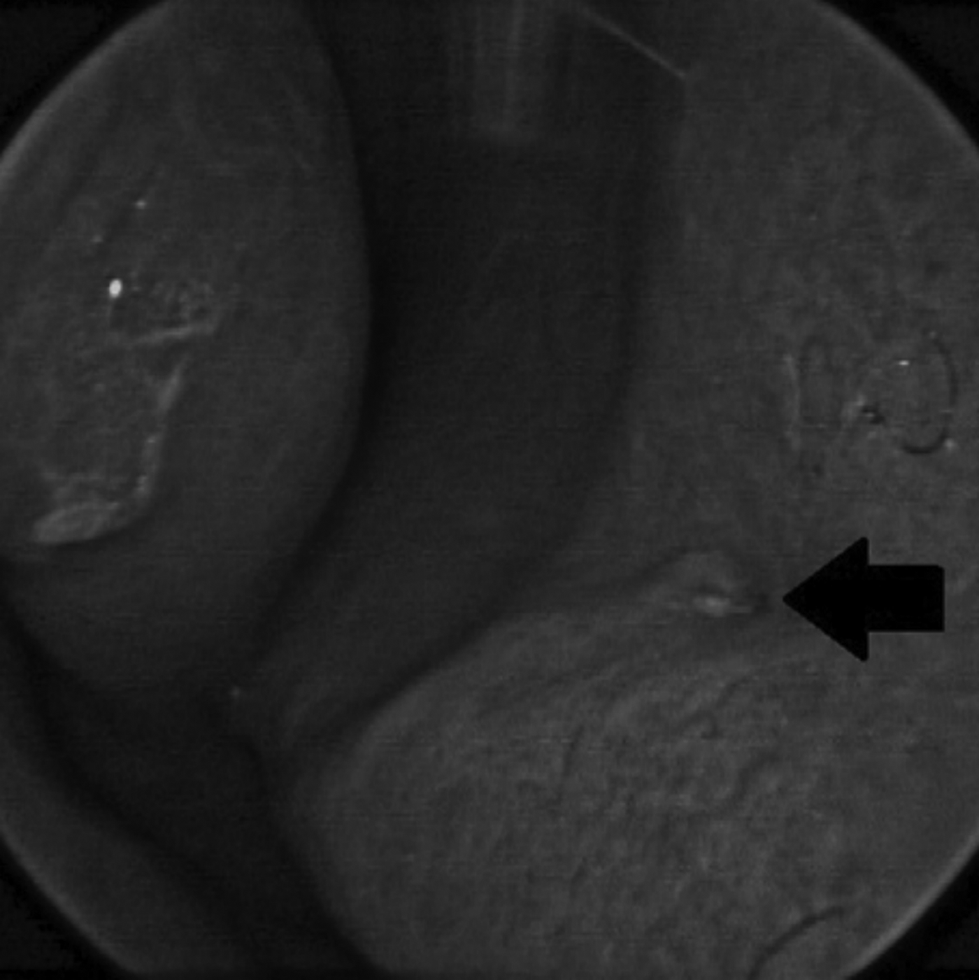
Evaluation of the recordings showed that the vomeronasal organ was evident in a considerable amount of subjects. Two major forms of the organ were observed: a pit-shaped formation (Figure 1) and a duct-shaped formation (Figure 2). Some of the vomeronasal organs showed evidence of mucus secretion, typical of the functioning of the organs in other animals.Reference Estes15, Reference Kratzing16 This suggests that the organ may very well have a mucus secreting function in humans too, aiding its main supposed function.
A pit-shaped vomeronasal organ (arrow) on the right of the nasal septum of a 52-year-old female, taken through a flexible nasopharyngolaryngoscope.

A duct-shaped vomeronasal organ (arrow) on the left side of the nasal septum of a 27-year-old male, taken through a rigid Hopkins rod lens ENT endoscope.
Data analysis
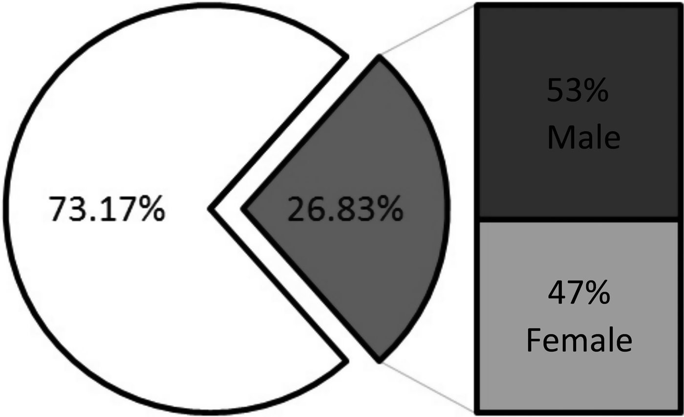
From the total of 205 cases selected as appropriate for review, a vomeronasal structure was identified in 55 cases. Calculation of the organ's occurrence compared to the total number of nasal halves reviewed showed that the vomeronasal organ was present in 14.93 per cent of the cases evaluable on the left side (20 out of 134 left nasal halves), 21.15 per cent of the cases evaluable on the right side (33 out of 156 right nasal halves) and in 2.35 per cent of the cases evaluable on both sides (2 out of 85 bilateral cases). Based on these findings, the total percentage of occurrence of the vomeronasal organ in the north-eastern Bulgarian population is 26.83 per cent (55 out of 205 cases), which equates to more than 1 in every 4 subjects.
In the 55 cases in which the vomeronasal organ was identified as a structure on the nasal septum, it was found predominantly unilaterally on the right side of the nasal septum, in 60 per cent of cases (33 out of 55 cases). There was a lower percentage of unilateral occurrence on the left side, 36.36 per cent (20 out of 55 cases), and a considerably lower percentage bilaterally, 3.64 per cent (2 out of 55 cases).
With regard to patient characteristics, patients' birth date and examination date information was included in the file indexing system, and the sex of each patient was contained in their personal identification number.Reference Sapundzhiev17 Analysis of the subjects' sex and age showed that the 55 patients in whom the organ was evident were aged 15–72 years at the time of the video recording. Twenty-nine of the 55 subjects were male, aged 17–72 years, and 26 were female, aged 15–64 years. From the total number of subjects in whom the organ was evident only on the left side, 11 were male, aged 17–69 years, and 9 were female, aged 35–64 years, at the time of the recordings. From the total number of subjects in whom the organ was evident on the right side only, 18 were male, aged 20–72 years, and 15 were female, aged 15–64 years, at the time of the recordings. Both of the subjects in whom the organ was evident bilaterally were female, aged 22 and 53 years at the time of the recordings. The total occurrence of the vomeronasal organ, and the male to female ratio of the subjects that have it, is shown in Figure 3.
Population distribution of vomeronasal organ incidence, showing subjects without a vomeronasal organ (clear), subjects with a vomeronasal organ (shaded) and their sex distribution.
Discussion
Our study involved a review of recorded endoscopies. The objective of the nasopharyngolaryngoscopies was not to identify and detect the vomeronasal organ; as such, not all recordings were started at the anterior part of the nasal cavity, nor was this area of interest examined with appropriate care in all patients. Thus, the number of cases considered as evaluable was significantly reduced. Therefore, it could be assumed that a significant number of cases might have been missed. This, together with the retrospective nature of our study, is a significant weakness. It is therefore possible, even probable, that the results reported underestimate the organ's true prevalence. The higher prevalence in prior reports further reinforces this possibility.Reference Won, Mair, Bolger and Conran10–Reference Gaafar, Tantawy, Melis, Hennawy and Shehata12
Based on the collected data, the incidence of the vomeronasal organ is not gender dependent, apart from those in whom the organ was evident bilaterally, where the results do not seem representative. Patients' age is not expected to play a role in the incidence of the vomeronasal organ.
The current study used only endoscopic techniques to evaluate the presence of the vomeronasal organ. Other research groups have used imaging modalities to study the organ, such as magnetic resonance imaging and computed tomography, following endoscopically confirmed findings, albeit with the direct application of contrast material to the nasal vestibule.Reference Abolmaali, Kühnau, Knecht, Kohler, Huttenbrink and Hummel18 These techniques are less specific to the organ's recognition, but in a greater aspect provide information about the length of the canal and the diameter of its lumen.
Though the organ remains relatively unexplored, there is some evidence that it may be functional in humans, and may hypothetically play a role in the formation of social and sexual behaviour.Reference Wessels, Hoogland and Vorster19–Reference Berliner, Monti-Bloch, Jennings-White and Diaz-Sanchez21 Therefore, more attention must be paid when surgical manipulations are carried out in the area of the organ, as this could lead to irreversible changes to a patients' personality if the organ is functional in humans and damaged during a procedure. According to some controversial studies, there have been signs of behavioural changes in humans who have undergone surgery in the region of the vomeronasal organ, such as a Le Fort I osteotomy. Such procedures have resulted in confusion in the process of selecting an appropriate mate and settling in a relationship.Reference Foltan and Sedy22 Nevertheless, these findings have not been supported by any significant data, and the explanation remains an educated guess.
The vomeronasal organ in humans may be functional, and its function may be associated with that of cranial nerve zero because they both carry out the same function: to modulate sexual and social behaviour.Reference Dakrory, Issa and Ali23–Reference Demski25 However, more research is needed to either confirm or dismiss these statements.
• The vomeronasal organ is an accessory olfactory organ present in nearly all vertebrates
• The incidence in humans is widely disputed
• The study findings indicate that the organ is present in more than a quarter of the adult population, similar to findings of studies on different populations
The histological components and the presence of the vomeronasal organ may explain the rare incidence of ectopic olfactory neuroblastoma, which has been shown to manifest in the topographical area of the organ.Reference Smith, Bhatnagar, Shimp, Kinzinger, Bonar and Burrows26–Reference Wormald, Lennon and O'Dwyer28
Conclusion
The present findings in adult humans, at least on a gross anatomical basis, are similar to and confirm some of the results of other studies carried out in similar conditions in different populations. For instance, Won et al. found that the vomeronasal organ was present in 28.2 per cent of a US population.Reference Won, Mair, Bolger and Conran10 Trotier et al. reported a rate of 26 per cent on first examination of subjects in France,Reference Trotier, Eloit, Wassef, Talmain, Bensimon and Doving11 and Johnson et al. reported a rate of 39 per cent in Canada.Reference Johnson, Josephson and Hawke29 However, Won et al.Reference Won, Mair, Bolger and Conran10 and Trotier et al.Reference Trotier, Eloit, Wassef, Talmain, Bensimon and Doving11 also reported higher estimates, with the former reporting 59.1 per cent on cadaver dissection, and the latter reporting 92 per cent on follow-up re-evaluation (based on at least four observations).Reference Trotier, Eloit, Wassef, Talmain, Bensimon and Doving11 Gaafar et al. found that the vomeronasal organ was present in 76 per cent of an Egyptian population on first examination using endoscopic evaluation only.Reference Gaafar, Tantawy, Melis, Hennawy and Shehata12
The histological, functional and modulating aspects of the vomeronasal organ are far from being fully explained. Nevertheless, the present study confirms that a vomeronasal structure is present, at least in some adult humans, while other aspects of the vomeronasal organ's morphology remain to be addressed.Reference Wysocki and Preti30, Reference Meredith31
Acknowledgements
The authors would like to thank the reviewers of The Journal of Laryngology & Otology for their comments and recommendations, which represent an important contribution to the final form of this article.