Introduction
Clinical anophthalmia is defined by the absence of visible ocular structures in the socket. In this condition, there is likely to have been a highly disordered environment for the development of the extraocular musculature. While eye and retinal development can be unaffected by defects in the nuclei innervating the muscles, denervation or abnormalities of the extraocular musculature [e.g., Duane retraction syndrome (Kang & Demer, Reference Kang and Demer2006; Demer et al., Reference Demer, Clark, Lim and Engle2007); congenital fibrosis of the extraocular muscles (Engle et al., Reference Engle, Kunkel, Specht and Beggs1994; Engle, Reference Engle2002; Traboulsi, Reference Traboulsi2004)], the effects of abnormal eye development on the muscles, and the cranial nerves innervating these muscles are unknown. Clinical anophthalmia provides an opportunity, therefore, to investigate the codependence of these developmentally separate events, which under normal conditions synergistically combine to create a system optimal for visuomotor function. If the development of the muscles were independent of the eye, it is likely that the muscles and cranial nerves that innervate them would be intact in anophthalmia. If there were little or no activity in the muscles, however, it is possible that both nerves and muscles would atrophy.
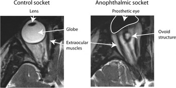
Although there is no “visible globe” in clinical anophthalmia, when observing the socket, it is often possible to see the outline of a remnant under the socket lining that, in cases of unilateral anophthalmia, may even show conjunctive movements with the normal eye. Furthermore, during surgery to place an implant in the socket of an anophthalmic individual, a vestigial globe complete with small extraocular muscles may be seen (N. Ragge, personal communication). Fig. 1 is an illustration of the normal eye socket and contents (left) and an example of the contents of an anophthalmic socket (right); the prosthetic eye is outlined in white. Postmortem studies of anophthalmic cases describe abnormal tissue in the orbit. One such case, a male infant who died at two months, had “no vestige of a globe” detectable macroscopically, but microscopic examination revealed a minute nodule of fibrous tissue in each orbit that contained no recognizable cell structure and was pigmented (Recordon & Griffiths, Reference Recordon and Griffiths1938). Similar nodules had been previously described in at least one of the orbits of three other cases (van Duyse, Reference van Duyse1899; Hanke, Reference Hanke1904; Gallemaerts, Reference Gallemaerts1924); cited in Recordon and Griffiths (Reference Recordon and Griffiths1938). More recent histological studies also described orbital contents comprising muscle, nerve, and adipose tissue with fibrous nodules but no recognizable eye elements (Duckworth & Cooper, Reference Duckworth and Cooper1966; Haberland & Perou, Reference Haberland and Perou1969; Brunquell et al., Reference Brunquell, Papale, Horton, Williams, Zgrabik, Albert and Hedley-Whyte1984; Pierson et al., Reference Pierson, Subtil, Taboada and Butler2002). In vivo, magnetic resonance imaging (MRI) and computerized tomography (CT) studies of cases with clinical bilateral anophthalmia describe ovoid nodules thought to be vestigial globes of varying size and typically isointense with muscle (Brunquell et al., Reference Brunquell, Papale, Horton, Williams, Zgrabik, Albert and Hedley-Whyte1984; Daxecker & Felber, Reference Daxecker and Felber1993; Albernaz et al., Reference Albernaz, Castillo, Hudgins and Mukherji1997; Aktekin et al., Reference Aktekin, Oz, Saygili and Kurtoglu2005). In most cases in which the presence of extraocular muscles was investigated, these were found to be present, typically attached to the nodules in the orbit and of variable organization and size (see references above and Bohnsack et al., Reference Bohnsack, Gallina, Thompson, Kasprick, Lucarelli, Dootz, Nelson, McGonnell and Kahana2011). The optic nerves and chiasm were described as “absent” (Recordon & Griffiths, Reference Recordon and Griffiths1938; Duckworth & Cooper, Reference Duckworth and Cooper1966; Haberland & Perou, Reference Haberland and Perou1969; Brunquell et al., Reference Brunquell, Papale, Horton, Williams, Zgrabik, Albert and Hedley-Whyte1984; Daxecker & Felber, Reference Daxecker and Felber1993) or hypoplastic (Pierson et al., Reference Pierson, Subtil, Taboada and Butler2002). The oculomotor, trochlear, and abducens nerves were described as present in all cases where they were investigated (Recordon & Griffiths, Reference Recordon and Griffiths1938; Duckworth & Cooper, Reference Duckworth and Cooper1966; Haberland & Perou, Reference Haberland and Perou1969; Pierson et al., Reference Pierson, Subtil, Taboada and Butler2002), with the exception of the case of Gallemaerts (Reference Gallemaerts1924; cited in Recordon & Griffiths, Reference Recordon and Griffiths1938), in which the trochlear and abducens nerves were noted to be absent. Previous neuroimaging studies in cases of clinical anophthalmia have not reported on the presence or absence of cranial nerves other than the optic nerve. Here, we summarize the findings from these previous studies of bilateral anophthalmia. The variability in the different patterns of abnormality described may reflect distinct genetic disruptions that have influence at different points during the eyes’ development. It should also be noted that nearly all the previously described cases of bilateral anophthalmia investigated at postmortem had other developmental abnormalities in addition to the anophthalmia.
Illustration of the normal and anophthalmic socket to indicate the different structures described in the text.

In this study, we used high-resolution MR of the brainstem and orbits to visualize the cranial nerves, extraocular muscles, and other orbital contents in five cases of isolated bilateral anophthalmia. We compared their scans with those performed in five age- and gender-matched controls. Also, to place our findings in the context of the published literature, we provide a summary of the contents of the orbits, presence or absence of extraocular muscles and cranial nerves as described in the cited studies and in our five cases.
Materials and methods
Subjects
The study was conducted under ethical approval from the Oxfordshire National Health Service Research Ethics Committee (07/Q1605/20) in accordance with the Declaration of Helsinki; all subjects provided informed written consent. In the case of the anophthalmic subjects, Braille versions of the documents were provided. Five anophthalmic subjects participated (age range 20–33 years; two females). The subjects are five of the six individuals participating in a previous study investigating the structure of the “visual” system (Bridge et al., Reference Bridge, Cowey, Ragge and Watkins2009), although they were scanned again 18–24 months after the prior data acquisition. To aid comparison, the subjects retain the same identities and are therefore referred to as Cases 2, 3, 4, 5, and 6. Cases 3 and 5 have bilateral orbital implants. Case 4 had an orbital cyst removed from the right side at age 2 years. None of the participants had any infundibular malformations that may have confounded imaging results. Imaging data were also obtained in five age-matched controls (age range 20–31 years; two females).
Image acquisition and analysis
Subjects were scanned in a Siemens 3-Tesla TIM Trio, with a 12-channel radio frequency coil. We used a heavily T2-weighted protocol, at a resolution of 0.52 × 0.52 × 0.6 mm3 (80 slices; echo time = 131 ms; repetition time = 1000 ms). Horizontal (axial) slices were acquired at an angle approximately parallel to the ventral surface of the frontal lobe. The small slice thickness (0.6 mm) and almost isotropic voxel dimensions reduced the influence of slice positioning on detection and measurement of structures in the images. This is important to note as the longitudinal axes of different cranial nerves occupy different planes, necessitating the reorientation and resampling of images to obtain the optimal visualization of each pair. Images were reorientated in OsiriX (www.osirixmac.com). To measure the maximum diameter of the optic and oculomotor nerves, the image was aligned in the plane of the longitudinal axis of the specific nerve. Cross-sectional measurements were made perpendicular to this axis where the nerve had its largest diameter.
Measurements of the lateral, medial, and inferior recti and the superior oblique muscles were made in all controls and in anophthalmic subjects where possible. The technique used was similar to that of Aydin et al. (Reference Aydin, Guven, Sencer, Cikim, Gul and Minareci2003), involving the measurement of both the long and short diameter of each of the above muscles at a location 6 mm posterior to the back of the globe in sighted subjects, and as close to this location as possible in the anophthalmic subjects.
Results
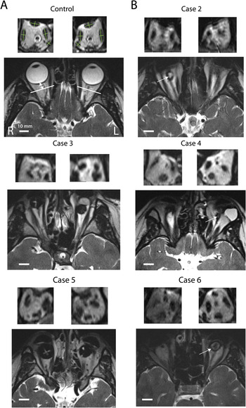
Examination of the images obtained in the five anophthalmic subjects revealed very small (6–12 mm diameter) ovoid tissue masses in the right orbit of Case 2 and in the left orbits of Cases 4 and 6 (see Fig. 2B). A dark nerve-like structure extends posteriorly from these small masses; this structure is likely to be a hypoplastic optic nerve or nerve sheath, similar to those described in previous studies. The dark spherical structures seen bilaterally in the orbits of Cases 3 and 5 are the implants; the hemispherical structures seen more anteriorly are prostheses (artificial eyes). In addition, an ovoid tissue mass can be seen in the right orbit of Case 3 lying between the implant and the prosthetic eye. The nerve-like structure extending posteriorly from the ovoid structures in Cases 2, 4, and 6 was not evident in Case 3, however. As Case 3 has orbital implants, we cannot rule out the possibility that the tissue mass in the right orbit is an ocular remnant that became detached from any remnant optic nerve or nerve sheath by the surgical insertion of the implant. Finally, in addition to the tissue mass in the left orbit of Case 4, there is an orbital cyst that is isointense with eyes seen in the control subjects.
The orbital contents and extraocular muscles. The nerves and muscles are dark and CSF is bright in these T2-weighted images. The morphology of the muscles is extremely variable in the anophthalmic subjects. They range from all present and identifiable (Case 6, left side) to indistinct (Case 6, right side). These images also indicate the ovoid structures that are present deep in the orbit in three subjects (Cases 2, 4, and 6; white arrows) and between the implant and prosthesis in Case 3, which are presumably vestigial globes. In Case 4, the bright white structure on the right side of the axial section lateral to the ovoid structure is a cyst. The scale bar shows 10 mm in each subject.

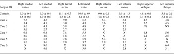
Fig. 2A shows the muscles in a control subject, the axial view showing the section through which the size of the muscle measurement was assessed. The slice used to measure the diameter of the muscle was perpendicular to the optic nerve, 6 mm posterior to the globe. The lateral, medial, inferior recti, and the superior oblique muscles were measured along the long and short axes in each control subject. The mean value for each measurement in control subjects is shown in Table 1. Similar measurements for each anophthalmic subject are in the table; an “X” indicates where it was not possible to define the axes of the muscle.
Size of the long (upper) and short (lower) axes of the medial, lateral, and inferior recti and superior oblique muscles in mm for all anophthalmic subjects and the mean and standard deviation for the controls

X, measurement was not possible as no clear tissue was evident; NS, region not included within scanned volume.
There is considerable variability in the size and integrity of the muscles in the anophthalmic subjects even between the two sides of the same individual. This is clearest in Case 6, in whom the left orbit appears to have almost normal musculature with the muscles appearing to attach to the ovoid structure described above. In contrast, the muscles of the right orbit are impossible to define due to the lack of distinct tissue. A similar pattern is evident in Case 2 in whom the muscles of the right orbit (in which an ovoid structure is present) are much larger than those in the left (where there is no obvious structure). However, it is also clear that a tissue mass is not necessary for muscles to be present as Case 5 lacks any obvious structure in either socket, but all recti muscles are present, albeit slightly reduced in size compared to controls. It should be noted, however, that any ocular remnant present in Cases 3 and 5 might have become detached from the muscles or nerve-like structures during the surgery to insert their implants.
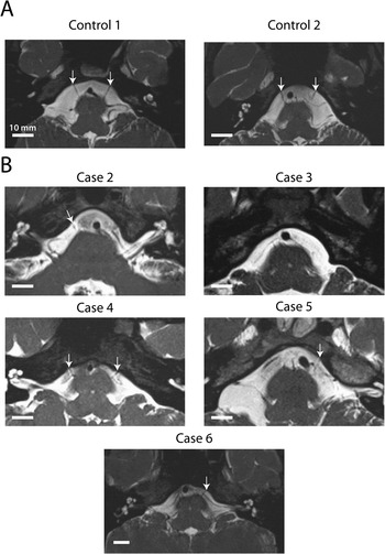
In all control subjects, the optic, oculomotor, trigeminal, and abducens nerves were clearly visible. The trochlear nerve, which is the thinnest cranial nerve, was sometimes difficult to distinguish from neighboring blood vessels where it exits from the dorsal aspect of the brainstem. Fig. 3 shows the optic nerve and chiasm in two controls subjects (A) and four of the anophthalmic subjects (B). The optic nerve is indicated by the black arrows and is prominent in each of the control subjects. Two of the anophthalmic subjects showed an extremely hypoplastic chiasm and optic nerves bilaterally (Cases 4 and 6), while Case 2 shows a single hypoplastic nerve unilaterally (on the right). These structures are continuous with the nerve-like structures described in the orbits (see above) and attach to the ovoid structures in Case 2 on the right and Cases 4 and 6 on the left. There are no ovoid structures in the orbits of Case 5 who also shows no chiasm and no evidence of optic nerve extending into the socket. The chiasm for Case 3 was unfortunately missed from the scan prescription and, although the more extensive T1 structural images showed no evidence of either optic nerves or chiasm, we cannot rule out the possibility that a highly atrophied optic nerve is present. Across the five controls subjects, the mean diameter of the optic nerve is 4.1 ± 0.6 mm compared to 1.4 ± 0.5 mm in the three anophthalmic subjects in whom either one or both optic nerves could be identified.
The optic nerves and chiasm. The nerves are dark and CSF is bright in these T2-weighted images. Black arrows indicate the optic nerves in controls (A) and anophthalmic subjects (B). No optic nerve can be identified in Case 5; the visible dark objects in Case 5 are parts of the prosthetic eyes. The scale bar shows 10 mm in each subject.

The oculomotor nerve has a smaller diameter than the optic nerve in control subjects; in the five control subjects, the mean diameter at the widest point is 2.1 ± 0.4 mm. In contrast to the optic nerve, the oculomotor nerve can be identified in all anophthalmic subjects as can be seen in Fig. 4. As in Fig. 3, (A) shows examples of the nerve in two controls subjects and (B) shows all five anophthalmic subjects. In Case 3, the oculomotor nerve appears only to be present unilaterally but is actually present bilaterally but the nerves run in slightly different planes and therefore cannot be viewed in the same slice. The mean diameter for the anophthalmic subjects is 1.9 ± 0.2 mm, slightly but not significantly smaller than in control subjects (t = 0.8; df = 9; P > 0.05). Each nerve is considered independently for the t-test as the presence of a particular nerve on one side does not guarantee that nerve will be present on the other side.
The oculomotor nerves. The nerves are dark and CSF is bright in these T2-weighted images. White arrows indicate the oculomotor nerves in all control (A) and anophthalmic subjects (B). The scale bar shows 10 mm in each subject.

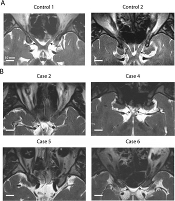
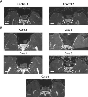
The abducens nerve, one of the thinner cranial nerves, was visible in all control subjects. Two examples are shown in Fig. 5A. The pair of nerves can be seen exiting anteriorly from the lower pons. In the anophthalmic subjects (Fig. 5B), the nerve was detectable in Cases 2 and 5 only unilaterally and in Cases 4 and 6 bilaterally. White arrows indicate the point at which the nerves protrude from the cerebrospinal fluid (CSF). There is no detectable nerve in Case 3.
The abducens nerves. The nerves are dark and CSF is bright in these T2-weighted images. White arrows indicate the abducens nerves as they exit the anterior aspect of the brainstem at the junction of the medulla and the pons. The nerves are visible bilaterally in all control subjects (A). In the anophthalmic subjects (B), the nerve is much more difficult to detect, although appears to be present bilaterally in Case 4, and possibly Case 6. One nerve was also detectable in each of Cases 2 and 5. The scale bar shows 10 mm in each subject.

The trigeminal nerve, included as a control structure to ensure that the results are not biased by image quality, does not differ in size between the two groups, with mean diameters of 5.0 ± 0.7 mm and 5.0 ± 0.9 mm for control and anophthalmic subjects, respectively.
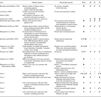
The results of our findings in five anophthalmic cases are summarized in Table 2 alongside those from previous reports of similar investigations carried out at postmortem or more recently with CT and MRI. In nearly all cases, there was some tissue in the orbit to which extraocular muscles might attach. Optic nerves were mostly absent at postmortem and described as absent or extremely hypoplastic in imaging studies, including our own; it is possible that the nerve-like structures observed in our images in the orbit and intracranially are residual nerve sheaths. The oculomotor nerves are typically present and often noted to be normal. In our imaging data, it was not possible to distinguish the trochlear nerve from surrounding vessels but the abducens nerve was observed in all but one of our cases. In summary, despite the varied etiology and clinical presentation of the cases described previously with clinical bilateral anophthalmia and our own cases, there are discernable patterns that presumably reflect the timing of disruptions in ocular development.
Summary of findings in anophthalmic cases from postmortem and imaging studies

Italicized cases are described and cited in Recordon and Griffiths (Reference Recordon and Griffiths1938).
II, optic; III, oculomotor; IV, trochlear; VI, abducens; Ch, chiasm; L, Left; R, Right; H, hypoplastic nerve; ✔, present; ✖, absent;?, undetermined.
Discussion
The imaging methods described here permit the visualization of the orbital contents and identification of the cranial nerves and extraocular muscles. The diameters of the nerves in control subjects are comparable to those found previously for the optic (3.5–4.0 mm) (Karim et al., Reference Karim, Clark, Poukens and Demer2004) and for the oculomotor (2.0 mm) (Kim & Hwang, Reference Kim and Hwang2005; Lim et al., Reference Lim, Engle and Demer2007) nerves. As might be expected, there is more variation in the size of the optic nerve in the anophthalmic subjects than in controls.
The oculomotor nerve was present in all the anophthalmic subjects and had a similar diameter to that of sighted subjects. The presence of the oculomotor nerve in the anophthalmic subjects is particularly interesting since this nerve provides the major innervation for eye movements and yet there is no functioning globe to move. However, it is regularly observed that there are ocular remnants embedded in the orbit that have no functional role in vision (see Table 2 and references therein). Such vestigial tissue can provide a structure to which the extraocular muscles insert and provide the developmental environment needed for growth. Moreover, the oculomotor nerve also plays a role in eyelid opening, which can occur normally in some anophthalmic subjects. The abducens nerve, which controls only the lateral rectus muscle, was not detectable in one of the anophthalmic subjects, present unilaterally in two and bilaterally in another two. The small diameter of this nerve makes quantification of the difference between control and anophthalmic subjects impossible at the image resolution of 0.5 × 0.5 × 0.6 mm3.
The presence of the extraocular muscles in our anophthalmic subjects and the variability in their number, size, and integrity are consistent with findings in postmortem studies (see Table 2) and more recent imaging studies of bilateral anophthalmia using either MRI or CT (O’Keefe et al., Reference O’Keefe, Webb, Pashby and Wagman1987; Albernaz et al., Reference Albernaz, Castillo, Hudgins and Mukherji1997). Interestingly, there is no clear link between the integrity of the muscles and the visibility of the optic, oculomotor, and abducens nerves. In Cases 2 and 6, all muscles are present and distinct unilaterally with hypoplastic optic and abducens nerves evident on the same side and normal-sized oculomotor nerves bilaterally (see Table 2). In contrast, Case 4, in whom there is evidence of optic, oculomotor, and abducens nerves bilaterally, has only three of the muscles in each side; these are possibly the medial and lateral recti and superior oblique muscles. Furthermore, Case 5 shows at least four visible muscles bilaterally, oculomotor nerves bilaterally, and an abducens nerve on the left, but the optic nerve or ocular remnant could not be visualized. As Case 5 has had reconstructive surgery for the prosthetic implants, however, the contents of the socket are likely to have been displaced. Nevertheless, it should be noted that even several years after unilateral enucleation, the extraocular muscles generally retain their bulk (Detorakis et al., Reference Detorakis, Engstrom, Straatsma and Demer2003).
A very recent study investigating the extraocular muscles in three young bilaterally anophthalmic children using clinical MRI orbital images (Bohnsack et al., Reference Bohnsack, Gallina, Thompson, Kasprick, Lucarelli, Dootz, Nelson, McGonnell and Kahana2011) and see also editorial (McLoon, Reference McLoon2011) found a range of muscle formation from “well-formed” to “indeterminable.” The main focus of the paper by Bohnsack et al. (Reference Bohnsack, Gallina, Thompson, Kasprick, Lucarelli, Dootz, Nelson, McGonnell and Kahana2011) was, however, an elegant illustration in two animal models of a correlation between time of ocular insult and extraocular muscle formation. Based on these findings, the authors proposed that the noninvasive investigation of the extraocular muscles in human subjects with anophthalmia could provide insight into the timing (and therefore cause) of the insults.
In our cases, and consistent with the animal model data referred to above, the best predictor of the integrity of both the extraocular muscles and the cranial nerves appears to be the presence of an ocular remnant in the socket. In particular, Cases 2 (right) and 6 (left) both have such remnants, as seen in Fig. 2. However, the muscles appear to be attached to the remnant in Case 6 but not in Case 2. Additionally, the lack of “normal” muscle organization in Case 4 bilaterally suggests that these structures are not sufficient for muscle preservation. However, it is clear from the images that the tissue composition of these structures is not consistent, as the center of the structure can be either dark or bright, with an opposite contrast perimeter indicating large variations in water and fat content. This small sample of clinically anophthalmic subjects is not sufficient to make any strong statement about the causes for the lack of development; however, the presence of orbital tissue and a hypoplastic optic nerve suggests that development was arrested after the globe had begun to develop. In contrast, the complete absence of rudimentary eye-like structures and optic nerve may mean that development was prevented at an even earlier stage possibly even a failure of optic stalk development.
Finally, in our previous study (Bridge et al., Reference Bridge, Cowey, Ragge and Watkins2009), it was determined by a clinical radiologist that there was evidence of the optic nerve in Cases 2 and 6, but not in Case 4, in whom we now report a hypoplastic optic nerve bilaterally. The increased resolution and use of T2 imaging has permitted improved detection, therefore. Additionally, the orbital contents evident in three of the anophthalmic subjects were not apparent on the T1-weighted scans acquired at 1 mm3 and were previously unknown.
Conclusions
Despite all the cases presented here being defined as clinically anophthalmic, there is considerable heterogeneity both in the extraocular musculature and the presence and integrity of the cranial nerves related to these muscles. Moreover, there is no consistent relationship between the visibility of the optic, oculomotor, and abducens nerves and the integrity of the extraocular muscles for reasons that remain obscure but are likely to involve both genetic and developmental variations. These T2-weighted high-resolution scans, which are quick to acquire, provide an opportunity to visualize the vestigial structures that remain in the eye sockets and may eventually provide clues to the stage at which the eye development was aborted.
Acknowledgments
This work was supported by the Medical Research Council, the Royal Society through a University Research Fellowship to H.B., a Senior Surgical Scientist Award from the Academy of Medical Sciences/Health Foundation to N.R., and a Leverhulme Trust Emeritus Fellowship to A.C.