Introduction
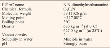
Trimethylamine (TMA) is a hygroscopic nitrogen-containing flammable organic gas that boils at 3°C. It has a characteristic fishy odour at low concentrations, which alters to an ammonia-like odour at higher concentrations (Ref. 1). (Table 1) Partly owing to TMA's low boiling point, up till the 1930s it was extensively used in vapour pressure studies (Ref. Reference Burg and Green2). The chemistry-centred focus on TMA shifted when it was associated with banishment of menstruating women from kitchens (Ref. Reference Wilson3). An increase in the levels of TMA excretion had been observed immediately prior to as well as during menstruation, which hence was associated with ruining of food articles such as ham, preserves, bread, wine and flowers (Ref. Reference Ashley-Montagu4). In the related work, menstruating rats were found to secrete 80–100 folds higher amounts of choline, which presumably was metabolised to TMA by bacterial enzymatic activity present on their skin surface (Ref. Reference Wastl5). Recently, human gut microbes have been implicated in metabolising choline to TMA, which we now know is subsequently oxidised to non-odorous trimethylamine-N-oxide (TMAO) by hepatic flavin-monooxygenase 3 (FMO3) before excretion via urine (Ref. Reference Wang6). TMA involvement in human physiology and disease was further highlighted when TMA was found in blood and cerebrospinal fluids of healthy as well as neurodegenerative disease patients (Ref. Reference Doree and Golla7). Since then, changes in the levels of TMA in urine, blood and sweat have been quantified and correlated to a number of disease pathologies. For example, trimethylaminuria (TMAU), viral infection, hormonal modulation, intake of competitive FMO3 substrates, kidney or liver damage all lead to the characteristic fishy odour arising from increased TMA levels (Ref. Reference Cashman8).
The chemical properties of TMA

Source: Pubchem.
TMA is also an air pollutant that is produced during chemical manufacturing, via vehicle exhausts, and by food-fishery industries (Refs Reference Rappert and Muller9, Reference Ho, Chung and Tseng10, Reference Rehbein11, Reference Diab12). Recently, various studies have highlighted roles for TMA's more toxic metabolites like N-nitrosodimethylamine (NDMA) and TMAO (Refs Reference Usunomena13, Reference Zeisel, daCosta and LaMont14). Specifically, TMAO has gained much attention lately because of its potential role in development of heart diseases (Ref. Reference Koeth15). In a recent study, inhibition of TMA lyase that is responsible for the production of TMA has been shown to prevent atherosclerosis in vivo (Ref. Reference Wang16). Here, we provide an in-depth overview of TMA's attributes that are of immediate relevance to human health.
TMA detection
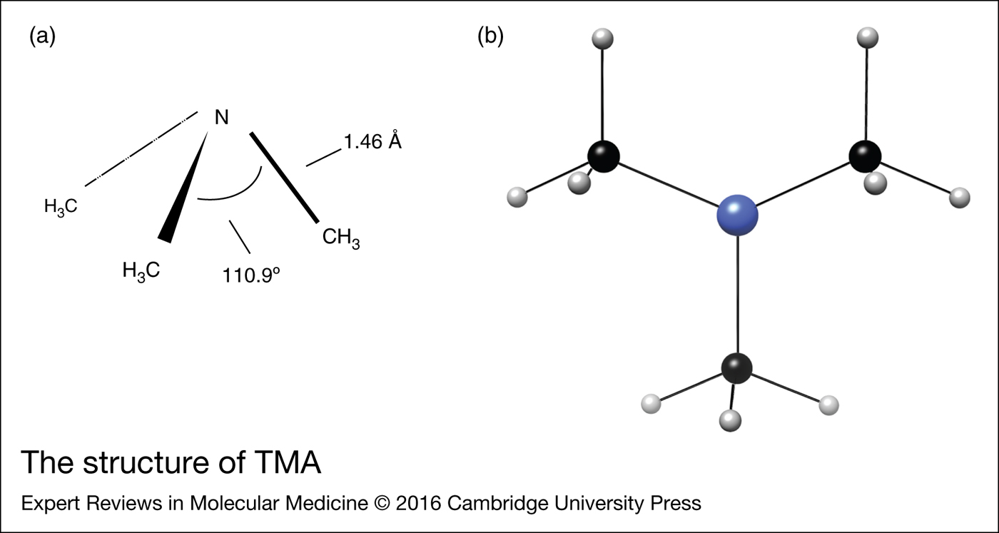
TMA is a tertiary amine with a chemical formula of C3H9N, molar mass of 59.11 g/m and good solubility in water. Its aqueous miscibility is attributed to the presence of amine nitrogen which forms hydrogen bond with water (Ref. 17) (Fig. 1). Since TMA has no native chromophore, its analysis requires specialised equipment. Methods used to measure TMA include proton nuclear magnetic resonance (1H NMR) spectrometry, electrospray ionisation tandem mass spectrometry, direct infusion electrospray quadruple time-of-flight mass spectrometry, headspace gas chromatography and matrix-assisted laser desorption/ionisation time of flight mass spectrometry (Refs Reference Lee18, Reference Mills, Walker and Mughal19, Reference daCosta, Vrbanac and Zeisel20, Reference Johnson21, Reference Mamer, Choinière and Lesimple22, Reference Hsu23). A number of new TMA quantification approaches have also been developed lately. A recent study has employed solid polymer substrates and coated fibres containing 2,4,6-trinitrobenzene motifs that react with amines in the atmosphere. The ambient amines diffuse into the polymer and form coloured Meisenheimer complexes leading to visual detection of biogenic amine vapours (Ref. Reference Pablos24). Independently, a bioelectronic nose based on a novel microfluidic system has been fabricated to detect gaseous TMA in real-time (Ref. Reference Lee25). Another approach has employed a recombinant cell line of Xenopus laevis melanophores where these cells were genetically modified to express human trace amine-associated receptor (TAAR5) (Ref. Reference Suska26). TAAR5 is an evolutionarily conserved olfactory receptor which is activated in a concentration-dependent manner by TMA (Ref. Reference Wallrabenstein27). This cell-based sensor detects TMA from 1 to 100 p.p.m. within 35 min (Ref. Reference Suska26). Hence, the bioelectronic nose and TAAR5 are few of the many methods by which TMA can be detected readily, and these hold promise for biomedical applications too.
The structure of TMA. (a) Stereochemistry of TMA with proposed bond angles and lengths indicated. (b) Structure of TMA as a ball-and-stick model (Blue-Nitrogen, Black-Carbon, Gray-Hydrogen). The figure was created using UCSF software chimera.

TMA synthesis and metabolism
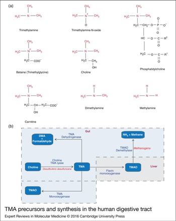
TMA precursors include dietary choline, betaine (trimethylglycine), phosphatidylcholine and L-carnitine (Ref. Reference Zhang, Mitchell and Smith28). Similar to the choline TMA lyase, and putative Rieske-type proteins that convert choline and L-carnitine to TMA, respectively, various other gut microbial enzymes may also catabolise TMA precursors (Refs Reference Zhu29, Reference Craciun and Balskus30). It is important to note that differences in gut microbiota among individuals, and even within the same individual over time can vastly influence both synthesis and levels of TMA (Ref. Reference Delgado31). TMA can either be oxidised to TMAO by the gut microbial enzyme TMA monooxygenase or by human hepatic enzyme FMO3 (Refs Reference Motika32, Reference Chen33). Additionally, gut microbiota-associated inflammation results in the secretion of inflammatory infiltrates and cytokines that in turn induce the expression of enzymes such as iNOS, NOX1, DUOX2, PHOX, SOD and MPO. These enzymes generate antimicrobial radicals such as superoxide, peroxide, hypochlorite, nitric oxide and peroxynitrite (Ref. Reference Winter, Lopez and Bäumler34), which are capable of oxidising TMA to TMAO (Refs Reference Balagam and Richardson35, Reference Schöneich36). Alternatively, TMA can be degraded to dimethylamine (DMA) and formaldehyde by the microbial enzyme trimethylamine dehydrogenase (TMADH) (Refs Reference Kim, Bae and Lee37, Reference Colby and Zatman38, Reference Shi, Mersfelder and Hille39). Similarly, TMAO can be acted upon by TMAO demethylase (TMADM) to form methane and ammonia by some methanogens (Ref. Reference Zhu40) (Fig. 2). Hence, diverse host or microbial enzymes can alter levels of both TMA and TMAO.
TMA precursors and synthesis in the human digestive tract. (a) Chemical formulae of TMA, its precursors trimethylamine-N-oxide, choline, phosphatidylcholine, carnitine, betaine, and its degradation products DMA and methylamine. (b) Schematic representation of the origin and fate of human gut TMA, which is synthesised using dietary precursors such as choline, carnitine by gut microbial enzymes. Choline TMA lyase acts upon choline leading to the synthesis of TMA (Ref. Reference Mirvish116). TMA is then oxidised to TMAO by either mammalian hepatic FMO3 or by the action of microbial TMM (Ref. Reference Chen33). TMAO can further be degraded to methane and ammonia by the action of TMADM (Ref. Reference Zhu40), whereas TMA is metabolised to DMA and formaldehyde by the action of TMADH (Ref. Reference Shi, Mersfelder and Hille39).

Environmental TMA
TMA is a universal air pollutant. Exogenous sources of TMA include fish-meal manufacturing plants, swine waste storage pits, animal husbandry operations, vehicle exhaust and anaerobic fermentation of food waste (Refs Reference Rappert and Muller9, Reference Ho, Chung and Tseng10, Reference Rehbein11, Reference Diab12). The primary production of agricultural TMA stems from interaction between excreted urine (which contains TMAO) and the microbes present in faeces (Ref. Reference Sintermann, Schallhart and Kajos41). The recommended average airborne TMA exposure limit over a 10-h work shift is 10 ppm, where it should not exceed 15 ppm over a 15 min period (Ref. 17). Hence, TMA level monitoring and control are important elements of air quality management schemes. In the presence of nitrate radicals in the air, TMA promotes secondary aerosol particle formation, which then determines its atmospheric lifetime (Refs Reference Silva42, Reference Murphy43). In a study carried out in five European cities [Ireland (Cork), France (Paris, Dunkirk and Corsica) and Switzerland (Zurich) between 2008 and 2013], it was reported that TMA was the most ubiquitously detected particle phase alkyl amine (Ref. Reference Healy44).
High levels of ambient TMA may lead to its increased concentration in humans via absorption through the skin. For instance to study the passage of TMA across skin, Kenyon et al. excised skin circles and revealed that exposure to TMA could lead to its percutaneous absorption in an in vitro diffusion cell apparatus (Ref. Reference Kenyon45). The authors observed slight but detectable oxidation of TMA to TMAO during its passage across the skin, which was not fully attributed to FMO activity detected in the animal skin (Ref. Reference Kenyon45). Along similar lines, it was reported that ambient molecules such as carbon dioxide increase the concentration of TMA that is adsorbed onto mineral surfaces and carpets indoors by acidifying the surface-bound water (Ref. Reference Ongwandee, Bettinger and Morrison46). On the other hand, NH3 seems to compete with TMA molecules for adsorption sites (Ref. Reference Ongwandee and Morrison47). It was also suggested that any adsorbed TMA may be released from the mineral surfaces over time into the breathing zone (Ref. Reference Ongwandee and Morrison47). Thus, TMA abundance and exposure is correlated not only to diet and host microbiome, but also to environmental pollution.
TMA in human biology
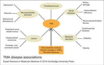
TMA in blood, urine and breath has been associated with a number of human conditions including various diseases. In the following sections, we discuss the significance of TMA in human biology (Fig. 3).
TMA disease associations. TMA may be a central biomarker for several human diseases and disorders.

Trimethylaminuria
TMA has been known to human civilisation for a long period. In regards to human sensitivity, ~7% of human population have specific anosmia for TMA (Ref. Reference Amoore and Forrester48), with young subjects being most sensitive to TMA and adults least so (Ref. Reference Solbu, Jellestad and Strætkvern49). TMAU is a TMA-related metabolic autosomal recessive disorder that exhibits variation in metabolic detoxification of xenobiotics by hepatic FMO3 (Ref. Reference Motika32). TMA as the underlying cause of fishy odour remained undetermined until the early 1970s. At the time, Dr J.R. Humbert described the first clinical case and provided a link between TMA and its associated metabolic disorder – then referred to as the fishy odour syndrome (FOS) but now as TMAU, characterised by an increase in TMA excretion in urine, sweat and breath (Ref. Reference Humbert50). Modern day diagnosis of TMAU requires specialised tests that include diet challenges and analysis of host FMO3 gene sequences for mutations (Refs Reference Zhang, Mitchell and Smith28, Reference Wise51). While common causes of fishy odour include poor hygiene, gingivitis, bacterial vaginosis and urinary tract infections, in individuals with TMAU malodours are produced despite their good hygiene (Ref. Reference Rehman52).
Renal diseases
TMA is a metabolic waste product that is excreted in the urine. Several research groups have reported increased levels of TMA in sweat, breath or blood as suggestive of kidney damage, potentially because of increased synthesis or reduced/no clearance from the human body. For instance, raised TMA was observed in patients with end-stage renal disease (ESRD) (Ref. Reference Bain53). A possible reason for this could be differential microbial flora in ESRD patients compared with individuals with normal renal function (Ref. Reference Simenhoff54). Additionally, low glomerular filtration rate and low TMAO plasma clearance was observed in ESRD patients (Ref. Reference Hao55). In animal models, elevated TMA precursors or its metabolites (e.g. choline and TMAO) can lead to progressive renal tubulointerstitial fibrosis and dysfunction (Ref. Reference Tang56).
Cancer
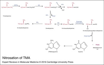
Elevated TMA is considered as a marker of proliferating tumour tissue. For instance in peripheral nerve sheath tumour, TMA seems to be a sensitive (100%), yet not specific (50%) marker of malignant disease (Ref. Reference Fayad57). Raised TMA is also a biomarker for renal cell carcinoma (Refs Reference Zira58, Reference Gao59), and mechanistically this is attributed to cells switching to glycolysis to maintain energy homeostasis following the loss of ATP caused by impaired TCA cycle (Ref. Reference Xia60). It has further been suggested that high concentration of TMA in gut can lead to nitrosation of TMA, which produces NDMA. Latter is a well-known poison, hepatotoxin and carcinogen (Ref. Reference Wang, Li and He61). Methylamine, DMA and TMA present in human gastric fluid are readily converted to NDMA in the presence of sodium nitrite (Ref. Reference Zeisel, daCosta and LaMont14). Sodium nitrite is a commonly used food additive in cured meat and poultry products (Ref. Reference Pierson and Smoot62) (Fig. 3). NDMA in human body is acted upon by liver microsomal membrane-bound enzyme cytochrome P-450, which generates the methyldiazonium ion, latter can then form DNA adducts such as O6-methylguanine that are carcinogenic agents (Ref. Reference Guengerich, Kim and Iwasaki63). Thus, endogenous TMA can be converted to more toxic metabolites that can drive abnormal cellular proliferation in humans (Fig. 4).
Nitrosation of TMA. TMA undergoes nitrosation leading to the formation of nitrosated amine – N-nitrosodimethylamine (NDMA). Latter is converted to methyldiazonium ion (MI) via action of liver microsomal membrane-bound enzyme cytochrome P-450. MI can form DNA adducts such as O6-methylguanine, a carcinogenic agent (Refs Reference Usunomena13, Reference Mirvish116).

Metabolic disorders
TMA-induced intracellular calcium mobilisation from endoplasmic reticulum (ER) calcium stores contributes towards ER stress (Ref. Reference Schönthal64). Interestingly, ER stress is emerging as an important mechanism that is common in metabolic diseases such as obesity and diabetes (Ref. Reference Cnop, Foufelle and Velloso65). ER stress can lead to aberrant transcriptional regulation, altered gene expression, ion channel failure, dysmetabolism, impaired signalling, oxidative stress and inflammation (Ref. Reference Cnop, Foufelle and Velloso65). Thus, raised TMA can result in downstream effects that contribute to increased ER stress. Increase in endogenous TMA is observed in both obesity and diabetes; it also positively correlates with the levels of TMAO and in turn with an increased risk of developing cardiovascular diseases (Ref. Reference Miao66).
So far, the increased incidence of metabolic disorders had been attributed in part to high-fat and high-carbohydrate Western style diets, lack of physical activity and genetic susceptibility (Ref. Reference Pitsavos67). However, recent advances in microbiome studies have highlighted the importance of gut microbes and the adverse effects of microbial dysbiosis on metabolic disorders (Ref. Reference Quigley68). Both microbial dysbiosis and higher TMA have been linked to inflammatory bowel syndrome (IBS). For example, TMA concentrations in normal mice with IBS correlated with those in IL-10 knockout mice with IBS. These mice showed distinct metabolic differences typical of IBS as disease progressed. However, IL-10 knockout mice raised in germ-free environment failed to develop IBS. Thus, highlighting the importance of microbial flora derived TMA in IBS progression (Refs Reference Swidsinski69, Reference Murdoch70). Further, while its metabolite TMAO has been directly correlated with risk of developing heart disease, TMA (and not TMAO) has been associated with atherosclerotic plaque formation in HIV patients (Ref. Reference Srinivasa71). Hence, higher TMA levels correlate with the presence of various metabolic disorders, and the cause/effect axis of this linkage needs further exploration.
Hepatic diseases
The liver is a major site for oxidation of TMA to non-odorous TMAO by hepatic FMO3, and high level of TMA has been correlated with liver dysfunction. For example, elevated TMA can be observed in breath of patients with alcoholic hepatitis, in nonalcoholic fatty liver disease (NAFLD) patients and in the biopsy samples from hepatocellular carcinoma (Refs Reference Shariff72, Reference Alkhouri73, Reference Hanouneh74). Further, in addition to being a potent carcinogen (Subsection ‘Cancer’), the TMA metabolite NDMA is also a well-known hepatotoxin that can cause liver fibrosis (Ref. Reference George75). Reactive metabolites of NDMA also trigger activation of apoptotic cell death in P450-expressing cells (Ref. Reference Hecht76). We speculate that liver damage may in turn reduce the oxidation of TMA to TMAO, leading to decreased clearance of TMA from the blood. Accumulated TMA can then feed into the synthesis of NDMA causing further organ damage.
Neurological diseases
Increased excretion of TMA is implicated in depression, seizures and behavioural disturbances (Refs Reference Pellicciari77, Reference McConnell78). These neuropsychological disorders have been attributed to the role of TMA as a gap-junction opener, where TMA reportedly increases thalamic modulation on cortical seizures (Ref. Reference Chang, Wu and Shyu79). Also, TMA seems to be involved in modulation of local cell discharges in hippocampus as seen in anaesthetised rats (Ref. Reference Bocian80). Thus, TMA may be correlated with propagation of seizures.
Beneficial roles of TMA
Contrary to conditions where high TMA levels correlate with disease conditions, high-altitude pulmonary oedema (HAPE) – a life threatening form of noncardiogenic pulmonary edema – is accompanied by a decrease in TMA (Ref. Reference Luo, Zhu and Gao81). Simultaneously, an increase in several biomolecules (valine, lysine, leucine, isoleucine, glycerol phosphoryl choline, glycine, glutamine, glutamic acid, creatinine, citrate and methyl histidine) is seen in HAPE patients. These metabolites are suggested to be putative biomarkers (Ref. Reference Paul82). Unrelatedly, decreased TMA levels are significantly associated with increased incidence of foetal growth rate and pre-term births (Ref. Reference Maitre83). Changes in the gut microbial composition throughout pregnancy are partly responsible for an increase in insulin resistance and greater adiposity during this phase (Ref. Reference Koren84). Although normally associated with disease, increased TMA may be of benefit during normal pregnancy in nonobese women to help support growth of the foetus and to prepare the body for energetic demands of lactation (Refs Reference Di Cianni85, Reference Lain and Catalano86, Reference Scott, Nelson and Poston87).
TMA–TMAO axis
The potential of TMA to modulate diverse physiological processes is evident from the wide range of TMA concentrations observed in healthy and diseased subjects. For instance, in healthy subjects TMA concentration is 0.42 ± 0.12 µm in blood and 2.5 (0.3–19.4) μm/mm creatinine in the urine, while in diseased subjects it is 1.38 ± 0.48 µm in blood and 22.2 µm/mm creatinine in urine (Human Metabolome Database).
However, most studies have focused on the measurement of TMAO and not TMA, possibly because of stability of TMAO (Ref. Reference Wang88). There are several key unresolved questions surrounding how circulating TMAO concentrations are sensed to elicit pathological responses (Ref. Reference Mark Brown and Hazen89). Table 2 highlights the diseases for which TMA concentrations have been monitored. Additionally, since TMA drives TMAO production, it remains undetermined, whether pathological responses observed are because of increased TMAO concentrations alone or because of increased TMA production that triggers higher concentrations of circulating TMAO.
Diseases for which TMA has been proposed as a biomarker

TMAO concentrations in blood were first correlated with early detection of myocardial infarction, where an increase in TMAO concentrations was observed in patients who had spontaneous myocardial infarction as compared with those who had planned myocardial infarction (Ref. Reference Lewis90). Latter refers to a planned destruction of heart muscle caused by alcohol injection. It is a successful treatment for hypertrophic cardiomyopathy and provides a carefully controlled human model of a spontaneous myocardial infarction – one in which each patient provides his/her own control samples, thereby minimising the impact of inter-individual differences (Ref. Reference Lewis90). An increase in choline, betaine and TMAO concentrations were observed amongst other 18 metabolites, and a correlation between TMAO concentrations and risk of developing cardiovascular diseases (atherosclerosis, myocardial infarction) was suggested (Refs Reference Mark Brown and Hazen89, Reference Hartiala91). A strong prognostic value of TMAO rather than choline/betaine in chronic systolic heart failure was suggested with no significant correlations between other metabolites and inflammatory biomarkers (Ref. Reference Tang92). Aside from TMAO linkage with heart disease, elevated serum TMAO has been correlated directly with markers of nutritional status and inversely with markers of inflammation in patients receiving haemodialysis (Ref. Reference Kaysen93). For instance, urinary TMAO acts as a marker associated with the quantity of nitrogen and thereby consumed protein (Refs Reference Rasmussen94, Reference Lloyd95). Elevated concentrations of TMAO have also been linked with aggressive prostate (Ref. Reference Mondul96), oral cancer (Ref. Reference Bag97), colorectal cancer (Ref. Reference Bae98), with paediatric renal transplant patients (Ref. Reference Klepacki99), and with lung granulomas in Mycobacterium Tuberculosis-infected guinea pigs (Ref. Reference Somashekar100). Thus, analysing the relative concentrations of TMA and TMAO may provide even more insightful linkages.
TMA removal from the environment and the human body
Via plants and plant-based diets
Plants remove pollutants from the air by adsorbing them either through leaves or roots. In some plants, biomaterial and leaf wax can adsorb ~70% of total gaseous TMA, while additional ~30% can be effectively adsorbed by cellulose (Ref. Reference Boraphech and Thiravetyan101). Leaf wax from these plants has been shown to be composed of short-chain fatty acids (SCFA, ≤C18, especially the C18 octadecanoic acid) and short-chain alkanes (C12–C18) along with other aromatic components, which together enhance TMA absorption (Ref. Reference Boraphech and Thiravetyan101). Pterocarpus indicus (new guinea rosewood) and Sansevieria trifasciata (snake plant or mother-in-law's tongue) are most effective in adsorbing TMA, and can also degrade TMA to methylamine and DMA via multiple metabolic pathways which are associated with carbon/nitrogen interactions (Ref. Reference Boraphech and Thiravetyan102).
Plant-based diets may be healthier in part because of their ability to maintain microbial symbiosis in the gut, while discouraging colonisation by microbes responsible for an increase in synthesis of metabolites such as TMA (Refs Reference Vieira and Pagovich103, Reference Zarrinpar104). Indeed, it may be of value to correlate microbiome differences in vegetarians and nonvegetarians to assess where specific populations of TMA-producing microbes are altered. It is noteworthy that TMAO production from dietary L-carnitine is found to be negligible in vegans, while changes in TMA levels in vegans still need to be assessed (Ref. Reference Glick-Bauer and Yeh105). It is probable that vegan diet leads to decrease in synthesis of TMA and subsequently negligible TMAO production. Thus, in addition to a decrease in consumption of red meat (which is a source of precursors that drive TMA production), increased consumption of plant-based foods may help to establish appropriate physiological levels of TMA and TMAO in the human body (Fig. 5).
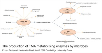
The production of TMA metabolising enzymes by microbes. Schematic representation of the various microbes involved in the TMA metabolic pathway (Refs Reference Craciun, Marks and Balskus117, Reference Chen33, Reference Dridi111, Reference Romano118).

Via microbes
Several microbial species have been used for efficient removal of TMA from atmosphere. For example, biofilters inoculated with Arthrobacter sp. or in combination with Paracoccus were tested on the exhaust air of swine waste storage pits, which led to simultaneous elimination of TMA from the air with high average removal efficiency (Refs Reference Chang106, Reference Ho107). Similarly, multiple approaches have been explored to decrease the fishy odour associated with TMAU patients. An unconventional approach is the use of laxatives which speeds up the passage of food through bowel thus decreasing the amount of TMA synthesised in gut (Ref. Reference Smits108). Brief courses of antibiotics such as neomycin and metronidazole have been used to suppress gut microbes and prevent FOS in cases where microbial dysbiosis is an important factor leading towards TMAU (Refs Reference Wang6, Reference Koeth15, Reference Wilson Tang109). More recently, it has been suggested that the production of the enzyme TMA monooxygenase (TMM), which is similar to FMO3, using microbes like Ruegeria pomeroyi and Methylocella silvestris could remove TMA and help TMAU patients (Ref. Reference Lidbury, Murrell and Chen110). Furthermore, TMM-expressing bacteria is available as part of a topical cream or spray could be a cheap and stable way to deliver therapeutic enzymes to TMAU patients. Additionally, TMAO can be metabolised by human gut methanogens that are restricted to utilising methyl compounds (including TMAO) as substrates to obtain energy under anaerobic conditions. Indeed, Methanomassiliicoccus luminyensis B10, Methanobrevibacter smithii, Methanosphaera stadtmanae are able to deplete TMAO by reducing it with hydrogen for methanogenesis (Refs Reference Dridi111, Reference Brugère112). Thus, investigations that aim to identify human commensal oral or gut microbes that possess enzymes capable of TMA/TMAO degradation into harmless metabolites may be helpful future additions to pro-biotic formulations.
Conclusion
Clinical and experimental evidence suggests that human microbiome plays an important role in development and progression of health disorders. The metabolites produced by resident host microbiome are considered to confer protective effects; however, microbial dysbiosis can lead to altered metabolite profiles. The generation of TMA by gut microbes using common diet precursors is an example of a metabolite that correlates with disease states, and requires extensive further profiling. Since the quantification of TMA is relatively cumbersome compared with TMAO, most studies have so far addressed TMAO disease correlations. We propose that TMA and TMAO can be together assessed in order to fully explore their potential as sets of biomarkers. Finally, the genomic and metabolomics bio-signatures of microbes that control the TMA/TMAO axis may be worth cataloguing in wider nets of diseased populations.
Acknowledgements
We would like to thank V.K. Aves and CB Indus for constant encouragement. This work was funded by DBT OSRP grant and DST JC Bose fellowship to AS.
Conflict of interest
The authors declare that they have no conflict of interest with the contents of this article. All authors reviewed and approved the final version of the manuscript.