Case report
A 7-year-old previously healthy boy was transferred to our emergency department from an outside facility after successful resuscitation from a documented ventricular fibrillatory arrest. The child was wrestling with his 18-year-old maternal uncle earlier that evening. As per the mother, they were “horsing around” and punching each other. After fighting his uncle, the child proceeded to the kitchen and was observed to be playing a game on his cell phone for approximately 10 minutes. The family then moved to another room. After a few minutes, the patient’s younger sister went to call her brother at which time he was found to be “not moving or breathing”. The mother denied a past history of chest pain, dizziness, seizures, fainting, or near-drowning, and there was no history of upper respiratory symptoms, cough, dyspnoea, abdominal pain, nausea, or diarrhoea in the weeks prior to presentation. In addition, there were no sick contacts, and the family history was negative for an inherited cardiomyopathy or channelopathy.
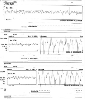
Emergency medical services were immediately called; however, since the patient lived only 2–3 blocks away from the hospital, a relative lifted the patient and ran down the street with the patient in his arms. On arrival in the emergency department, the patient was found to be pulseless and unresponsive. He was connected to a cardiac monitor which revealed ventricular fibrillation as the underlying rhythm. Patient was defibrillated twice and received a dose of epinephrine before return of spontaneous circulation was achieved (Fig 1). He was thereafter intubated and ventilated. Post-arrest electrocardiogram showed a narrow QRS rhythm with mild prolongation of corrected QT interval. The QT interval normalised within a few hours and subsequent electrocardiograms have not revealed any abnormality.
The tracings show ventricular fibrillation which recurs after the first shock. The patient reverts back to a stable perfusing rhythm after the second shock.

The patient was transferred to our emergency department for further evaluation where no evidence of external injury was found on clinical examination and no fractures were noted on radiographic imaging. A contrast-enhanced CT scan of chest, upper abdomen, and pelvis showed no evidence of solid organ injury, bony injury, or free air.
The patient was admitted to Pediatric Intensive Care Unit (PICU) for post-cardiac arrest care. After admission, child protective services and local police were involved to elicit the precise details of the event as one of the concerns was commotio cordis. As per the child protective services investigation, there was a clear lucid interval (almost 10 minutes) between the fight between the patient and his maternal uncle and the event which ruled out commotio cordis as a cause for his ventricular fibrillatory arrest.
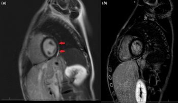
Investigations in PICU revealed moderately diminished left ventricular function and mildly diminished right ventricular function on the initial echocardiogram which rapidly improved within the next 24 hours. A cardiac magnetic resonance scan, obtained 3 days later, showed epicardial late gadolinium enhancement involving the basal, mid inferolateral, and anterolateral left ventricular walls (Fig 2a). The late gadolinium enhancement was also noted to involve the adjacent pericardium and pleura. Biventricular size and function appeared normal.
(a) Epicardial late gadolinium enhancement (red arrows) in the basal, mid inferolateral, and anterolateral left ventricular walls. (b) Repeat Cardiac Magnetic Resonance (CMR) imaging performed 9 months later shows complete resolution of these changes.

Telemetric monitoring during the hospital stay did not reveal any dysrhythmia. The patient’s total leucocyte count, serum troponin concentration, and erythrocyte sedimentation rate were normal at admission; however, his respiratory viral panel was positive for rhinovirus/respiratory enteroviruses. A urine drug screen was also obtained and was only positive for benzodiazepines which were administered at the time of intubation. A loop recorder was implanted, a life vest (wearable cardioverter-defibrillator) was fitted (single ventricular fibrillation zone >250 bpm), and appropriate teaching carried out for the same prior to discharging the patient to home.
Given the uncertainty surrounding the diagnosis and possibility of left-dominant arrhythmogenic right ventricular cardiomyopathy masquerading as myocarditis, genetic testing was obtained (CardioNext Panel, Ambry genetics, CA). The testing showed several variants of unknown significance including p.L2845F variant in Alstrom syndrome (ALMS1) 1 gene, mutations in which are inherited in an autosomal recessive manner and have been associated with Alstrom syndrome which is associated with dilated cardiomyopathy, hypertension, and congestive heart failure; p.E1111K variant in myosin binding protein C3 (MYBPC3) gene, mutations in which have been associated with hypertrophic and left ventricular non-compaction cardiomyopathies and are inherited in an autosomal dominant fashion with reduced penetrance and variable expressivity; p.T1004A, p.V5766I, and p.N22897D variants in titin (TTN) gene mutations in which are difficult to interpret due to reduced penetrance, age-dependent expression, and genetic heterogeneity but have been associated with dilated and hypertrophic cardiomyopathies and skeletal myopathies. However, none of these detected variants have been associated with any human pathology till date.
The mother, younger brother, and a younger sister were evaluated, and no abnormality was found on electrocardiogram, echocardiogram, or exercise stress testing (mother and younger sister).
Cardiac MRI was repeated 9 months after the initial event and showed normal chamber sizes with normal biventricular systolic function without any segmental or regional wall motion abnormalities. The previously seen left ventricular myocardial late gadolinium enhancement had completely resolved. Normal native left ventricular myocardial T1 and T2 times were noted in the sampled regions (interventricular septum and free wall). No appreciable pericardial late gadolinium enhancement or pericardial effusion was seen (Fig 2b).
The patient has done well without any cardiovascular symptoms. Serial loop recorder interrogations have not revealed any tachy or bradyarrhythmia. He recently underwent an exercise stress test which showed normal heart rate and blood pressure response to exercise. No ectopy or arrhythmias was noted. Given the reassuring findings on extensive testing and complete resolution of abnormal cardiac MRI findings, he has been taken off the life vest.
Discussion
Myocarditis, an inflammatory disease of the myocardium caused by infectious and noninfectious triggers which include viral infections and post-viral immune mediated phenomena,Reference Kindermann, Barth and Mahfoud1 can be asymptomatic or can present with a broad range of symptoms including chest pain, cardiac arrhythmias, and acute or chronic heart failure.Reference Kindermann, Barth and Mahfoud1
In the absence of clear signs and symptoms, diagnosing myocarditis can be challenging. Inflammatory markers such as C-reactive protein and erythrocyte sedimentation rate can be elevated in patients with myocarditis, but normal values do not exclude the diagnosis.Reference Kindermann, Barth and Mahfoud1 Biomarkers (such as troponin or creatine kinase) lack sensitivity and specificity but may help confirm the diagnosis if elevated.Reference Rodriguez-Gonzalez, Sanchez-Codez, Lubian-Gutierrez and Castellano-Martinez2 Our patient had a positive respiratory viral panel for rhinovirus/respiratory enteroviruses which have been associated with myocarditis.Reference Chow, Murphy, Subedi and Jyothidasan3,Reference Kandolf, Klingel and Zell4 Mahfoud et al compared viral serology to viral genome detection in endomyocardial biopsy specimens in patients with clinically suspected myocarditis. Only 5 of 124 patients (4%) had serological evidence of an infection with the same virus that was detected by polymerase chain reaction assay in endomyocardial biopsy specimens. These findings indicate that viral serology is not helpful in diagnosis in a vast majority of patients with suspected myocarditis. A viral serology panel was therefore not obtained.Reference Kindermann, Barth and Mahfoud1,Reference Mahfoud, Gartner and Kindermann5
Electrocardiography has been widely used as a screening tool but also suffers from low sensitivity.Reference Morgera, Di Lenarda and Dreas6–Reference Fung, Luo, Qiu, Yang and McManus9 Echocardiography has also been utilised in patients with myocarditis but suffers from a lack of specific features; however, it does allow the evaluation of cardiac chamber sizes and wall thickness as well as systolic and diastolic function. More importantly, it helps in ruling out other causes of myocardial dysfunction. Patients with fulminant myocarditis often have normal cardiac chamber sizes with an increased septal thickness secondary to acute myocardial oedema, whereas patients with acute myocarditis have marked left ventricular dilation and normal wall thickness and carry a worse long-term prognosis.Reference Kindermann, Barth and Mahfoud1,Reference Felker, Boehmer and Hruban10 Cardiac MRI has emerged as an important imaging modality in evaluation of myocarditis. Our patient had epicardial late gadolinium enhancement involving the basal, mid inferolateral, and anterolateral left ventricular walls, adjacent pericardium, and some of the adjacent pleura which resolved on follow-up. Cardiac MRI findings in our patient were consistent with common patterns of myocardial involvement that have been described in myocarditis. These include an intramural, rim-like pattern in the septal wall which is associated with worse prognosisReference Aquaro, Perfetti and Camastra11 or a subepicardial (patchy) distribution in the left ventricular inferior or lateral walls.Reference Kindermann, Barth and Mahfoud1 Despite advances in cardiac MRI, endomyocardial biopsy remains the gold standard in diagnosis of myocarditis. According to the Dallas criteria, acute myocarditis is defined by lymphocytic infiltrates in association with myocyte necrosis.Reference Kindermann, Barth and Mahfoud1 Given the acute presentation with ventricular fibrillation, rhinovirus/respiratory positivity, and characteristic changes on cardiac MRI, endomyocardial biopsy was not carried out in our patient.
Post-mortem data identify myocarditis in 8.6–12% of cases of sudden death in young adults. The prevalence has been found to be higher (40%) in certain selected populations such as air force recruits who had sudden cardiac death.Reference Kindermann, Barth and Mahfoud1,Reference Phillips, Robinowitz, Higgins, Boran, Reed and Virmani12
Sudden cardiac death in patients with myocarditis can be mediated by both brady and tachyarrhythmias. Potential mechanisms for these arrhythmias include myocardial replacement fibrosis leading to reentry, myocyte necrosis, proarrhythmic effects of cytokines, altered function at myocardial gap junctions, altered calcium handling, infarction, microvascular ischaemic insult, protease release resulting in cleavage of dystrophin with consequent cytoskeletal abnormality, unmasking of an underlying cardiomyopathy such as dilated or arrhythmogenic right ventricular cardiomyopathy, and a concomitant channelopathy.Reference Baksi, Kanaganayagam and Prasad13
Reported bradyarrhythmias in patients with myocarditis include sinus arrest, sinoatrial blocks, and atrioventricular blocks (1st degree, 2nd degree, advanced, or complete atrioventricular block).Reference Caforio, Pankuweit and Arbustini14,Reference Buttà, Zappia, Laterra and Roberto15 Both transient and permanent atrial standstill has also been reported.Reference Larsen, Maleszewski and Edwards16,Reference Prabhu, Srinivas Prasad, Thajudeen and Namboodiri17 Atrioventricular blocks tend to be more common with Lyme carditis, giant cell myocarditis, and fulminant myocarditis.Reference Buttà, Zappia, Laterra and Roberto15
Supraventricular arrhythmias such as sinus tachycardia, atrial flutter, and atrial fibrillation are also common in patients with myocarditis and are associated with worse prognosis.Reference Buttà, Zappia, Laterra and Roberto15
Patients with myocarditis and ventricular arrhythmias may present with a wide spectrum of symptoms ranging from palpitation to syncope. Ventricular arrhythmias are frequent in fulminant myocarditis and also in infiltrative myocarditis, where they can occur even in patients with normal ventricular function.Reference Granér, Lommi, Kupari, Räisänen-Sokolowski and Toivonen18 Prevalence of ventricular arrhythmias during the initial hospitalisation in giant cell myocarditis has been reported to be between 14 and 22%, and the risk of life-threatening ventricular arrhythmias exceed 50% at 5 years.Reference Cooper, Berry and Shabetai19,Reference Ekström, Lehtonen, Kandolin, Räisänen-Sokolowski, Salmenkivi and Kupari20 Ventricular arrhythmias at presentation predict the occurrence of sudden cardiac death and ventricular tachycardia during long-term follow-up in patients with giant cell myocarditis;Reference Ekström, Lehtonen, Kandolin, Räisänen-Sokolowski, Salmenkivi and Kupari20 as a consequence, the presence of malignant ventricular arrhythmias warrants earlier consideration of an implantable cardioverter-defibrillator in these patients.Reference Priori, Blomström-Lundqvist and Mazzanti21,Reference Al-Khatib, Stevenson and Ackerman22 In contrast, acute-phase ventricular arrhythmias in viral myocarditis tend to be self-limiting and, if promptly managed, do not bear significant adverse long-term prognosis.Reference Peretto, Sala and Rizzo23 In the absence of a dilated cardiomyopathy phenotype, the prognosis for individuals surviving cardiac arrest due to ventricular arrhythmia in the setting of acute myocarditis seems favourable if resuscitation is prompt and effective.Reference Baksi, Kanaganayagam and Prasad13,Reference Chau, Chow, Chiu and Wang24 Therefore, a permanent implantable cardioverter-defibrillator should be deferred until resolution of the acute episode; bridging the critical period to full recovery by a wearable cardioverter-defibrillator vest is an appropriate therapeutic option in this setting as was done in this patient.Reference Priori, Blomström-Lundqvist and Mazzanti21
Cardiac MRI offers prognostic information in patients with myocarditis. A normal cardiac MRI confers a good prognosis regardless of symptoms or other findings. Arrhythmic events on follow-up are only noted in patients with persistent abnormal cardiac MRI findings.Reference Baksi, Kanaganayagam and Prasad13 However, it is important to note that complete resolution of cardiac MRI findings was only seen in 28% in a small paediatric series (n = 18); the rest had ongoing active inflammation (28%) or persistent scars (44%).Reference Małek, Kamińska and Barczuk-Falęcka25 A persistent scar or ongoing inflammation on follow-up cardiac MRI scan would have prompted us to consider an implantable cardioverter-defibrillator.
We strongly considered cardiac contusion, which is associated with severe blunt chest trauma and is difficult to diagnose due to a lack of a gold standard diagnostic test as a differential in this patient. Chest radiography, chest CT scan, electrocardiogram, and echocardiogram have been shown to have poor sensitivity (<15%) in patients with cardiac contusion. In addition, troponin was only elevated in 38% of patients with myocardial contusion at presentation in a large series suggesting that commonly used biomarkers also lack adequate sensitivity.Reference Duclos, Scemama, Leone and Zieleskiewicz26 The presenting symptoms in patients with cardiac contusion mimic those in patients with myocarditis and include malignant arrhythmias, heart failure, and myocardial infarction. Cardiac CT imaging has been used to assess myocardial contusion with regions of hypoenhancement (easier to visualise in the left ventricle than the right due to thicker walls) suggesting this diagnosis.Reference Hammer, Raptis and Cummings27 Cardiac MRI findings similar to myocardial infarction, including delayed gadolinium hyperenhancement and visualisation of necrotic tissue and myocardial oedema on T2 black blood imaging, have been reported in patients with cardiac contusion.Reference Vago, Toth and Apor28–Reference Kaneda, Yamamura, Yamamoto, Matsumoto, Ehara and Mizobata30 Regional wall motion abnormality involving anterior, anterolateral, anteroseptal left ventricular walls or septum, right ventricular wall oedema, pericardial effusion, tricuspid valve damage, and intramural haemorrhage have also been reported.Reference Burrell, Kaye and Fitzgerald31 No external or internal trauma was noted on clinical examination and extensive radiological investigations in our patient. In addition, subepicardial late gadolinium enhancement in the inferior and lateral left ventricular walls, as seen in this patient, is considered consistent with a diagnosis of myocarditis.Reference Zeldetz, Greenberg, Zeller, Zahger and Shalev32
A distinct left-dominant variant of arrhythmogenic right ventricular cardiomyopathy has been described. This variant is characterised by late gadolinium enhancement in left ventricular subepicardial/mid wall distribution on cardiac MRI, corresponding to areas of fibrofatty replacement, and fibrosis on histopathology. These patients are often misdiagnosed with viral myocarditis, dilated cardiomyopathy, hypertrophic cardiomyopathy, or idiopathic ventricular tachycardia. Arrhythmic events, including initial presentation with ventricular fibrillatory arrest, are common in these patients. This entity should be suspected in patients of any age with unexplained arrhythmia of left ventricular origin, inferolateral/lateral T-wave inversion on electrocardiogram, apparent dilated cardiomyopathy with arrhythmic presentation, or myocarditis (chest pain and enzyme rise with unobstructed coronary arteries).Reference Sen-Chowdhry, Syrris and Prasad33 Our patient had a negative family history, and evaluation of family members did not reveal any electrical or structural cardiac abnormality. Genetic testing was also obtained, and no pathogenic genetic alterations were found. Most importantly, complete normalisation of cardiac MRI findings in our patient upon follow-up favoured a diagnosis of myocarditis over left-dominant arrhythmogenic right ventricular cardiomyopathy.
To conclude, our patient had no obvious symptoms but presented with ventricular fibrillatory arrest. Cardiac MRI revealed epicardial late gadolinium enhancement in the basal, mid inferolateral, and anterolateral left ventricular walls which completely resolved on follow-up. Myocarditis is an important cause of arrhythmogenic sudden cardiac arrest in the young. However, a strong index of suspicion is required as arrhythmia can be the only clinical manifestation. Cardiac MRI is immensely helpful in making this diagnosis as these patients can have normal cardiac biomarkers, electrocardiographic and echocardiographic findings, and inflammatory markers. Patients with ventricular arrhythmias in the setting of viral myocarditis, especially the ones in whom cardiac MRI imaging findings normalise upon follow-up, tend to do well in the long run, and an implantable cardioverter-defibrillator should be avoided in these patients.
Acknowledgements
None.
Financial Disclosure
This research received no specific grant from any funding agency or from commercial or not-for-profit sectors.
Conflict of Interest
None.
Ethical Statement
Informed consent was obtained from all individual participants included in the report. This report does not include any human or animal experimentation.