Introduction
The vast majority of oral cavity cancers in the head and neck are squamous cell carcinomas (SCCs). Squamous cell carcinoma is the sixth most common cancer worldwide and its incidence is rising in industrialised nations.Reference Sankaranarayanan, Masuyer, Swaminathan, Ferlay and Whelan1, Reference Duvvuri and Myers2 Head and neck cancer is a major cause of morbidity and mortality, with many cancers of this region arising from pre-malignant lesions. Such lesions, characterised in general descriptive terms as leukoplakia on gross physical examination, are quoted as having up to a 10 per cent chance of cancerous transformation in some series, with the rates being even higher for lesions exhibiting erythroplakia. Other benign lesions of the oral cavity, such as lichen planus, have a prevalence of 0.5–2 per cent in the general population and have a reduced risk of malignant transformation of approximately 1 per cent.Reference Upile, Jerjes, Kafas, Angouridakis and Hopper3 Therefore, prompt detection and diagnosis of suspicious mucosal lesions provides an important opportunity to intervene and cure at an early stage of disease.
Nevertheless, many oral mucosal lesions are not cancerous, which presents a clinical dilemma to the physician. Furthermore, various benign pathologies presenting as leukoplakia may exhibit mild structural alterations in the mucosa that can be difficult to distinguish from normal healthy tissue. Currently, obtaining histopathology via biopsy is the ‘gold standard’ of diagnosis; however, this procedure can pose significant morbidity to the patient, such as the risk of bleeding, wound infection and tissue scarring leading to impairment of oral cavity function if multiple biopsies are performed.
A further clinical challenge relates to the monitoring of patients with wide areas of dysplastic field change for progression to invasive SCC. The discomfort of biopsy and compromise of tissue integrity can lead to problems with future biopsy interpretation, or in the case of laryngeal biopsy, considerable problems in individuals with high vocal demands.Reference Cikojević, Gluncić and Pesutić-Pisac4 The reliable pathological evaluation of lesions in these instances without the requirement for invasive biopsy could potentially offer improved patient comfort and functional outcomes.
Detailed examinations of the texture, colour, contour and extent of mucosal lesions have been performed using many instruments, such as the Hopkins rod-lens telescopes, flexible endoscopes, direct laryngoscopes and advances in microlaryngoscopic visualisation techniques. However, these methods are limited by their inability to provide pathological data during the clinical examination. Over the last decade, technological advances in optical imaging detection techniques have emerged, with a variety of methods used to facilitate detailed examination and provision of pathological information of mucosal lesions. Contact endoscopy is one such imaging technique.
Contact endoscopy is a minimally invasive technique whereby a rigid endoscope contacted onto stained mucosa produces highly magnified images that demonstrate cellular architecture. The use of the contact endoscope as a method of in vivo cytological evaluation has been in clinical use for over 30 years, with the first description in the literature provided by Hamou in 1979.Reference Hamou5 The usefulness of contact endoscopy in otolaryngology was first described by Andrea et al. in the 1990s.Reference Andrea, Dias and Santos6 Despite this potential for clinical application, published data are lacking and its use in routine clinical practice appears to be limited. We aim to report our institution's experience with contact endoscopy, and endeavour to add to the current body of literature evaluating its use and potential clinical application.
Materials and methods
Study population
With the permission of the Research Ethics Board of Western University (London, Ontario, Canada) and after obtaining written informed consent, 34 consecutive, eligible and consenting subjects with oral cavity/oropharyngeal lesions requiring biopsy were recruited from the Otolaryngology – Head and Neck Surgery clinic at Victoria Hospital, London Health Sciences Centre and the Oral Pathology clinic at University Hospital, London Health Sciences Centre and actively enrolled in the study between the dates of 1 January 2010 and 31 December 2011.
Patients who were 18 years old and above, and who presented to the oral pathology clinic with oral cavity mucosal lesion requiring biopsy, were eligible for inclusion in the study. Exclusion criteria were as follows: previous biopsy or surgery in the area of interest; patients with limited mouth opening; and patients who were taking anticoagulation medications.
Contact endoscopy and biopsy
The technique of contact endoscopy used was as described by Andrea et al. Reference Andrea, Dias and Santos6 The mucosal surface was cleaned where necessary with gentle suction or using a swab. The area to be examined was stained with 1 per cent methylene blue by applying a soaked swab onto the mucosa for 5 min. Any excess dye was swabbed off. The tip of the contact endoscope (Karl Storz Contact Micro-Laryngoscope 0 degrees (8715AA) or 30 degrees (8715AB)) was gently contacted against the mucosal surface and video recordings of images were taken at magnifications of ×60 and ×150 using a xenon light source (Welch Allyn, Mississauga, Ontario, Canada) and a Panasonic CD1 micro video camera (Panasonic, Osaka, Japan) and endoscopic system (AKAI VF 440, Tokyo, Japan). Figures 1 and 2 show the typical findings of benign and malignant lesions. After completion of the contact endoscopy assessment, biopsies of the area examined by contact endoscopy were taken for paraffin section histopathology.
Contact endoscopy image of pathologically confirmed benign oral mucosa. (Methylene blue; ×150)

Contact endoscopy image of pathologically confirmed squamous cell carcinoma. (Methylene blue; ×150)
Contact endoscopy and pathological evaluation
In each case, contact endoscopy was performed by the investigating surgeon or surgical fellow. Each surgeon performing contact endoscopy undertook five cases on control patients prior to examining the study participants to ensure a thorough understanding of the equipment and endoscopic process. Although the time taken to perform contact endoscopy was not prospectively collated, all imagery was collated within our standard out-patient setting without implications for either the number of patients reviewed per clinic or the staff required.
All contact endoscopy images and histopathological specimens were evaluated by a single head and neck pathologist to eliminate the potential inter-observer variation in interpretation, given that the aim of the study was to determine accuracy. Contact endoscopy images were de-identified and presented to the pathologist separately from their corresponding histopathology specimens. The contact endoscopy images were then reviewed and a diagnosis was made based on the published description of the contact endoscopy findings.Reference Duvvuri and Myers2, Reference Cikojević, Gluncić and Pesutić-Pisac4, Reference Andrea, Dias and Santos6–Reference Paczona, Temam, Janot, Marandas and Luboinski13
Data management and statistical analysis
Patient data were entered into a spreadsheet created in Microsoft Excel (Redmond, Washington, USA) and the statistical analysis was carried out using version 12.2.7 of the software. The results obtained from the contact endoscopy examination were compared with the paraffin section histopathology results. To examine the relationship between contact endoscopy and final histopathology, a two-by-two contingency table was constructed. Sensitivity, specificity, negative predictive value, positive predictive value and accuracy for contact endoscopy were calculated.
Results
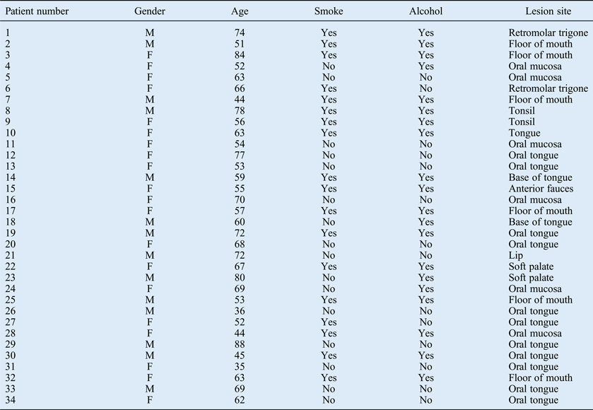
Table I shows the demographic characteristics of the 34 patients enrolled in the study. The average age across the entire cohort was 61.5 years with a predominance of women (20 women, 14 men). All patients were white. Most of the patients had a history of smoking or alcohol consumption or both.
Patient demographics and lesion characteristics
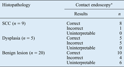
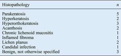
Tables II and III show the histopathological results obtained from the biopsy. Nine patients had biopsy-proven SCCs while of the remaining 25 lesions, 80 per cent were benign (20/25). Of these 20 benign cases, there were various types of pathology including keratotic, inflammatory and infectious lesions. The remaining five cases were biopsy-proven dysplastic lesions of varying degrees.
Contact endoscopy results by lesion type
*For malignant lesions: sensitivity = 89 per cent, specificity = 100 per cent. SCC = squamous cell carcinoma
Histopathology of benign lesions
Contact endoscopy correctly identified the invasive SCC lesions in 8 of 9 cases, demonstrating a sensitivity of 89 per cent for malignant lesions. There were no false-positive cases for diagnosis of malignancy (specificity 100 per cent). Contact endoscopy demonstrated features suggestive of dysplasia in 10 cases but dysplasia was confirmed on histopathology in only 5 of these cases. In the remaining five false-positive cases for dysplasia, the final histopathology revealed benign pathology in four and invasive SCC in the remaining one case.
In the remaining 20 cases of benign pathology the correct diagnosis was made only 50 per cent of the time. In four cases, contact endoscopy imaging was interpreted as representing dysplasia and the final six cases had images that were uninterpretable.
Discussion
The results of our prospective study reflect an experience that differs to some of the reports within the wider literature at present and demonstrates our challenges with overcoming some well-recognised difficulties with both image collection and interpretation.Reference Pelucchi, Bianchini, Travagli and Pastore14, Reference Warnecke, Averbeck, Leinung, Soudah, Wenzel and Kreipe15 We have shown excellent sensitivity and specificity for the assessment of invasive malignant lesions, and this is consistent with the existing literature (sensitivity 89 per cent, specificity 100 per cent). However, our results with dysplastic lesions and benign lesions are less accurate. Furthermore, our data demonstrate a high rate of uninterpretable images captured across the entire cohort (6/34 patients).
It has now been nearly two decades since the initial descriptions by Andrea et al. first introduced contact endoscopy to the field of otolaryngology and head and neck surgery.Reference Andrea, Dias and Santos6 Subsequently, multiple studies examining the use of contact endoscopy in the oral cavity,Reference Upile, Jerjes, Kafas, Angouridakis and Hopper3, Reference Pelucchi, Bianchini, Travagli and Pastore14, Reference Saeki, Tsuzuki, Negoro, Nin, Sagawa and Uwa16 pharynx and larynx,Reference Andrea, Dias and Santos6, Reference Warnecke, Averbeck, Leinung, Soudah, Wenzel and Kreipe15, Reference Andrea, Dias and Santos17–Reference Sone, Sato, Hayashi, Fujimoto and Nakashima22 nasal cavity,Reference Andrea, Dias, Macor, Santos and Varandas23 in cases of nasopharyngeal carcinoma (NPC),Reference Pak, Vlantis, Chow and van Hasselt24–Reference Wai Pak, To, Lee, Liang and van Hasselt27 and even parathyroid tissue identification,Reference Dedivitis and Guimarães28, Reference Guimarães, Brandão and Dedivitis29 have been described. However, there have been few well-designed clinical studies to subsequently support the use of contact endoscopy over the current gold standard of biopsy for histopathological diagnosis.
Warnecke et al. published a prospective clinical study of 42 patients, comparing contact endoscopy with histopathology; they reported sensitivity, specificity and overall accuracy rates of 90, 93.75 and 88 per cent respectively.Reference Warnecke, Averbeck, Leinung, Soudah, Wenzel and Kreipe15 The largest published study is that by Cikojević et al., who demonstrated similar sensitivity and specificity rates in 142 patients with laryngeal pathology.Reference Cikojević, Gluncić and Pesutić-Pisac4 They concluded that contact endoscopy was preferable to frozen section and that contact endoscopy improved diagnostic accuracy. Pak et al. evaluated the use of contact endoscopy in 64 patients with NPC treated with radiotherapy and reported sensitivity and specificity of 100 per cent for the detection of persistent and recurrent disease, and overall accuracy of 92.1 per cent when compared with histopathology.Reference Pak, To, Leung and van Hasselt26 They concluded that contact endoscopy is an accurate and reliable office-based procedure allowing for rapid in vivo diagnosis of persistent and recurrent NPC in post-irradiated patients. An earlier study by Pak et al. also demonstrated the potential role of contact endoscopy as a minimally invasive diagnostic tool for patients with NPC.Reference Pak, Vlantis, Chow and van Hasselt24
There are many potential advantages of contact endoscopy. It is a minimally invasive means of in vivo assessment of cytological structures, with the potential to avoid the need for an invasive biopsy and its associated risks. It allows the examination of larger areas of mucosa than would be feasible with biopsy or excision and is potentially helpful in improving the accuracy of targeted biopsies, should this be required.Reference Pak, To, Leung and van Hasselt26 It also avoids the possibility of damage or alteration of cellular structure in the process of tissue sampling and preparation.Reference Cikojević, Gluncić and Pesutić-Pisac4 Contact endoscopy has a potential clinical role in the minimally invasive diagnosis of benign and malignant mucosal lesions in the clinic or operating theatre setting, real-time intra-operative margin assessment, guided biopsies and surveillance. It also allows for the assessment of mucosal microvasculature that can aid in the diagnosis of malignant mucosal lesions.
The major obstacle to the wider clinical use of contact endoscopy is its inability to assess more than the superficial epithelial cell layers. Mucosal staining is required to provide contrast between the cell nuclei and the cytoplasm, and the accepted method of staining with methylene blue only penetrates superficially into the mucosa. Combined with the restrictive capacity of the endoscope to assess to depths of 80 µm and 30 µm at magnifications of ×60 and ×150 respectively, the ability to evaluate deeper tissue planes is limited. Vertical assessment of malignant or dysplastic cells is crucial in the differentiation between different grades of dysplasia, carcinoma in situ and invasive carcinoma, and although reports exist that describe techniques used to help make these distinctions, we have found this difficult to replicate. Contact endoscopy does not appear to be suitable for the assessment of submucosal pathology.
Further hindering the wider application of contact endoscopy in head and neck oncology are real and perceived technical limitations. The practicality of performing contact endoscopy on oral lesions without general anaesthetic remains difficult. Other authors have also reported technical difficulties with some lesions being inaccessible with the rigid contact endoscope and problems with consistent image quality due to artefact, staining techniques and scope positioning.Reference Pelucchi, Bianchini, Travagli and Pastore14, Reference Warnecke, Averbeck, Leinung, Soudah, Wenzel and Kreipe15 Our experience is certainly one of difficulty in obtaining consistent images and this is reflected in our high rate of uninterpretable images (17.6 per cent, 6/34 images).
Salivary flow can impede successful image capture, particularly when the lesion is adjacent to major salivary duct orifices. The scopes are generally heavy when combined with the video attachments required to record the contact endoscopy examination, and the design is such that adjusting the focus is difficult with a one-person technique. This generally means that the scope needs to be removed from the lesion for these adjustments to be made, making methodical mapping of the lesions difficult. This also lengthens the duration of the procedure and increases the discomfort for the patient. Being able to accurately image the whole lesion is crucial to the safe and effective implementation of this technique, particularly if the proposition is that it be used as a replacement to gold standard histopathological examination. Due to patient movement (particularly when imaging the oral tongue), abundant salivary flow across the field and the difficulties with instrument control, we believe the technology requires further development to facilitate practical clinical use.
• Contact endoscopy provides high sensitivity and specificity when evaluating malignant lesions
• The accurate characterisation of benign lesions presents a significant challenge and reduces the overall accuracy rate of contact endoscopy compared to tissue diagnosis
• Our results demonstrate a relatively high rate of capturing uninterpretable images secondary to limitations in instrumentation. There is a significant learning curve for the endoscopist
Future directions to help address issues with the subjective interpretation of contact endoscopy images include the development of computer-assisted programs to aid contact endoscopy evaluation.Reference Tarnawski, Fraczek, Jeleń, Krecicki and Zalesska-Krecicka30 This would aim to improve the consistency and objectivity of image interpretation and would be useful in ensuring quality control for future research endeavours. In addition, the significance of intra-rater assessment needs further investigation to determine the correlation between contact endoscopy experience and long-term patient outcomes. Further patients currently being recruited to assess this relationship are under review and will be reported in a follow-up study.
Ultimately, the wider application of contact endoscopy may require an improvement in the usability of the equipment to make it more user-friendly for the investigator and, importantly, more comfortable for the patient.
Conclusion
In summary, the in vivo assessment of head and neck oral mucosal pathology may be useful in the (1) early detection of pre-malignant and malignant lesions, (2) serial follow-up examinations of suspicious lesions, such as leukoplakia and lichen planus and (3) the assessment of resection margins.
Our study has demonstrated that although contact endoscopy is a simple, repeatable, minimally invasive examination performed with standard equipment, there is a learning curve associated with its use. In general, the effective capturing of images has required a two-person technique to help with the appropriate placement and adjustment of the endoscope and assist the patient in their positioning to improve comfort. Despite its limitations, contact endoscopy represents a promising optical technology that may afford reliable, accurate and minimally invasive in vivo assessment of malignant mucosal pathology that needs close collaboration between otolaryngologists and pathologists. Its diagnostic accuracy is lessened in the assessment of non-malignant lesions.
Acknowledgements
We would like to thank Dr Mercedes Ceron, Research Assistant, for her help. This study was supported by a Regional Cancer Program Small Grant (#LHR F8119).