Introduction
Ascites, or pulmonary hypertension syndrome, is a metabolic disorder in birds resulting in fluid accumulation in the abdominal region due to different factors influencing the function of the heart, lungs and liver (Bamidel et al., Reference BAMIDEL, VAN AS and ELFERINK2012). The chicken is an important animal in breeding systems and is used as animal model in biomedical research fields. The size of the chicken genome is less than half of a human or mouse’ genome, and includes 38 autosomes and two sex chromosomes; Z and W. Chicken chromosomes, like other bird species, show widespread variation in chromosome sizes between 50 to 200 Mb length (Axelsson et al., Reference AXELSSON, WEBSTER, SMITH, BURT and ELLEGREN2005).
Chickens are often used as an animal model to study diseases such as pulmonary arterial hypertension (PAH) in parallel with human research, and for achieving progress in the poultry industry for meat production because of their relatively short generation interval, breeding feasibility and distinct phenotypes. Furthermore, ascites disease is common in high growth rate, meat type chickens, because, with increasing growth rate, metabolic rate increases and the need for O2 increases. As selection in poultry has been intensive for growth rate traits, the incidence of ascites has increased in broiler chickens over recent decades, with about 8% of annual mortality being due to this metabolic disorder, making it a major economic disease in the poultry industry now a days. Major efforts have been made to link genotype and phenotype for several different traits associated with ascites to find the effective genes. Therefore, accurate and successful selection to prevent ascetic susceptibility is possible based on the indicator traits and bio-markers associated with this disease, so the risk of ascites in poultry can be reduced (Rowland, Reference ROWLAND2013). There are diverse indicator traits correlated with ascites, such as body weight, carcass quality, respiratory gases, fluid accumulation in the ventricular cavity, heart rate ratio and right ventricle ratio to total heart weight (Closter et al., Reference CLOSTER, VAN AS, GROENEN, VEREIJKEN, VAN ARENDONK and BOVENHUIS2009; Tarrant et al., Reference TARRANT, DEY, KINNEY, ANTHONY and RHOADS2017; Pakdel et al., Reference PAKDEL, VAN ARENDONK, VEREIJKEN and BOVENHUIS2005; Liu, Reference LIU2009; Closter et al., Reference CLOSTER, ELFERINK, VAN AS, CROOIJMANS, GROENEN and BOVENHUIS2010). The main reason for ascites is the imbalance between oxygen required and the potential of the cardiovascular system to supply oxygen. Studies have shown that the incidence of ascites has increased in recent years due to intensive selection for growth related traits achieving greater meat production in the poultry industry (Wood et al., Reference WOOD, MILLER and DILLON2015).
In silico modelling of ascites: source material
A systematic study was conducted on the basis of the PRISMA protocol (Wood et al., Reference WOOD, MILLER and DILLON2015). Literature search was performed using Google Scholar, NCBI, PubMed and animal QTLdb to find articles related to ascites. In this search, certain keywords were used such as: ascites, pulmonary hypertension syndrome, broilers, single-nucleotide, QTL mapping, GWAS, and candidate genes to find published papers and in order to make the study repeatable. The search was limited to poultry, and the most of the articles were in English. After collecting the articles in order to create a dataset, editorial and reviewed articles were excluded from the final list avoiding double counting. Then a comprehensive network for gene interaction pathways of identified genes were drawn by the online software geneMANIA (https://genemania.org).
The systematic review of articles
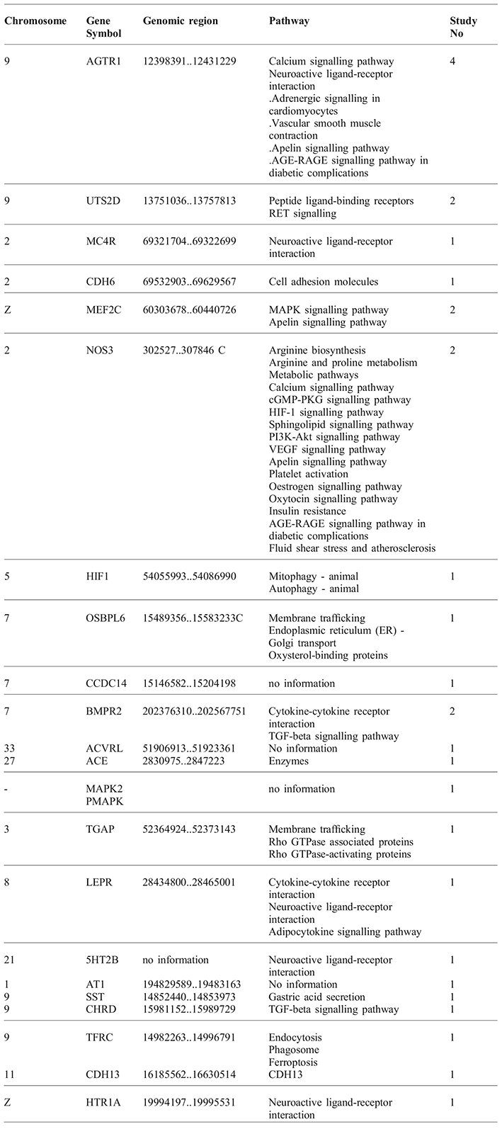
In total, 36 articles were collected, and after deleting repetitive and reviewed articles, 25 articles were included for further assessment. The included articles reported 21 different genes on different chromosomes which are mostly associated with ascites. Various studies have shown different and heterogeneous results. Such that, in one QTL study, five genomic regions on five chromosomes (chromosomes 2, 4, 8, 10 and 28) were associated with ascites development in sensitive chickens (Closter, Reference CLOSTER2004). In another study, using segregation pattern analysis and comparing ratios of 1:1 to 3:1, it was found that two genes with an epistasis effect were effective in the incidence of ascites (Druyan and Cahaner, Reference DRUYAN and CAHANER2007). The result of genome-wide association study (GWAS) in 2014 reported that several genomic regions were associated with ascites syndrome, but no area with a major effect was found. These studies showed that ascites appears to be affected by multiple genes with small effects which act as a polygenic trait (Closter, Reference CLOSTER2014). Also the study examining the amount of oxygen saturation (SaO) as an indicator of ascites in broilers, indicated that major genes play a meaningful role in the genetic control of SaO (Navarro et al., Reference NAVARRO, VISSCHER, CHATZIPLIS, KOERHUIS and HALEY2006). Moreover a QTL mapping study using microsatellites based on 24 autosomal chromosomes found three highly significant QTLs on chromosome 2 and other QTLs were considered important, found on chromosome 5, 8, 10, 27, and 28 (Rabie et al., Reference RABIE, CROOIJMANS, BOVENHUIS, VEREIJKEN, VEENENDAAL, VAN DER POEL, VAN ARENDONK, PAKDEL and GROENEN2005). In this regard, it was suggested that ascites was affected by high performance or major genes, so it was considered possible to find specific genes and implementation of certain selection criteria based on significant genes in breeding program for decreasing the sensitivity of poultry to ascites (Navarro et al., Reference NAVARRO, VISSCHER, CHATZIPLIS, KOERHUIS and HALEY2006; Wideman et al., Reference WIDEMAN, RHOADS, ERF and ANTHONY2013; Krishnamoorthy et al., Reference KRISHNAMOORTHY, SMITH, AL-RUBAYE, ERF, WIDEMAN, ANTHONY and RHOADS2014). The whole extracted results showed in Table 1 are discussed as follows.
List of genes affecting ascites incidence and their involved biological pathways.

The Angiotensin gene
Angiotensin II is a strong vasopressor hormone and a primary regulator of aldosterone secretion. It is an important effector regulating blood pressure and volume in the cardiovascular system. It acts through at least two kinds of receptors. This gene encodes the type 1 hormone receptor which is thought to be able to differentiate the major effects of angiotensin II in the cardiovascular system. This gene may play an important role in the development of ischemic myocardial infarction in humans. It has previously been thought that there was a related gene called AGTR1B; however, it is now believed that there is only one receptor gene in humans. The biological pathways of this gene are the calcium signalling pathway, the neuroactive ligand-receptor interaction, adrenergic signalling in cardiomyocytes, vascular smooth muscle contraction, apelin signalling pathway and the advanced glycation end products and receptors (AGE-RAGE) signalling pathway in diabetic complications (NCBI, 2018, KEGG, 1995-2018). In the sequence analysis of several single nucleotide polymorphisms in the AGTR1 gene in resistant and susceptible groups of Asian chickens, a SNP observed in exon3 converted proline 82's codon into the serine codon, and, since this amino acid is outside of the hydrophobic domain of the encoded protein, it cannot affect the hydrophobicity of the protein, but it will change the second structure of the protein (Burks, Reference BURKS2011). Another study identified the AGTR1 gene that is responsible for the production of angiotensin-converting enzyme from the liver. This enzyme causes an increase in blood pressure, and expression of this gene in susceptible and resistant poultry were significantly different (Ezzulddin and Ali, Reference EZZULDDIN and ALI2012). In a different study, using VNTR markers in an F2 generation of broilers, ventricular fluid, cardiac index, and heart hypertrophy were used as indicator traits. The results showed a significantly related region on chromosome 9, and it is reported that the AGTR1 gene is located in this same region (Rhoads et al., Reference RHOADS, KRISHNAMORTHY, AL-RUBYE and ANTHONY2013; Closter, Reference CLOSTER2014).
The UTS2D gene
UTS2D is a protein coding gene and its associated biologic pathways are ligand peptide binding receptors and RET (rearranged during transfection gene) signalling. The gene ontology (GO) annotation of this gene includes the activity of the hormone and the G-protein binding receptor (KEGG, 1995-2018, NCBI, 2018). In a study, using microsatellites (VNTR) in an F2 generation of chickens with indicators such as ventricular fluid, cardiac index and hypertrophy, a significant genomic region was found on chromosome 9, and the UTS2D gene was located in this area as a candidate gene. In association studies, a genomic region was determined with a significant association with ascites, the recognised region was placed on chromosome 9 containing two UTS2D and AGTR1 genes. The P value for the UTS2D was significant at P=0.003 (Rhoads et al., Reference RHOADS, KRISHNAMORTHY, AL-RUBYE and ANTHONY2013; Krishnamoorthy et al., Reference KRISHNAMOORTHY, SMITH, AL-RUBAYE, ERF, WIDEMAN, ANTHONY and RHOADS2014).
The MC4R gene
The encoded protein by this gene is a membrane bound receptor which belongs to the family of the melanocortin receptors. This protein interacts with the adrenocorticotropic and melanocyte stimulating hormones (MSH) and acts as a mediator of G proteins. MC4R is one of the Intron less genes and has one known biological pathway, which is the neuroactive-ligand-ligand interaction (KEGG, 1995-2018; NCBI, 2018). Consequently, studies reported that the flawed MC4R gene triggers autosomal dominant obesity. In addition a GWAS study was performed to detect genomic regions associated with ascites, which found a 70 Mb region on chromosome 2 which had significant association with the heart ratio (RV/TV) index (P=0.02) and this region contained the MC4R gene (Tarrant et al., Reference TARRANT, DEY, KINNEY, ANTHONY and RHOADS2017).
The CDH6 gene
This gene encodes for a member of the super family of cadherin proteins. Cadherin is a membrane glycoprotein that interferes with the adhesion of haemophilic cell-cell which plays an important role in cell differentiation and morphogenesis. The encoded protein is a type II cadherin and may play a critical role in the endometrium and placenta formation and kidney development. Lower expression of this gene may be accompanied by tumour growth and metastasis (KEGG, 1995-2018; NCBI, 2018). Additionally, in a GWA study, an area of 70 Mb in chromosome 2 was characterised based on the ratio of RV/TV (P=0.03) to identify the genomic regions associated with ascites, which contained the CDH6 gene (Tarrant et al., Reference TARRANT, DEY, KINNEY, ANTHONY and RHOADS2017).
The MEF2C gene
This gene encodes for the MADS transcription enhancer factor 2(MEF2), which plays an important role in myogenesis. The encoded protein, the MEF2 polypeptide C, has both trans-activating and DNA binding properties. This protein can play an important role in maintaining the status of muscle cells. Mutations and deletions in this place have been associated with severe cognitive impairment, brain malformations, stereotypic movements and epilepsy (KEGG, 1995-2018; NCBI, 2018). In research to identify the genomic region associated with ascites by the GWAS method, a 60 Mb region was found on the Z-chromosome related to RV/TV and heart attribute value (P=0.02) which contains the NEF2C gene (Tarrant et al., Reference TARRANT, DEY, KINNEY, ANTHONY and RHOADS2017; Wideman et al., Reference WIDEMAN, RHOADS, ERF and ANTHONY2013). In another investigation, using the marker assisted selection (MAS) method, studied homozygous progeny compared with heterozygous ones, which displayed much more resistance to the ascites (Catron et al., Reference CATRON, MENDIOLA, SMITH, BORN and WALKER2001).
The NOS3 gene
Nitric oxide is a reactive free radical involved in several processes, including neurotransmitters, antimicrobial and anti-tumour activity. Nitric oxide is generated from L-arginine by the nitric oxide synthesis enzyme. Hence, variation in this gene shows association with susceptibility to coronary spasms. Different splicing processes and using alternative promoters has led to multiple isoforms of this protein. The NOS3 gene involves many biological pathways, such as arginine biosynthesis, arginine and proline metabolism, metabolic pathways, calcium signalling, cGMP-PKG signalling, HIF-1 signalling, sphingolipid signalling, PI3KAkt signalling, VEGF signalling, apelin signalling pathways, platelet activation, oestrogen signalling pathway, oxytocin signalling pathway, insulin resistance, AGE-RAGE signalling pathway in diabetic complications, fluid shear stress and atherosclerosis (KEGG, 1995-2018; NCBI, 2018). One study examined the association between the polymorphism of the NOS3 and HIF1a genes with the susceptibility of commercial broilers to ascites, whereby the cardiac index (right ventricular to total heart ratio) was analysed as an indicator trait. Some SNPs were found in NOS3 and HIF1a genes with significant association with ascetic sensitivity (P<0.001, LOD> 6) (Moncaleano-Vega et al., Reference MONCALEANO-VEGA, ARIZA and HERNÁNDEZ2013; Kreider et al., Reference KREIDER, ANTHONY, WHITING, ERF and HAMAL2010) These two genes can regulate blood pressure and maintain oxygen homeostasis at a cellular level, similar to corresponding genes in human (AmiGO2, 2018; Moncaleano-Vega et al., Reference MONCALEANO-VEGA, ARIZA and HERNÁNDEZ2013). The NOS3 gene is located on chromosome 2 in birds. There is a special sequence (ACGTG) in the first intron of this gene and, if the first allele changes the risk of ascites incidence increases by about 41%. If both first and second alleles change simultaneously, the susceptibility percentage of disease increases, which is probably due to the fact that it acts as a regulatory Cis mediator with a transcription factor such as HIF1a (Moncaleano-Vega et al., Reference MONCALEANO-VEGA, ARIZA and HERNÁNDEZ2013).
The HIF1 gene
This gene encodes an alpha subunit of the hypoxia inducible transcription factor (HIF-1), consisting of a heterodimer with an alpha and a beta unit. Actually, HIF-1 acts as the main regulator of systemic and cellular histopathicity in hypoxia by activating the transcription of many genes, including those involved in energy metabolism, angiogenesis, apoptosis, and other genes which increase protein products or facilitate metabolic matching with hypoxia conditions. Therefore, HIF-1 plays an important role in the development of embryonic coronary arteries, angiogenesis tumours, and ischemic pathophysiology. For this gene, different transcriptomes have been encoded for different isoforms, and their biological pathways in animals are mythophagic and autophagic (KEGG, 1995-2018; NCBI, 2018). A lack of oxygen (hypoxia) controls the expression of the HIF1 gene which is located on the chromosome 5 in the poultry genome. In fact, the expression of this gene in terms of artificial hypoxia has been reported to increase by about 400% (Catron et al., Reference CATRON, MENDIOLA, SMITH, BORN and WALKER2001). One study analysed the association between polymorphisms of NOS3 and HIF1a genes with the susceptibility of commercial broiler chickens to ascites by considering the involvement of the cardiac index. This revealed that SNPs in NOS3 and HIF1a genes have significant association with the sensitivity of this disease (P<0.001, LOD> 6) (Moncaleano-Vega et al., Reference MONCALEANO-VEGA, ARIZA and HERNÁNDEZ2013).
The OSBPL6 gene
This gene is located on chromosome 7 and encodes a member of the oxyterol binding protein (OSBP) family, a group of intracellular lipid receptors. Most members of this family have an N-terminal pleckstrin homology domain and a highly conserved C-terminal OSBP-sterol binding domain. Furthermore, transcription variants and encoding of various isoforms have been identified in this gene. Their biological pathways include membrane trafficking, endothelial reticulum (ER) Golgi transport and oxyterol binding proteins (KEGG, 1995-2018; NCBI, 2018). A GWAS was conducted to find the association of this gene in F2 broilers resulting from the crossbreeding of an Iranian native breed and Aryan line with ascites syndrome, which lead to the discovery of a genomic region that carries OSBPL6 (P=0.0025). In this study, the attribute of the right ventricle to total heart ratio was used as an indicator trait (Catron et al., Reference CATRON, MENDIOLA, SMITH, BORN and WALKER2001).
The CCDC141 gene
The CCDC141 is an encoding protein gene. The GO annotation of this gene contains a nucleotide linkage. This gene plays a major role in radial migration and centrosomal function (KEGG, 1995-2018; NCBI, 2018). Through the study of the whole genome scan in an F2 generation of Iranian native birds and Aryan lines using the genomic best linear unbiased prediction (GBLUP) method, the association between SNPs and related traits to ascites was evaluated and the two candidate genes: CCDC141 and OSBPL6, which were previously reported as associated genes in heart failure in humans, were revealed as important, but this genomic region is located on the bird's chromosome 7 (Ahmadpanah et al., Reference AHMADPANAH, HOSSEIN-ZADEH, SHADPARVAR and PAKDEL2017).
The BMPR2 gene
This gene codes for one member of the bone marrow protein receptor (BMP) family of serine / threonine kinases. The ligands of this receptor are BMPs, which are considered to be the main family of TGF-beta. BMPs are involving in endocardia formation of bone and embryogenesis. These proteins transmit their signals through the formation of heteromeric complexes of two different types of serine (threonine) kinase receptors. Type I receptors are about 50-55 kDa and type II receptors are 70 to 80 kDa. Type II receptors bind ligand receptors in the absence of I type receptors, but require corresponding type I receptors for signalling, whereas I type receptors require the corresponding type II receptors for ligand binding. A mutation in this gene has been associated with primary pulmonary hypertension. Its biological pathways include two routes of interaction between cytokine-cytokine receptors and the TGF-Beta signalling pathway (KEGG, 1995-2018; NCBI, 2018). This gene in chicken cells is located on chromosome 7, and has been known as a major gene in relation to ascites in humans (Soubrier et al., Reference SOUBRIER, CHUNG, MACHADO, GRÜNIG, ALDRED, GERACI, LOYD, ELLIOTT, TREMBATH, NEWMAN and HUMBERT2013; Catron et al., Reference CATRON, MENDIOLA, SMITH, BORN and WALKER2001). Expression differential has been reported in susceptible and resistant poultry to ascites (Anthony, Reference ANTHONY2006).
The ACVRL gene
This gene encodes the Type I cell-surface receptor of the TGF-beta superfamily of ligands. It has another type I receptor with a high degree of similarity under the secondary serine-threonine kinase subdomains, with a glycine-serine (GS) region preceding the C-terminal short tail and kinase side. The encoded protein, called ALK1, shares similar domain structures with other proteins associated with ALK or activin-receptor like proteins, which form a subfamily of serine/threonine kinase receptors. From the information gathered on general databases, a mutation in this gene was associated with type2 haemorrhagic talangiectasia, which was reported as Rendu-Osler Weber syndrome 2 (KEGG, 1995-2018; NCBI, 2018). In humans, this gene is known as one of the major genes affecting ascites susceptibility, moreover, genetically sensitive individuals had about 75% of single-nucleotide polymorphisms in this genomic region (Soubrier et al., Reference SOUBRIER, CHUNG, MACHADO, GRÜNIG, ALDRED, GERACI, LOYD, ELLIOTT, TREMBATH, NEWMAN and HUMBERT2013). This gene is located in a different genomic region in chickens, i.e. in chromosome 33.
The ACE gene
This gene encodes the enzyme which involves in catalysing and converting angiotensin I into the psychoactive peptides of angiotensin II. Angiotensin II is a potent vasopressor and aldosterone stimulant that controls blood pressure and electrolyte fluid balance. In addition, this enzyme plays a key role in the renin-angiotensin system. There are many scientific reports that have linked the presence or absence of 287 bp Alu repeat element in this gene, with the level of circulating enzymes or cardiovascular pathophysiology. Multiple glycosylated transcripts have identified different isoforms, and the two most frequent spliced variants encode somatic and testicular forms that are equally active (KEGG, 1995-2018; NCBI, 2018). In a QTL examination, a significant association with ascites was found in a genome wide association study in broiler chickens. Considering the characteristic of the vascular response to oxygen deficiency, the location of the ACE gene has been reported on the chromosome 27 in poultry (Rhoads et al., Reference RHOADS, KRISHNAMORTHY, AL-RUBYE and ANTHONY2013).
Mitochondria protein kinase
Mitochondria protein kinase (MAPK) is linked to certain amino acids like threonine and tyrosine, particularly in serine/threonine as a specific protein kinase. MAPKs are involved in conducting cellular responses to different stimulations, such as anti-inflammatory cytokines, mitosis, osmotic stress and heat shock. They regulate many cellular activities, including gene expression, proliferation, mitosis, cell survival, differentiation, and apoptosis (KEGG, 1995-2018; NCBI, 2018). Their gene is located on chromosome 1 in poultry, in a genomic region of 18.3-21.48 Mb, which is promoted by means of the angiotensin-2 enzyme influencing heart muscle growth, which is associated with ascites (Ezzulddin and Ali, Reference EZZULDDIN and ALI2012).
The LEPR gene
This protein coding gene belongs to the gp130 family of cytokine receptors which is a transcription stimulator, activating STAT cytosol protein. Moreover, this protein is a receptor for leptin that regulates body weight and it is involved in regulation of fat metabolism as well as in the haematopoietic pathway, which is essential for normal lymphopoiesis. In addition, there is transcriptional splicing that produces different isoforms of this gene. The reported mutation in this gene is associated with obesity and pituitary dysfunction. It is noteworthy that LEPR and the LEPROT gene have the same promoter, and their first two exons will encode different proteins. It is involved in three biological pathways; cytokine-cytokine receptor interaction, neuroactive ligand receptor interaction and the adipocytokine signalling pathway (KEGG, 1995-2018; NCBI, 2018) A study conducted by Rosmond et al. (Reference ROSMOND, CHAGNON, HOLM, CHAGNON, PÉRUSSE, LINDELL, CARLSSON, BOUCHARD and RNTORP2000) showed that blood pressure regulation directly depends on the LEPR gene, and blood pressure can influence the development of ascites. If the mutation takes place in a receptor region, given the fact that body weight and ascites are associated, this gene cannot control the body weight. In one QTL region examination, the LEPR gene was considered as a candidate gene in ascites (Wang et al., 2006). In previous studies, an SNP was found in exon 9 of this gene, related to abdominal fat. However, a similar study was carried out through the sequencing of nucleotides, which found no genetic variants associated with susceptibility to ascites (Bamidel et al., Reference BAMIDEL, VAN AS and ELFERINK2012). Another informative study performed in broiler chicks reported that 67 SNPs were found to be significant in a whole genome association study that covered chromosomes 1 to 28, and that there is a correlation between two-week body weight and ascetic susceptibility. In that research, right ventricular to total heart ratio was taken as an indicator trait, which confirmed a significant negative relationship between SNP and body weight (Closter et al., Reference CLOSTER, ELFERINK, VAN AS, CROOIJMANS, GROENEN and BOVENHUIS2010).
The 5HT2B gene
This encodes one of several receptors for serotonin belonging to the G-protein coupled receptor family. Serotonin is a biogenic hormone that acts as a neurotransmitter, and acts both like a hormone and a mitogen. These receptors are involved in many of the central and peripheral physiological functions of serotonin, including cardiovascular regulation and heart rate. Population and family analysis based on minor alleles (substitution of glutamine codon with stop codon, designated Q20*) ceased the expression of 5HT2B, and a gene mutation study in mice suggested an important role for this gene. However, other factors, such as high level of testosterone, may be involved, and alternatively spliced transcript variants have been found for this gene. The only biological pathway of this gene is the binding pathway for the receptor neuroactive ligand (KEGG, 1995-2018; NCBI, 2018). Previous studies have proven that serotonin receptors play a key role in the development of ascites in poultry (Krishnamoorthy, Reference KRISHNAMOORTHY2012; Dey et al., Reference DEY, KRISHNA, ANTHONY and RHOADS2017). In a study in which poultry was used as an animal model for the assessment of similar conditions in humans, a GWAS showed that the genomic region on chromosome 9 is associated with the incidence of ascites, and this region carries the 5HT2B gene. In view of SNP detection, polymorphisms on the upstream region of the promoter of this gene have shown significant alteration in expression of the gene and an association with resistance to ascites (Alzahrani et al., Reference ALZAHRANI, RHOADS, WIDEMAN and ANTHONY2015).
The AT1 gene
ADP-ribosyltransferase catalyses the ADP-ribosylation and arginine residuals in proteins. Mono-ADP-ribosylation is a post-transcriptional alteration of the protein that happens due to the activity of a variety of different bacterial toxins, including coluria and E. coli thermophilic enterotoxins. The sequence of amino acids consists of dominant hydrophobic N- and C-terminal hydrophobic regions, which are characterised by enhanced glycosyl phosphatidylin stole (GPIs). The previous name of this gene was ART2 (KEGG, 1995-2018; NCBI, 2018). In a study based on microsatellite markers, the genomic region containing the AT1 gene was located on chromosome 1 and was related to an indicator trait of the right ventricle to the left ventricle of the heart and has a significant relationship with the incidence of ascites in the poultry (Tarrant et al., Reference TARRANT, DEY, KINNEY, ANTHONY and RHOADS2017).
The SST gene
The somatostatin (SST) hormone has an active form, which is produced by intermittent breakdown of the pre-protein encoded by this gene. Somatostatin is expressed throughout the body, and the release of secondary hormones is inhibited by binding to somatostatin receptors along with G-protein. This hormone is an important regulator of the endocrine system that interacts with the pituitary growth hormone, thyroid stimulating hormone and most of the gastrointestinal hormones. Somatostatin affects the amount of neurotransmitter in the central nervous system and the proliferation of normal cells and germline tumours. Its biologic pathway is via gastric acid secretion (KEGG, 1995-2018; NCBI, 2018). Genomic analysis has confirmed the region containing somatostatin 14 on chromosome 9 for the highest body fat and the body weight in chickens (P =0.07 and 0.06 respectively) (Krishnamoorthy et al., Reference KRISHNAMOORTHY, SMITH, AL-RUBAYE, ERF, WIDEMAN, ANTHONY and RHOADS2014; Wideman et al., Reference WIDEMAN, RHOADS, ERF and ANTHONY2013).
The CHRD gene
This gene encodes a secretory protein that perforates early vertebrate embryonic tissues by binding to TGF-beta-like bone morphogenetic proteins, sequestering them in latent complexes. The encoded protein may have roles in organogenesis and during adulthood. It has been suggested that this gene could be a candidate gene for Cornelia de Lange syndrome in humans. Research showed that reduced expression of this gene leads to enhanced bone regeneration, and there are multiple transcript variants due to alternative splicing in this gene (KEGG, 1995-2018; NCBI, 2018). This gene is located on chromosome 9, and its significance was in association with fluid accumulation in the ventricular area, which is one of the attributes of ascites (Krishnamoorthy et al., Reference KRISHNAMOORTHY, SMITH, AL-RUBAYE, ERF, WIDEMAN, ANTHONY and RHOADS2014; Wideman et al., Reference WIDEMAN, RHOADS, ERF and ANTHONY2013).
The TFRC gene
This gene encodes for a cell surface receptor which is necessary for iron uptake by the process of receptor-mediated endocytosis and is required for erythropoiesis and neurological development. Multiple, alternatively spliced variants have been identified in this gene, so that there are different transcriptions (Gene-cards, 1996-2018). The transferrin receptor gene is located on chromosome 9, and, in one study, it was related to ascetic-fluid accumulation in the abdominal cavity (Krishnamoorthy et al., Reference KRISHNAMOORTHY, SMITH, AL-RUBAYE, ERF, WIDEMAN, ANTHONY and RHOADS2014; Wideman et al., Reference WIDEMAN, RHOADS, ERF and ANTHONY2013).
The CDH13 gene
The CDH13 gene encodes a member of the cadherin superfamily. The encoded protein is localised on the surface of the cell membrane and is anchored by a GPI moiety, rather than by a transmembrane domain. The protein lacks the cytoplasmic domain characteristic of other cadherins, and is not thought to be a cell-cell adhesion glycoprotein. This protein acts as a negative regulator of axon growth during neural differentiation. It protects vascular endothelial cells from apoptosis due to oxidative stress, and is associated with resistance to atherosclerosis. The gene becomes hypermethylated in many types of cancer. Alternative splicing results in multiple transcript variants encoding different isoforms (KEGG, 1995-2018; NCBI, 2018). Due to induction of ascites and the developing of artificial disease in broiler chickens using a single nucleotide polymorphism analysis in multiple-generations, GWAS found that the CDH13 gene had a significant association with ascites disorder with the P-value for SNP in both resistant and susceptible groups being 0.212 and 0.069, respectively (Tarrant, Reference TARRANT2016).
The HTR1A gene
This gene encodes a G protein-coupled receptor for 5-hydroxytryptamine (serotonin), and belongs to the 5-hydroxytryptamine receptor subfamily. Serotonin has been implicated in a number of physiological processes and pathological conditions. Inactivation of this gene in mice causes behaviour consistent with increased irritation and stress. Moreover, mutation in the promoter region of this gene has been associated with menstrual cycle-dependent periodic fevers (KEGG, 1995-2018; NCBI, 2018). Additionally, a genome wide association study in broiler chickens was conducted to detect ascetic associations with SNPs, and, consequently, a genomic region was located on chromosome Z. There was a three-state polymorphism that had a total SNP effect of 0.235, 0.041, 0.493, respectively, which indicated a relationship with ascites, and this significant region carries the protein coding gene HTR1A (Tarrant, Reference TARRANT2016).
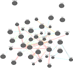
Additionally, comprehensive network for gene interactions of the identified genes were drawn by the online software geneMANIA and its schematic picture showed as follows (Figure 1).
Conclusions
By identifying the genes that are effective and relevant to the indicator traits of ascites in poultry, the genes were separated based on chromosomes to determine the most effective region governing ascites. Chromosome 9, with the presence of six related genes (chromosomes 1, 2 and 7 with three related genes and chromosome Z with two genes) have the most influence on the sensitivity of the bird to ascites syndrome. Likewise, because of the presence of two genes on the sex chromosome Z, the hypothesis of greater sensitivity in the male poultry might be acceptable. The results of previous studies based on the simulation of ten discrete generations of randomised cohorts have shown that the incidence of ascites in poultry increased from 5% to 30% when genetic parameters and heritability was estimated by using two maternal models compared with one without maternal effects. Ascites syndrome’ heritability was increased by REML and Bayesian methods, being 0.013 and 0.005 to 0.110 and 0.162 respectively (Ahmadpanah et al., Reference AHMADPANAH, HOSSEIN-ZADEH, SHADPARVAR and PAKDEL2017).
Interaction networks of related genes with ascites syndrome (https://genemania.org).

Additionally, by using VNTR markers and microsatellites to allow access to genotype information, it was found that a region on chromosome 9 (12 to 13 Mb) is associated with ascites syndrome in several commercial lines. There are two candidate genes in this area that have been determined in four studies; AGTR1 (a type 1 angiotensin II receptor) and UTS2D (region containing protein 2) associated with hypertension and hypoxic responses in mammals (Closter et al., Reference CLOSTER, VAN AS, GROENEN, VEREIJKEN, VAN ARENDONK and BOVENHUIS2009; Ahmadpanah et al., Reference AHMADPANAH, SHADPARVAR, GHAVI-HOSSIEN‐ZADEH and PAKDEL2016; Wideman et al., Reference WIDEMAN, RHOADS, ERF and ANTHONY2013; Ezzulddin and Ali, Reference EZZULDDIN and ALI2012; Krishnamoorthy et al., Reference KRISHNAMOORTHY, SMITH, AL-RUBAYE, ERF, WIDEMAN, ANTHONY and RHOADS2014). Using microsatellites, a study indicated the 5HT2B candidate gene, placed on chromosome 9, as another gene that affects ascites incidence (Krishnamoorthy, Reference KRISHNAMOORTHY2012; Dey et al., Reference DEY, KRISHNA, ANTHONY and RHOADS2017; Alzahrani et al., Reference ALZAHRANI, RHOADS, WIDEMAN and ANTHONY2015). Individuals with the highest phenotype for FCR had the highest sensitivity of ascites, with genes identified in the region that contains the somatostatin 14 gene on chromosome 9, whereby the traits of highest body fat and maximum body weight were 0.07 and 0.06 respectively. Furthermore, the CHRD and TFRC genes on chromosome 9 are associated with ascites indicator traits (Krishnamoorthy et al., Reference KRISHNAMOORTHY, SMITH, AL-RUBAYE, ERF, WIDEMAN, ANTHONY and RHOADS2014). Chromosome 9, with a size of 24.09 Mb, contains 615 genes, and six of these are associated with ascites syndrome. Hence, the most involved chromosome for this metabolic disease appears to be chromosome 9. The available gene interaction network has been predicted based on human (81.4) and mouse (18.63) information which represents 36.87% physical interaction, 24.29% Co-expression between related genes (Figure 1). Therefore, this synergy might confirm that tabulated genes work together to regulate negatively or positively the path of occurrence of ascites disorder.