Introduction
Fertilization in mammals is an unique process of fusion of haploid, highly specialized gametes (sperm and egg), resulting in the production of totipotent zygotes with a predetermined potential for the creation of genetically distinct individuals (Florman and Ducibella, Reference Florman and Ducibella2006). The transformation of gametes with a singular function to a totipotent zygote requires complete molecular rearrangement (Hemberger et al., Reference Hemberger, Dean and Reik2009), including massive degradation of parentally inherited transcripts, genomic reprogramming and initiation of embryonic transcript production. The process of embryonic genome activation (EGA) is accompanied by the gradual activation of RNA polymerase (RP) I, II, and III transcription, which in a well coordinated manner enables the start of embryo development. This event is also reflected at the cellular level by e.g. transformation of dense nucleolar precursor bodies (NPBs) into functional reticulated nucleoli. Active nucleoli consist of fibrillar centres (FCs), a dense fibrillar component (DFC) and a granular component (GC) (Lavrentyeva et al., Reference Lavrentyeva, Shishova, Kagarlitsky and Zatsepina2015).
Maternal and paternal factors passed on from the germ cells to the embryo play a crucial role in preimplantation development. Previous studies have reported the presence of maternal proteins and mRNAs in early embryos, as well as describing their role in several significant EGA events; e.g. histones mRNA (Labrecque et al., Reference Labrecque, Lodde, Dieci, Tessaro, Luciano and Sirard2015). Maternal mRNAs are regulated and stabilized by small non-coding RNAs, also originating from sperm and oocyte, in order to avoid premature degradation and enabling proper translation into proteins (Lykke-Andersen et al., Reference Lykke-Andersen, Gilchrist, Grabarek, Das, Miska and Zernicka-Goetz2008; Suh and Blelloch, Reference Suh and Blelloch2011; Liebers et al., Reference Liebers, Rassoulzadegan and Lyko2014). While maternal mRNAs are extensively degraded before the EGA, maternal miRNAs are still active during the post-implantation stages of development, multiple days after fertilization (Suh et al., Reference Suh, Baehner, Moltzahn, Melton, Shenoy, Chen and Blelloch2010; Suh and Blelloch, Reference Suh and Blelloch2011). However, the course of maintenance of maternal rRNA or its role in regulation of maternal-to-embryonic transition has not yet been explored.
Ribosomal RNA represents a specific nucleic acid category, in means of being not translated into proteins, having a specific polymerase (RPI) for synthesis and high content of GC pairs in the gene, making it extra sensitive to actinomycin D inhibition (Kovalska et al., Reference Kovalska, Hruska-Plochan, Østrup, Adamkov, Lehotsky, Strejcek, Statelova, Mikuskova, Varga and Petrovicova2012). The rRNA genes, i.e. the rDNA, transcription and initial processing are spatially organized within the FCs and DFC. Initiation of rDNA transcription requires the formation of the so-called transcription initiation complex, comprised of RPI, upstream binding factor (UBF), species-specific promoter selectivity factor (SL-1) and topoisomerase I. The unprocessed rRNA is predominantly localized at the periphery of FCs and the inner part of DFC together with proteins of early processing (fibrillarin) (Shishova et al., Reference Shishova, Lavrentyeva, Dobrucki and Zatsepina2015). The final rRNA processing, ribonucleoprotein (RNP) assembly and pre-ribosomal particles are localized to the GC (Svarcova et al., Reference Svarcova, Strejcek, Petrovicova, Avery, Pedersen, Lucas‐Hahn, Niemann, Laurincik and Maddox‐Hyttel2008). The ultrastructural and functional organization of transcriptionally active nucleoli is closely correlated with cell growth and its cyclicity. In somatic cells the inhibition of rRNA gene transcription occurs during mitosis and during specific periods of meiosis accompanied by separation of rRNA synthetic and processing mechanisms. The cell growth-dependent control of rRNA gene transcription is initially regulated by post-translational modifications of crucial transcription factors as e.g. UBF and its interaction with pocket proteins pRb and p130 (Hannan et al., Reference Hannan, Hannan, Smith, Jefferson, Lun and Rothblum2000a; Reference Hannan, Kennedy, Cavanaugh, Hannan, Hirschler-Laszkiewicz, Jefferson and Rothblum2000b). Similar mechanisms of repression of nucleolar function were observed towards the end of the oocyte growth phase in pig and cattle accompanied with the co-localization of UBF and p130 in the periphery of the nucleolus (Baran et al., Reference Baran, Pavlok, Bjerregaard, Wrenzycki, Hermann, Philimonenko, Lapathitis, Hozak, Niemann and Motlik2004; Bjerregaard and Maddox-Hyttel, Reference Bjerregaard and Maddox-Hyttel2004).
The reestablishment of functional nucleoli occurs at late telophase in cycling somatic cells (Angelier et al., Reference Angelier, Tramier, Louvet, Coppey-Moisan, Savino, De Mey and Hernandez-Verdun2005) and during EGA in early embryos (Fulka and Aoki, Reference Fulka and Aoki2016) distinguished by two major events, the activation of RPI and the formation and subsequent reticulation of NPBs. Preview studies have indicated that transformation of NPBs into fully functional tripartite nucleoli was conditioned by activation of rDNA transcription machinery (Dousset et al., Reference Dousset, Wang, Verheggen, Chen, Hern andez-Verdun and Huang2000). However, more detailed studies on different mammalian cells, Xenopus laevis, and porcine and bovine embryos using more differentiated transcription inhibition experiments (e.g. α-amanitin, DNA topoisomerase I inhibitor, actinomycin D) demonstrated that the co-localization of maternally inherited molecular structures, e.g. rRNA processing proteins, nascent or partly processed rRNAs, with NPBs can induce the formation of DFC-like and GC-like structures independently of RPI transcription (Verheggen et al., Reference Verheggen, Almouzni and Hernandez-Verdun2000; Viuff et al., Reference Viuff, Greve, Holm, Callesen, Hyttel and Thomsen2002; Svarcova et al., Reference Svarcova, Strejcek, Petrovicova, Avery, Pedersen, Lucas‐Hahn, Niemann, Laurincik and Maddox‐Hyttel2008).
The present study was designed to explore the role of RPI transcription, maternally inherited rRNAs and RPII mediated de novo transcription in the early embryogenesis. Based on the observations from several molecular levels, we have proved the presence of parentally inherited rRNA and defined its interactions with nucleolar proteins. Using the approach of selective actinomycin D inhibition of RPI, RPII and RPIII in early porcine embryos processed for reverse-transcription polymerase chain reaction, autoradiography, transmission electron microscopy (TEM), fluorescence in situ hybridization (FISH), silver staining and immunofluorescence (for RPI), we provided strong evidence for the autonomous role of parental rRNA, as well as the requirements for de novo transcription for nucleolar formation.
Materials and methods
Production of porcine embryos in vivo
In vivo porcine embryos were collected using a protocol described previously by Bjerregaard et al. (Reference Bjerregaard, Wrenzycki, Strejcek, Laurincik, Holm, Ochs, Rosenkranz, Callesen, Rath, Niemann and Maddox-Hyttel2004). Pre-pubertal gilts were injected intramuscularly with 1500 IU of eCG (Intergonan; Intervet, Tonisvorst, Germany) and 500 IU of hCG (Ekluton; Intervet) 72 h later. The gilts were inseminated 24 h and 36 h after hCG injection with 3 billion spermatozoa of a fertility-tested boar. Subsequently, the gilts were killed at the Institute´s abattoir (FLI Mariensee, Germany) at 70–74 h after hCG injection and the oviducts and uteri were flushed to obtain presumptive late 2-cell embryos. Collected embryos were then cultured in vitro in NCSU 23 medium (Petters and Wells, Reference Petters and Wells1993) at 39°C under an atmosphere of 5% O2, 5% CO2 and 90% N2 into the subsequent cell cycles. The embryos were examined every second hour to detect the onset of cleavage. Embryos that did not progress to the subsequent cell cycle were excluded from further experiment.
Subsequently, two groups of late 4-cell stage embryos obtained at 30 h post cleavage (hpc) were cultured for 3 h with different concentrations of actinomycin D (AD): (i) 0.2 μg/ml (AD-0.2); and (ii) 2.0 μg/ml (AD-2.0) (Verheggen et al., Reference Verheggen, Almouzni and Hernandez-Verdun2000). Additionally, one group of late 2-cell stage embryos was cultured for 36 h in the presence of 0.2 μg/ml AD (AD-LT) until the late 4-cell stage. Control embryos were cultured continuously in the absence of AD (Ctrl). Late 4-cell stage embryos (33–34 hpc) from all four groups were fixed and processed for RT-PCR, light microscopic autoradiography, TEM, FISH, silver staining and immunocytochemistry.
3H-Uridine incubation for autoradiography
Embryos from each experimental and control group were labelled with 3H-uridine (sp. act. 962 GBq/mmol; Amersham Pharmacia Biotech Europe GmbH, Freiburg, Germany) at a final concentration of 4 MBq/mmol (Laurincik et al., 2000a) for 20 min in gas-equilibrated culture medium. After incubation with the radioactive precursor, the embryos were repeatedly washed in 3H-uridine-free culture medium, fixed and processed for further analyses.
Processing for light microscopic autoradiography and transmission electron microscopy
After incubation, the embryos were fixed in 3% glutaraldehyde in 0.1 M Na-phosphate buffer (pH 7.2). Subsequently, the specimens were washed in buffer, post-fixed in 1% OsO4 in 0.1 M Na-phosphate buffer, embedded in Epon (Sigma–Aldrich, Steinheim, Germany) and serially sectioned into semi-thin sections (2 μm). Every second section was stained with basic toluidine blue and evaluated by bright field light microscopy. Selected semi-thin sections were re-embedded according to Hyttel and Madsen (Reference Hyttel and Madsen1987) and processed for ultra-thin sectioning (70 nm). The ultra-thin sections were contrasted with uranyl acetate and lead citrate and examined on a Philips CM100 transmission electron microscope. Selected non-stained semi-thin sections were processed for autoradiography for detection of de novo RNA synthesis. The sections were coated with Ilford K5 liquid nuclear emulsion (Ilford, Basildon, Essex, UK) and exposed for 6 weeks at 4°C. Finally, the specimens were developed in Kodak D 19 at 18°C, stained with toluidine blue and evaluated by bright field light microscopy.
Whole-mount immunolabeling of RNA polymerase I
Human anti-RNA polymerase I primary antibody (1:500; Reimer et al., Reference Reimer, Raska, Tan and Scheer1987), a gift from Dr Ochs, was used. This antibody has previously been demonstrated to distinctly label the fibrillar component of nucleoli in murine (Baran et al., Reference Baran, Fléchon and Pivko1996) bovine (Laurincik et al., 2000b), and porcine (Hyttel et al., Reference Hyttel, Laurincik, Rosenkranz, Rath, Niemann, Ochs and Schellander2000) embryos.
For indirect immunofluorescence, the previously described protocol by Svarcova et al (Reference Svarcova, Strejcek, Petrovicova, Avery, Pedersen, Lucas‐Hahn, Niemann, Laurincik and Maddox‐Hyttel2008) was used. Briefly, 10 or 11 late 4-cell stage embryos in each group were harvested and fixed in a mixture of 4% paraformaldehyde and 0.1% Triton X-100 for 40 min at room temperature (RT). Subsequently the embryos were washed in 1% Triton X-100 in PBS, and preincubated for 2 h with 5% normal goat serum (NGS, Dako, Glostrup, Denmark) in PBS at RT. After fixation, the specimens were incubated with the primary antibody diluted in PBS containing 5% NGS for 1 h at RT. Unbounded primary antibody was removed by washing in PBS before a 4 h (at 4°C) and a 1 h (at RT) incubation in goat anti-human IgG conjugated with Texas Red® (Molecular Probes, Eugene, OR, USA), diluted in PBS containing 5% NGS. Finally, the embryos were mounted on glass slides using Dako fluorescent mounting medium (Dako, Glostrup, Denmark) and examined on a Leica fluorescence microscope. Control immunostaining was performed by omitting the primary antibody.
Nuclear extraction and fluorescence in situ hybridization
Nuclei from embryos were extracted by short incubation in lysis buffer (0.01 N HCl, 0.1% Tween 20) and immediately fixed in 3:1 methanol:glacial acetic acid at 4°C for at least 24 h, air dried, incubated at 60°C overnight and then stored at –80°C until hybridization (Viuff et al., Reference Viuff, Hyttel, Avery, Vajta, Greve, Callesen and Thomsen1998).
Embryonic nuclei were hybridized in situ with a rDNA probe using the previously described protocol (Viuff et al., Reference Viuff, Greve, Holm, Callesen, Hyttel and Thomsen2002) with slight modifications. In this study, the porcine genomic cosmid clone F6 used was a rDNA probe. The F6 rDNA digoxigenated probe recognized the chromosomal region of the 18S/5.8S/28S rRNA gene clusters on pig chromosomes 8p12 and 10p12-p13 (Yerle et al., Reference Yerle, Lahbib-Mansais, Pinton, Robic, Goureau, Milan and Gellin1997). The rDNA probe was labelled using digoxigenin-11-dUTP by nick-translation and the denatured probe was then hybridized at 37°C overnight to the denatured chromosomal DNA at a final concentration of 20 ng/ml hybridization mixture containing 50% deionized formamide, 10% dextran sulfate, 2× saline sodium citrate (SSC), 0.1% salmon sperm DNA, 0.1% porcine genomic DNA and stored until use at −20°C.
Specimens were washed twice at RT in 0.15 M PBS, pH 7.4 for 2 min, fixed in 1% formaldehyde for 2 min and washed twice in PBS for 2 min. Chromosomal DNA was denatured by immersing slides in 70% formamide, 2× SSC (pH 7) for 2 min at 71–72°C, immediately dehydrated in an ice-cold ascending ethanol series and air dried. The rDNA probe was denatured at 75°C for 5 min, left to pre-anneal at 37°C for 30–60 min and centrifuged at 13,000 rpm. Then 3-µl aliquots of the hybridization mix were applied to the area where the nuclei were concentrated, and a coverslip was placed over the slide, sealed with vulcanized glue and hybridized overnight at 42°C. Slides were then washed three times in 0.05× SSC for 10 min and twice for 3 min at 37°C. After washing, slides were preincubated at 37°C for 10 min in 4× SSC, 0.1% Tween 20 containing 5% skimmed milk powder in order to reduce non-specific antibody binding.
The sites of rDNA probe hybridization were visualized using anti-Dig−fluorescein (FITC) (Boehringer Mannheim, Germany), after one round of amplification using FITC−avidin (Boehringer Mannheim, Germany). DNA was counter-stained with diamidinophenylindole (DAPI, 1 µg/ml) from Vectashield (Sigma, Germany). The slides were examined and recorded using an epifluorescence microscope equipped with a charged coupled device (CCD) camera (Leica DM 4000B, Wetzlar, Germany). Subsequently the slides were sent for silver staining of the nucleolar proteins.
Silver staining
Silver staining of argentophilic nucleolar proteins was performed according to the method of Lindner (Reference Lindner1993). After FISH evaluation, the slides were incubated in 1% dithiothreitol for 12 min at RT, followed by careful rinsing with distilled water. Slides were then covered with 100 µl of a freshly prepared 3:1 mixture of 50% (w/v) AgNO3 (Merck, Darmstadt, Germany):2% (w/v) gelatine (Sigma, Germany) and 1% formic acid (Merck, Darmstadt, Germany), a coverslip was placed on the slide, which was then incubated for 1 h at 37°C. After a rinse in distilled water, slides were mounted in DABCO anti-fade solution (pH 8). Nuclei for which the FISH-labelling pattern had been previously recorded were relocated and the silver-stained nucleolar proteins were subsequently photographed using bright field microscopy (Leica).
Reverse-transcription polymerase chain reaction
Before RNA isolation, 1 pg of rabbit globin RNA (Sigma, Germany) was added to the samples as an internal standard. Subsequently, total RNA was isolated from pools (triplicates) of five embryos at the late 4-cell stage from each group (control; AD-0.2; AD-2.0; AD-LT) using an Absolutely RNA NanoPrep Kit® (Agilent, California, USA) followed by reverse transcription using a SuperScript First-Strand Synthesis kit (Invitrogen, Carlsbad, USA) in a final volume of 20 µl using 2.5 µM random hexamers. Reverse transcription was carried out at 25°C for 10 min, 42°C for 1 h, denaturation at 99°C for 5 min and fast cooling to 4°C followed by cDNA storage at −20°C.
Specific primers for porcine 45S rRNA gene (5´-CGATCCTCTTCAGCGCCTGT-3´ and 5´-GCCGGCGCACAGGCCCAGGC-3´) were designed for detection of de novo synthesis of the short-life span 160-bp rRNA sequence. Primers with sequences 5´-GTACGGCTGTCATCACTTAGACCTCA-3´ and 5´-GCTAGTGAACACAGTTGTGT-3´ were used to amplify a 162-bp β-globin gene fragment.
PCR was performed using the Platinum Taq DNA polymerase kit (Invitrogen, Carlsbad, USA) containing 1.5 mM MgCl2, 200 μM of each dNTP, 1 μM of each primer (sequence specific and globin) and 1 IU Platinum Taq DNA polymerase. PCR conditions were as follows: initial denaturation of Taq DNA polymerase at 97°C for 2 min, followed by 35 cycles of 95°C for 15 s, annealing for 30 s and extension at 72°C for 30 s. Amplified PCR products were separated by electrophoresis on a 2% agarose gel in 1× TBE buffer containing 0.2 μg/ml ethidium bromide (EtBr) and visualized using a gel documentation system (Gel Doc 2000, Bio-Rad, Hercules, California, USA).
Results
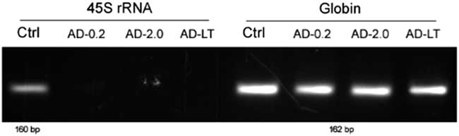
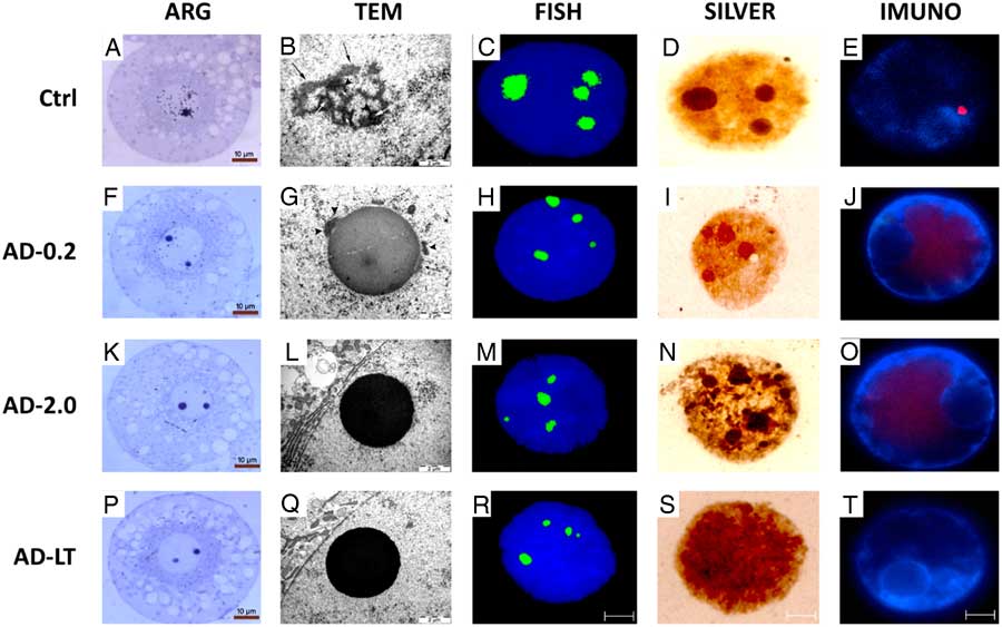
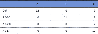
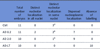
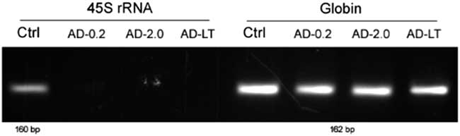
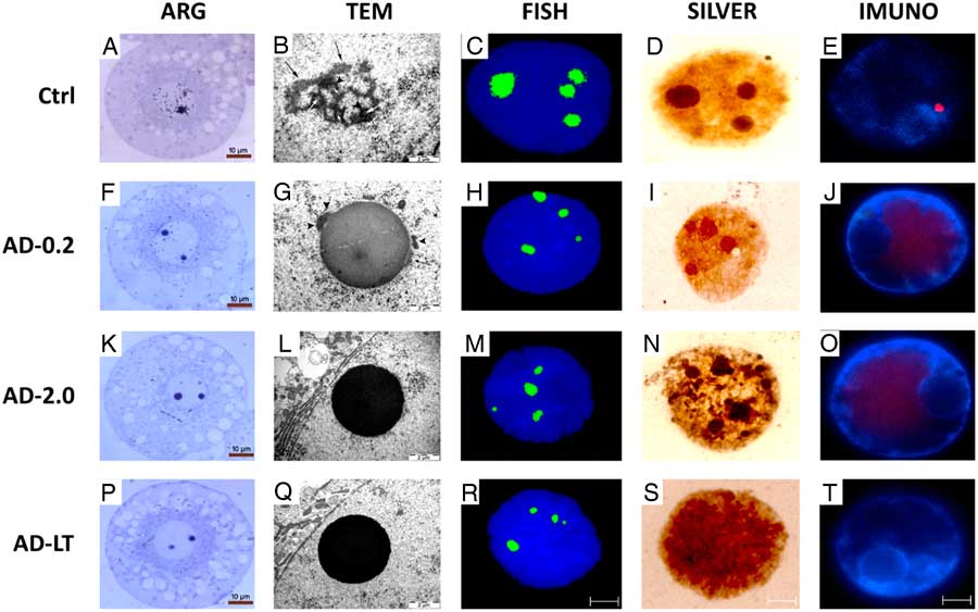
In the normally developing embryo, genome activation is accompanied by morphological transformation of NPBs into functional reticulated nucleolus. We observed normal nucleologenesis in in vivo fertilized embryos that were cultured under in vitro conditions from the 2-cell to 4-cell stages, i.e. during the genome activation period. These events were manifested by active de novo rRNA synthesis (Fig. 1, Ctrl), incorporation of uridine during transcription (Table 1, Fig. 2 A), distinct localization of RNA polymerase I in the rim of the developing nucleoli (Table 2, Fig. 2E ), and large, decondensed ribosomal RNA that co-localized with large foci of silver-stained nucleolar proteins (Table 3, Fig. 2C, D ). These events were reflected at the ultrastructural level, in which morphological transformation of nucleolar precursors into active compartments was observed in the vast majority of analyzed blastomeres (Fig. 2B ).
Representative electrophoretogram of PCR products obtained from RT-PCR using the specific primers designed for detection of de novo synthesis of the short-life span 160-bp rRNA sequence (45S rRNA). Each lane represents the PCR product derived from total RNA isolated from pools (triplicates) of five embryos at late 4-cell stage from each group (Ctrl=control; AD-0.2; AD-2.0; AD-LT). The internal standard (globin) is shown next to these lines (162 bp).
Autoradiography (ARG), transmission electron microscopy (TEM), fluorescence in situ hybridization (FISH), silver staining (SILVER) and immunofluorescence (IMUNO) of nuclei in 4-cell stage porcine embryos cultured with (AD-0.2, AD-2.0, AD-LT) or without (Ctrl) actinomycin D. In control embryos (Ctrl) active incorporation of uridine during transcription (A), fibrillogranular nucleolus (B), large decondensed foci of ribosomal RNA co-localized with large foci of silver-stained nucleolar proteins (C, D) and distinct localization of RNA polymerase I in the rim of developing nucleoli was observed (E). Embryos from the AD-0.2 group displayed extranucleolar ARG labelling (F), formation of dense fibrillar and granular component in the periphery of the nucleolus-related structure (G), clusters of silver-stained nucleolar proteins co-localized with rRNA foci (H, I) and dispersed RPI labelling in the nucleoplasm (J). Short-term inhibition of total transcription (AD-2.0) resulted in absence of ARG labelling (K) and presence of nucleolar precursor bodies (NPBs) (L). The large rRNA foci were observed in 39% of analyzed nuclei mainly co-localized with nucleolar proteins (M, N). Nuclei of AD-2.0 embryos displayed dispersed nucleoplasmic localization of RPI (O). Embryos from AD-LT group did not present any ARG (P) labelling and displayed NPBs (Q). Forty per cent of the analyzed nuclei displayed the accumulation of rRNAs molecules into large foci (R). Recruitment of nucleolar proteins was completely abolished (S). Immunofluorescent labelling of RPI was not detectable (T). In TEM micrographs the nucleolar components are indicated by black arrows (GC), arrowheads (DFC) and white arrows (FC). For the FISH, SILVER, IMUNO methods scale bars indicate 5 µm.
Transcriptional activity in nuclei in late 4-cell porcine embryos

Numbers of labelled nuclei in 4-cell porcine embryos cultured with (AD-0.2, AD-2.0, AD-LT) or without (Ctrl) actinomycin D.
A: Autoradiographic labelling over nucleoplasm and nucleoli.
B: Autoradiographic labelling over nucleoplasm, but not over nucleolus-related structures.
C: Lack of autoradiographic labelling.
Localization of RNA polymerase I in late 4-cell porcine embryos

Numbers of nuclei in 4-cell stage porcine embryos cultured with (AD-0.2, AD-2.0, AD-LT) or without (Ctrl) actinomycin D.
a Distinct nucleolar localization in at least three blastomeres of 4-cell embryo.
b Distinct nucleolar localization in a maximum two blastomeres of 4-cell embryo.
Proportion of nuclei in the late 4-cell stage porcine embryos from control group (Ctrl), group with inhibited RNA polymerase I transcription (AD-0.2), and groups with short-term (AD-2.0) and long-term (AD-LT) total transcriptional inhibition

a FITC clusters and deposits of silver staining with diameter d>1 µm.
b Determined from total number of nuclei with large deposits of silver staining.
Maternally inherited rRNA is sufficient for partial nucleolar assembly
Several studies to date have evaluated the role of maternally inherited RNAs in embryonic development (Lavrentyeva et al., Reference Lavrentyeva, Shishova, Kagarlitsky and Zatsepina2015; Shishova et al., Reference Shishova, Lavrentyeva, Dobrucki and Zatsepina2015;). Many doubts, however, have been raised about the stability of RNAs beyond the first cell cycle. To assess the role of maternally inherited ribosomal RNAs after the first division, activation of RNA polymerase I occurring at the late 4-cell stage was inhibited by a short-term incubation of embryos at the late 4-cell stage with low doses of actinomycin D (AD-0.2). Interestingly, despite the lack of de novo synthesized rRNA as shown by RT-PCR and ARG (Fig. 1, AD-0.2; Fig. 2F ), large foci of rRNA were observed in 57.1% of analyzed nuclei (Table 3, Fig. 2H ). Furthermore, in 90.9% of cases the clusters of silver-stained nucleolar proteins were co-localized with these foci, indicating active recruitment of nucleolar machinery (Fig. 2I ). On the ultrastructural level, FCs harbouring rDNA transcription were absent as expected from RPI inhibition (Table 2, Fig. 2J ). However, as seen during the formation of DFC and GC (Fig. 2G ), functional interactions between nucleolar proteins co-localized with rRNA were not abolished by the lack of active rRNA synthesis and led to processing of rRNA into ribosomal subunits. Therefore, nucleolar transformation and activation of ribosome assembly are partially independent of de novo rDNA transcription and can be initiated by, presumably, maternally inherited rRNA.
Recruitment of maternally inherited nucleolar factors is independent of de novo transcription but does not trigger activation of ribosome biogenesis
For the role of maternal rRNA in the recruitment of nucleolar proteins, we wanted to determine to what extent this process is independent of transcriptional activity. Our group showed previously that de novo transcription is a prerequisite for the supply of nucleolar proteins and therefore nucleolar activation in bovine embryos (Laurincik et al., Reference Laurincik, Schmoll, Mahabir, Schneider, Stojkovic, Zakhartchenko, Prelle, Hendrixen, Voss, Moeszlacher, Avery, Dieleman, Besenfelder, Muller, Ochs, Wolf, Schellander and Maddox-Hyttel2003). However, bovine and porcine embryos displayed two different models of nucleologenesis, and in vitro conditions used in the bovine study can, to some degree, influence genome activation. Therefore, short-term inhibition of total transcription by high doses of actinomycin D (AD-2.0) was used to assess the role of active transcription in in vivo fertilized porcine embryos. Surprisingly, despite the total block in transcription (Fig. 1, AD-2.0; Table 1, Fig. 2K ) and recruitment of RPI (Table 2, Fig. 2O ), the co-localization of rRNA with nucleolar proteins was only moderately affected. Large rRNA foci were observed in 39.4% of nuclei (Table 3, Fig. 2M ) and were seen in most silver-stained nuclei, co-localized with nucleolar proteins (85.7%) (Table 3, Fig. 2N ). Hence, the triggering role of maternal rRNA was independent of transcriptional activity. However, the initiation of ribosome assembly, and therefore morphological formation of DFC and GC, failed (Fig. 2L ), indicating the need for de novo synthesized factors in the activation of nucleolar functions.
Genome activation determines spatial and temporal interactions between maternally inherited rRNA and nucleolar proteins
Previous studies have raised the role of the minor genome activation in porcine embryos (Østrup et al., Reference Østrup, Hall, Petkov, Wolf, Hyldig and Hyttel2009), therefore active transcription took place before the short-term inhibition in our study. To eliminate biological bias due to minor genome activation, embryos were cultured from the late 2-cell stage until the late 4-cell stage in the presence of AD (0.2 µg/ml; AD-LT) to validate the presence of maternal, pre-processed rRNA. As the embryos divided from the 2- to 4-cell stage at the same rate as in the other experimental groups, cultivation conditions were assessed to be not toxic.
Embryos from the AD-LT group exhibited a total block of transcription activity, as shown by RT-PCR and ARG (Fig. 1, AD-LT; Table 1, Fig. 2P ), as well as a complete lack of immunofluorescence labelling of RPI (Table 2, Fig. 2T ). At the ultrastructural level, the nucleolus remained in the form of NPBs (Fig. 2Q ). Despite long-term transcriptional inhibition, almost half the number of the analyzed nuclei (40%) (Table 3) displayed the accumulation of rRNAs molecules into large foci (Fig. 2R ). Therefore, these molecules were of maternal origin and their recruitment represented a solitary process regulated by maternal factors. However, recruitment of nucleolar proteins was completely abolished (0%) (Table 3, Fig. 2S ), indicating the essential role of transcriptional activity during de novo nucleolar formation.
Discussion
Complete embryonic nucleolar reassembly requires an association of the RPI transcription machinery with rDNA and its activation, mediated by formation of the transcription initiation complex (RPI, PAF53, SL-1 and UBF), together with re-grouping and co-localization of rRNA processing proteins (fibrillarin, nucleolin and nucleophosmin). Previous studies on somatic cells and embryos of different animal species have shown that nucleologenesis is a very intricate and step-wise process, prearranged in maternal cells or oocytes (Verheggen et al., Reference Verheggen, Le Panse, Almouzni and Hernandez-Verdun1998; Dousset et al., Reference Dousset, Wang, Verheggen, Chen, Hern andez-Verdun and Huang2000; Viuff et al., Reference Viuff, Greve, Holm, Callesen, Hyttel and Thomsen2002; Bjerregaard et al., Reference Bjerregaard, Pedersen, Jakobsen, Rickords, Lai, Cheong, Samuel, Prather, Strejcek, Rasmussen, Laurincik, Niemann, Maddox-Hyttel and Thomsen2007).
To reveal the contribution of maternally inherited material to embryonic nucleolar formation, transcription inhibition experiments were performed using actinomycin D (AD). Low-dose AD (0.2 μg/ml) treatment of in vivo-derived porcine 4-cell stage embryos for a short time (3 h) resulted in selective inhibition of RPI transcription, and therefore nucleolar ARG labelling and de novo rRNA synthesis (RT-PCR) was not detected. At the ultrastructural level, NPBs did not exhibit FC formation. Instead, small segregated areas of dense fibrillar and granular structures were observed. These DFC-like and GC-like components appeared to correspond to those observed in somatic cells (HeLa) treated with 0.04 μg/ml of AD directly after telophase and allowed progression to the early G1 phase (Dousset et al., Reference Dousset, Wang, Verheggen, Chen, Hern andez-Verdun and Huang2000), as well as in Xenopus laevis embryos during the EGA (Verheggen et al., Reference Verheggen, Le Panse, Almouzni and Hernandez-Verdun1998) and in in vitro derived 8-cell stage bovine embryos cultured in 0.2 μg/ml of AD (Svarcova et al., Reference Svarcova, Strejcek, Petrovicova, Avery, Pedersen, Lucas‐Hahn, Niemann, Laurincik and Maddox‐Hyttel2008). Moreover, based on the co-localization of silver-stained nucleolar proteins with pre-processed maternal rRNA to the rDNA and dispersed RPI in nucleoplasm, it can be assumed that the activation of nucleologenesis and the rRNA processing machinery itself is independent of RPI transcription. These suggestions are in agreement with proteomic and immunofluorescence studies on NPB composition and corroborate the maternal origin of key nucleolar proteins (or at least their subsistent mRNA) involved in early (fibrillarin) and late (B23, C23) rRNA processing (Fair et al., Reference Fair, Hyttel, Lonergan and Boland2001; Lawson et al., Reference Lawson, Larentowicz, Laury-Kleintop and Gilmour2005; Ogushi et al., Reference Ogushi, Palmieri, Fulka, Saitou, Miyano and Fulka2008; Lavrentyeva et al., Reference Lavrentyeva, Shishova, Kagarlitsky and Zatsepina2015; Fulka and Aoki, Reference Fulka and Aoki2016) along with low amounts of unspecified long-lived RNA (Kopecny et al., Reference Kopecny, Landa and Pavlok1995) and 28S rRNA (mouse; Lavrentyeva et al., Reference Lavrentyeva, Shishova, Kagarlitsky and Zatsepina2015).
From these presented observations, new questions have arisen on the importance of processing components and factors of non-maternal, embryonic origin involved in nucleologenesis. Embryos incubated in the presence of AD with higher concentrations (2.0 µg/ml) for a short period (RPI, II and III block) displayed co-localization of argyrophilic proteins with rRNA clusters of maternal origin. Ultrastructurally, only NPBs consisting of densely packed fibrillar material were noticed. The lack of DFCs and GCs in NPBs indicated that maternal rRNA is incapable of activating the processing machinery and additional embryonic factors are required for coordination of this process. Surprisingly, despite the short-term total transcriptional inhibition, diffuse localization of RPI in the nucleoloplasm was observed. In agreement with previous studies, the RPI (or mRNA encoding it) is not inherited from the oocyte and therefore must be synthesized de novo in the embryos during EGA (Bjerregaard et al., Reference Bjerregaard, Wrenzycki, Strejcek, Laurincik, Holm, Ochs, Rosenkranz, Callesen, Rath, Niemann and Maddox-Hyttel2004; Svarcova et al., Reference Svarcova, Laurincik, Avery, Mlyncek, Niemann and Maddox‐Hyttel2007). Therefore, the preceding minor EGA, including the transcription of RPI messengers, had to be activated before the transcriptional block was observed.
In addition, in all evaluated embryos submitted to long-term total transcription inhibition, no immunocytochemical labelling of RPI was detected. Conversely, rRNA and rDNA targeted FISH in transcriptionally silenced embryos still demonstrated the appearance of maternal rRNA in the vicinity of NORs. Detected rRNA in embryos in which RPI, II and III were blocked cannot be of embryonic origin (Viuff et al., Reference Viuff, Greve, Holm, Callesen, Hyttel and Thomsen2002; Laurincik et al., Reference Laurincik, Schmoll, Mahabir, Schneider, Stojkovic, Zakhartchenko, Prelle, Hendrixen, Voss, Moeszlacher, Avery, Dieleman, Besenfelder, Muller, Ochs, Wolf, Schellander and Maddox-Hyttel2003) and therefore it might be assumed that they were synthesized during oocyte maturation and stored together with nucleolar proteins in nucleolus-like bodies (NLBs), morphologically similar to NPBs in zygotes. The material stored in NLBs is used in the processes of early embryogenesis including the rapid onset of rDNA transcription and processing of pre-rRNA. So far it has not been reported that rRNA of maternal origin is engaged in activation of nucleologenesis independently on rDNA transcription in porcine embryos. However, recruitment of silver-stained proteins to rRNA was not detected, suggesting the importance of different key factors regulating their RNA and DNA binding properties. These indications are in good agreement with previous studies describing the formation of so-called nucleolar accessory bodies (also called coiled bodies, CBs) accompanied with huge trafficking and accumulation of the proteins and RNAs from different nuclear compartments (Zatsepina et al., Reference Zatsepina, Baly, Chebrout and Debey2003). As many immunocytochemical studies on various early embryos have shown, most of these interacting proteins and RNAs, including phosphoproteins (p80-coilin, Nopp 140), snRNPS, snoRNPs or La proteins and snoRNAs must be transcribed de novo from the embryonic genome to complete the formation of a transcriptionally active, tripartite nucleolus (Baran et al., Reference Baran, Brochard, Renard and Fléchon2001; Kiss, Reference Kiss2001; Viuff et al., Reference Viuff, Greve, Holm, Callesen, Hyttel and Thomsen2002; Morimoto et al., Reference Morimoto, Ozaki, Okamura, Yoshida, Amorim, Tanaka, Kitamura and Haneji2007).
In conclusion: (i) maternally inherited rRNA co-localized with rDNA and nucleolar proteins can initiate partial nucleolar assembly, resulting in the formation of fibrilogranular structures (DFC and GC) in porcine embryos; (ii) initiation of nucleolar assembly does not require the activation of rDNA transcription; and (iii) for complete transformation of the NPB to a fully functional nucleolus, major EGA and de novo protein synthesis are required.
Financial support
This work was supported by the Slovak Research and Development Agency under contract no. APVV-14-0001, and also by the project ‘EXCELLENCE in Molecular Aspects of the Early Development of Vertebrates’, CZ.02.1.01/0.0/0.0/15_003/0000460 from Operational Programme Research, Development and Education and by the Danish Council for Independent Research/ Natural Sciences (FNU) 8021-00048B and was co-funded by projects VEGA 1/0022/15, VEGA 1/0327/16 and 1/0001/19.
Conflicts of interest
The authors declare that there are no conflicting interests.
Ethical standards
Farm animals were housed and handled according to Institutional Guidelines, and experimental procedures were approved by the Friedrich Loeffler Institute (FLI) board, and full accordance with EU Directive 2010/63/EU for animal experimentation.