Introduction
Wright Valley is a geographically diverse dry valley in southern Victoria Land, Antarctica. It contains the Onyx River (the largest and longest river in Antarctica), running inland (west) for 30 km from the Wright Lower Glacier and Lake Brownworth near the coast. The river is fed by streams draining from snowfields and alpine glaciers, mainly on the southern side of the valley. It terminates in the closed basin of Lake Vanda, which separates the eastern and western halves of the valley. Lake Vanda is perennially ice-covered, highly stratified, saline at depth and the site of a former small research station. The western half of the valley divides into the North and South Forks, both leading to the Labyrinth, an extensive system of bedrock channels below Wright Upper Glacier, which bounds the western end of Wright Valley.
Algae have been sporadically studied from Wright Valley. The first report appears to be that of Holm-Hansen (Reference Holm-Hansen1964), who studied a single sample from each of Lake Vanda, an unnamed shallow pond and dry sand. Seaburg et al. (Reference Seaburg, Parker, Prescott and Whitford1979) carried out a more systematic study, sampling soils, plankton and benthic mats associated with Lakes Brownworth, Vanda and Canopus, and the Onyx River, and reported 15 species of cyanobacteria, 15 species of Chlorophyta and 14 species of Chrysophyta (in which they included diatoms). Other studies have included those on endolithic algae from Linnaeus Terrace (e.g. Darling et al. Reference Darling, Friedmann and Broady1987), hypoliths (Cowan et al. Reference Cowan, Khan, Pointing and Cary2010), benthic cyanobacterial mats from Lake Vanda (Hawes & Schwarz Reference Hawes and Schwarz2001), ponds (Wood et al. Reference Wood, Mountfort, Selwood, Holland, Puddick and Cary2008b) and soils (Aislabie et al. Reference Aislabie, Chhour, Saul, Miyauchi, Ayton, Paetzold and Balks2006).
The taxonomic depth to which these organisms have been studied varies greatly. Knowledge of cyanobacteria that incorporates molecular data appears to be limited to automated ribosomal intergenic spacer analysis (ARISA) profiles (Wood et al. Reference Wood, Mountfort, Selwood, Holland, Puddick and Cary2008b) and clone libraries (Aislabie et al. Reference Aislabie, Chhour, Saul, Miyauchi, Ayton, Paetzold and Balks2006, Cowan et al. Reference Cowan, Khan, Pointing and Cary2010). Older studies from which identifications were based on morphology (Holm-Hansen Reference Holm-Hansen1964, Seaburg et al. Reference Seaburg, Parker, Prescott and Whitford1979) may be vulnerable to the problem of convergent evolution of simple morphotypes, the identification and classification of which requires the incorporation of molecular data (Komárek Reference Komárek2012). In addition, type material is often inaccessible or non-existent. This problem is not unique to cyanobacteria. The yellow-green alga Heterococcus endolithicus Darling, Friedmann & Broady from Linnaeus Terrace has received detailed investigation using cultures (Darling et al. Reference Darling, Friedmann and Broady1987), but from molecular evidence was recently discovered to be a green alga and was renamed Desmococcus antarctica (Darling & Friedmann) Rybalka, Wolf, Andersen & Friedl (Rybalka et al. Reference Rybalka, Wolf, Andersen and Friedl2013). There is a clear need to document the diversity and distribution of algae in the valley for biogeographic comparison with other valleys and to draw conclusions about dispersal, and to investigate the identity of operational taxonomic units (OTUs) reported from environmental sequencing studies.
This study aimed to determine the diversity of dominant algae (including cyanobacteria but excluding diatoms) throughout Wright Valley, to gain insight into dispersal of biota within and between valleys, and to examine range restrictions of taxa. Thirty-five sites from soil, pond and stream habitats in which growths were detectable by the naked eye were sampled throughout the valley. Samples were subjected to a combination of culturing and environmental cloning approaches. This revealed 12 strains of cyanobacteria and 25 strains of eukaryotes, representing four and eight species, respectively, or eight and 11 phylospecies taking both culture and clone information into account. The results demonstrate that dispersal is probably common within the valley and to and from sites further afield.
Methods
Sampling sites
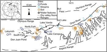
Samples were collected between 21 December 2010 and 19 January 2011 where algae were visible to the naked eye, or at least suspected by aggregated material (e.g. soil particles) (Fig. 1). Details of samples and locations are provided in Table S1 (found at http://dx.doi.org/10.1017/S0954102015000164). Samples of a few grams were collected into sterile Whirl-pak bags (Nasco, Fort Atkinson, WI, USA) and refrigerated in a chilly bin with ice until return to Scott Base, where samples were frozen until arrival at the laboratory in New Zealand in April 2011.
Map of Wright Valley, southern Victoria Land, Antarctica. Collection sites are shown and colour coded by the three site types (ponds, soils and streams). Inset map shows location within Antarctica. Site numbers correspond to those in Table I.

Some well-studied sites in the Wright Valley were not surveyed. These included Lake Vanda, which is well known to contain extensive benthic cyanobacterial mats (e.g. Hawes & Schwarz Reference Hawes and Schwarz2001). However, at accessible locations the marginal moat lacked evidence of benthic mats. Linnaeus Terrace (known for its endolithic communities) and the hypersaline Don Juan Pond (which is debatably lifeless) were not visited. Nor were ponds in the Lake Brownworth area, at the end of the valley nearest McMurdo Sound.
Culturing
Initial culturing was undertaken by suspending a small amount of sample in sterile deionized water and spreading on 1.5% agarized 10% BG-11 medium (Rippka et al. Reference Rippka, Deruelles, Waterbury, Herdman and Stanier1979). Cultures were grown at c. 5°C, under 16:8 hours (light:dark) of illumination of c. 50 µmol photons m-2s-1. Successive subculturing using standard aseptic techniques resulted in 25 unialgal strains of eukaryotes, but no cyanobacterial growth. Consequently, small amounts of samples known to contain cyanobacterial growths were suspended in 50 ml of liquid MLA medium (Bolch & Blackburn Reference Bolch and Blackburn1996) and cultures grown on an orbital shaker at 50 rpm at c. 18°C. The resulting growths were used to inoculate 1.5% agarized MLA culture plates. Successive subculturing as above resulted in 12 unialgal strains of cyanobacteria.
Microscopy
Collected and cultured material was examined on a Leica DMLB light microscope with Nomarski differential interference contrast optics (Leica, Solms, Germany) and images taken with a Canon DS126271 digital camera (Canon, Lake Success, NY, USA). Putative species identified during direct examination are not included here, as identifications are well known to be doubtful without molecular data. For instance, the cyanobacterial species name Leptolyngbya foveolarum has been applied to many distantly related strains.
DNA extraction, PCR, cloning and sequencing
Nucleic acids were extracted from samples as described by Barrett et al. (Reference Barrett, Virginia, Wall, Cary, Adams, Hacker and Aislabie2006). Extracts were further purified by passing through Zymo Clean and Concentrator columns (Zymo Research, Irvine, CA, USA) according to the manufacturer’s instructions, eluting in 30 µl. Extractions from cultures were undertaken as follows. Cells were harvested from a Petri plate by scraping or pipetting and suspending in sterile molecular-biology-grade water, and pelleted in the centrifuge. The water was replaced with lysis buffer from a Maxwell automated DNA extraction cartridge and manually disrupted with a small plastic pestle. The resulting mixture was transferred back to the cartridge and the extraction was completed in an automated Maxwell I6 instrument (Promega Corporation, Madison, WI, USA). The DNA solution was further purified by phenol-chloroform extraction and finally passed through a Zymo Clean and Concentrator column according to the manufacturer’s instructions, eluting in 40 µl.
Two fragments were amplified by PCR. For cyanobacteria, a fragment containing a partial sequence of the 16S rRNA gene, the 16S–23S internally transcribed spacer (ITS) and a partial sequence of the 23S gene was amplified (between positions 304 in the 16S rRNA gene and 45 in the 23S gene of Synechococcus sp. PCC 7002, GenBank accession NC_010475, i.e. a fragment of 1726 bp in that strain) using the primers 2F and 1R of Boyer et al. (Reference Boyer, Flechtner and Johansen2001). The PCR conditions were 94°C for 4 min, followed by 35 cycles of 94°C for 45 s, 50°C for 30 s and 72°C for 2 min. For eukaryotes, a fragment containing a partial sequence of the rbcL gene was amplified, corresponding to positions 1–1197 in the Chlamydomonas reinhardtii rbcL gene (GenBank accession NC_005353) in chlorophytes and 25–1218 in heterokonts. Primers used were primer 1 of Nozaki et al. (Reference Nozaki, Itoh, Sano, Uchida, Watanabe and Kuroiwa1995) and Pr3R (Novis et al. Reference Novis, Lorenz, Broady and Flint2010) for chlorophytes, and NDrbcL2 and NDrbcL9 for heterokonts (Daugbjerg & Andersen Reference Daugbjerg and Andersen1997). The PCR conditions were as above, except that the annealing temperature was 46°C.
Fragments that were amplified in sufficient quantity from environmental DNA extracts were cloned into the pCR4-TOPO TA vector (Life Technologies New Zealand, Auckland, New Zealand), which was then used to transform Escherichia coli One Shot TOP10 chemically competent cells according to the manufacturer’s instructions (with the most conservative method being employed). Twenty colonies were PCR-screened from each transformation using the T7 and M13R primers, with up to 12 of appropriate size being sequenced.
Sequencing reactions of diluted PCR products using BigDye 3.1 were carried out by Landcare Research, Auckland, New Zealand. Reads were checked and assembled using Geneious 7.0.5 (www.geneious.com), and the resulting sequences were accessioned to GenBank under the accession numbers provided in the figures herein. Heterokont rbcL sequences of some samples were dominated by diatoms (KM052754–6, KM052758–60, Pinnularia sp.; KM052757, Hantzschia sp.; and KM052761–8, Navicula sp.) and will not be considered in this paper.
Analysis of sequence data
Individual sequences were used to interrogate the GenBank database (http://www.ncbi.nlm.nih.gov/) using Megablast to identify close relatives. These were compiled into separate 16S rRNA gene and rbcL alignments using MEGA version 6 (Tamura et al. Reference Tamura, Stecher, Peterson, Filipski and Kumar2013), and checked by eye. Regions of ambiguity were deleted back to the nearest fully conserved base position on either side. The alignment of the 16S rRNA gene was 1178 bp long (with ITS removed), with 392 variable sites (286 parsimony-informative). The rbcL alignment was 1221 bp long, with 724 variable sites (684 parsimony-informative). On the grounds that close relatives were of interest rather than deep relationships, phylogenies were constructed via the neighbour-joining method using distances corrected with the Kimura 2-parameter model including gamma-distributed rate variation (chosen by the model-testing function) in MEGA version 6, with all substitutions included and gamma-distributed rate variation. Robustness of clades was tested using 1000 bootstrap replicates. Four reliable full-length sequences of oscillatorialean cyanobacteria selected from the Ribosomal Database Project (S000014358, S000387818, S000244894 and S000244897; http://rdp.cme.msu.edu), which were used to check the alignment, were removed prior to analysis.
The ITS region length has frequently been used in techniques such as ARISA to investigate the distribution of organisms in environmental samples (Kovacs et al. Reference Kovacs, Yacoby and Gophna2010). Therefore, the ITS sequences were used to investigate the distribution of Wright Valley organisms in nearby valleys. The ITS regions were omitted from the 16S rRNA gene dataset for tree building due to alignment issues, but the length of each was recorded for comparison against the data of Wood et al. (Reference Wood, Rueckert, Cowan and Cary2008a) from Miers and Beacon valleys. In our case, the ITS region was measured from the start of the conserved sequence GATCACCTCC at the 5’ end of the spacer region to the conserved sequence GGTCAAG at the start of the 23S gene, based on the 16S–23S operon sequences in the complete genome of Synechococcus sp. PCC 7002 (GenBank accession NC_010475), and retaining tRNA inserts as part of the measured length. Sequences from the two studies were made comparable by downloading the sequences of Wood et al. (Reference Wood, Rueckert, Cowan and Cary2008a) and measuring the same region as in our sequences. Sequences from the study of Michaud et al. (Reference Michaud, Šabacká and Priscu2012) from Taylor Valley could not be compared in this way, as their cloned sequences were trimmed to 500 bp within the 16S rRNA gene.
Interrogation of 454 data generated from soils in Wright Valley
As part of a related study, sequences of the 16S rRNA gene V4 variable region were generated from 23 different soil types in Wright Valley, each with four replicate soil pits. Three replicate soil samples were collected from different sides of each pit, except for Felsenmeer samples, in which one bulk sample was collected from each pit. The locations and characteristics of the sampling sites are given in Table S2 (found at http://dx.doi.org/10.1017/S0954102015000164). Soil samples were analysed for water content, pH, electrical conductivity (EC) and total phosphorus using standard methods (Blakemore et al. Reference Blakemore, Searle and Daly1987). Total organic carbon and nitrogen were determined in a Leco FP-2000 analyser at 1050°C. Numbers of colony forming units (CFU) of bacteria were determined by plating soil (c. 0.1 g) or soil dilutions (10-1 to 10-4) in phosphate buffered saline on R2A (Difco) agar plates with incubation at 15°C for up to 3 months. Soil chemical analyses were carried out in the Environmental Chemistry Laboratory, Landcare Research, Palmerston North, New Zealand.
All 454 sequencing and pipeline analysis was performed by the MR DNA Next Generation Sequencing Service (Shallowater, TX, USA). The 16S rRNA gene V4 variable region PCR primers 515/806 (Caporaso et al. Reference Caporaso, Lauber, Walters, Berg-Lyons, Lozupone, Turnbaugh, Fierer and Knight2011) were used in a single-step 30-cycle PCR using HotStarTaq Plus Master Mix Kit (Qiagen, Valencia, CA, USA) under the following conditions: 94°C for 3 min, followed by 28 cycles of 94°C for 30 s, 53°C for 40 s and 72°C for 1 min, after which a final elongation step at 72°C for 5 min was performed. Following PCR, all amplicon products from different samples were mixed in equal concentrations and purified using Agencourt Ampure beads (Agencourt Bioscience, MA, USA). Samples were sequenced using Roche 454 FLX titanium instruments and reagents following the manufacturer’s guidelines.
Sequence data were processed using a proprietary analysis pipeline (MR DNA, Shallowater, TX, USA). In summary, sequences were depleted of barcodes and primers, then sequences <150 bp removed, sequences with ambiguous base calls and with homopolymer runs exceeding 6 bp were also removed. Sequences were denoized, OTUs generated and chimeras removed. The OTUs were defined by clustering at 3% divergence (97% similarity). The OTUs were then taxonomically classified using BLASTn against a curated GreenGenes database. DNA samples extracted from four of the cultured cyanobacterial strains (Microcoleus vaginatus and one of Leptolyngbya cf. foveolarum) were subjected to the same procedure; the number of sequences recovered can be used as an indication of cyanobacterial population size in field DNA samples.
In order to determine the distribution of the eight cyanobacterial species identified within the soils of Wright Valley, all OTUs classified as Oscillatoriales were extracted, combined with sequences for the eight species and the pairwise uncorrected P-distances between them were calculated using MEGA version 6. These were used to group the species and OTUs according to a 98% similarity criterion (Michaud et al. Reference Michaud, Šabacká and Priscu2012) using a script written in R (R Core Team 2014) that placed all sequences in the alignment into groups according to the chosen similarity criterion; these groups were then used to delimit OTUs on the phylogeny. Counts of the matching sequences from every soil sample were then compiled into a dataset with soil chemical characteristics for graphical presentation. Two additional site characteristics were added: the distance of each site from the valley head and the distance of each soil site from the nearest site from which the relevant species had been identified in our study (Table I).
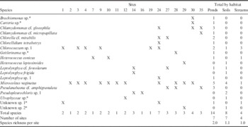
Distribution of species reported from Wright Valley (arranged alphabetically). Site numbers correspond to those in Fig. 1. Sites producing no results are omitted. Collections from Lake Canopus (sites 24–26) are pooled.

* Taxa found only in clone library data.
Table includes results from all methods except 454 data.
Results
Diversity of cultured strains
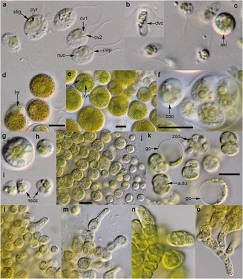
Twelve strains of cyanobacteria and 25 strains of eukaryotes were purified in culture, representing four oscillatorialean cyanobacterial species, and four yellow-green and four green algal species. Descriptions, relevant literature and biogeographic details for each of these species are given in the supplemental material (S2 found at http://dx.doi.org/10.1017/S0954102015000164). Illustrative material (images and phylogenies) is arranged alphabetically, by eukaryotes (Figs 2 & 3) followed by cyanobacteria (Figs 4–6). Table I summarizes the distribution of taxa using information obtained from both culturing and cloning experiments.
a. Chlamydomonas cf. micropapillata LCR-Ant35_2. b. Chlamydomonas cf. gloeophila LCR-Ant35_3. c. Chlamydomonas cf. gloeophila LCR-Ant24_1. d. Chlorococcum sp. 1 LCR-Ant9_2. e. & f. Chlorococcum sp. 1 LCR-Ant25_1. g. Chlorella cf. mirabilis LCR-Ant24_4. h. & i. Chlorella cf. mirabilis LCR-Ant27_3. j. Pseudopleurochloris sp. 1 LCR-Ant16_1. k. Chlorellidium tetrabotrys LCR-Ant26_3. l. Heterococcus conicus LCR-Ant7_1. m. Heterococcus conicus LCR-Ant10_2. n. & o. Heterococcus leptosiroides LCR-Ant29_2. Scales=10 µm (use scale in d. for l.–o. and in f. for a.–c., g.–i.). Abbreviations: aki=akinetes, auto=autospores, cv1/cv2=contractile vacuoles, dvc=dorsiventral curve, gc=giant cells, nsdc=non-spherical daughter cells, nuc=nucleus, pap=papilla, pyr=pyrenoid, stig=stigma, tw=thickened wall, zoo=zoospore.

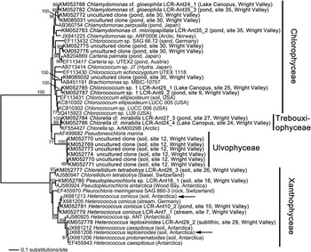
Neighbour-joining tree constructed from rbcL sequences of Wright Valley eukaryotes. Corrected distances were generated using the Tamura–Nei model with gamma-distributed rate variation in MEGA version 6. Bootstrap support values are shown when >75%. Sequences contributed in this study are in bold.

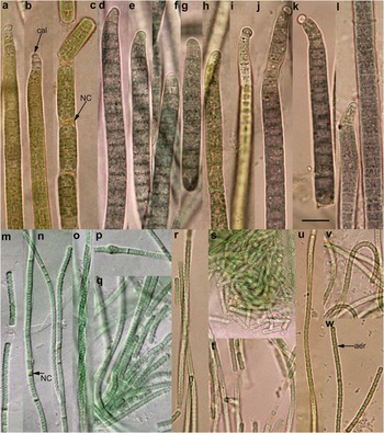
a.–c. Microcoleus vaginatus LCR-Cyant3a. d.–g. M. vaginatus LCR-Cyant10. h.–i. M. vaginatus LCR-Cyant11. j.–l. M. vaginatus LCR-Cyant30. m.–q. Leptolyngbya cf. foveolarum LCR-Cyant27; note necridic cells. r.–t. Leptolyngbya sp. 1 LCR-Cyant25. u.–w. Pseudanabaena cf. amphigranulata LCR-Cyant35. Scale=10 µm (use scale in k. for all). Abbreviations: aer=aerotopes, cal=calyptra, NC=necridic cells.

Distribution of cultured strains
Chlamydomonas cf. gloeophila Skuja 1948 (Fig. 2b & c) has been reported previously from Austria, the Czech Republic, Germany and Spain. No close relatives have been sequenced from elsewhere. In Wright Valley, the species appeared in culture from sites 24 (Lake Canopus) and 35 (an ice-covered pond in the Labyrinth, co-occurring with C. cf. micropapillata), and was detected in clones from site 30 (pond, South Fork).
Chlamydomonas cf. micropapillata Moewus 1931 (Fig. 2a) is otherwise known from Britain and Germany, but not with molecular data. No close relatives have been sequenced from elsewhere. In Wright Valley, the species was found in site 35 (an ice-covered pond in the Labyrinth).
Chlorella cf. mirabilis Andreeva 1973 (Fig. 2g–i) is closely related to Chlorella sp. ARF0006, cultured from the Norwegian Arctic. The species is otherwise known from soil in Russia/Ukraine and King George Island, Antarctica. It was found in sites 24 (Lake Canopus) and 27 (pond at head of North Fork) in Wright Valley.
Chlorellidium tetrabotrys Vischer & Pascher 1937 (Fig. 2k) is well known from Europe, where it first appeared in greenhouses and was thought to be an introduced tropical alga. It was found in Wright Valley at site 26 (Lake Canopus). It does not appear to have been documented in Antarctica previously.
Chlorococcum sp. 1 (Fig. 2d–f) also lacks sequences from close relatives and is difficult to identify according to morphology. Similar species have been documented world wide, including from Antarctica and the Dry Valleys specifically. This species was widespread in Wright Valley, appearing in cultures from sites 1 (stream near Wright Lower Glacier), 3 (soil below Denton Glacier), 4 (stream below Denton Glacier), 9 (pond below Bartley Glacier), 19 (Bull Lake inflow) and 24–26 (Lake Canopus).
Heterococcus conicus Pitschmann 1963 (Fig. 2l & m) occurs elsewhere in Antarctica and in Germany, as shown by molecular data (Fig. 3). It was found in sites 7 (stream opposite Meserve Glacier) and 10 (pond below Bartley Glacier) in Wright Valley.
Heterococcus leptosiroides Pitschmann 1963 (Fig. 2n & o), identified using molecular data (Fig. 3), appears to be known only from Antarctica to date. In Wright Valley, it appeared in cultures from site 29 (sublithic soil near pond in South Fork).
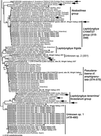
Leptolyngbya cf. foveolarum (Rabenhorst ex Gomont) Anagnostidis & Komárek 1988 (Fig. 4m–q) has been reported from sites elsewhere in Antarctica and around the world; however, molecular data demonstrate that these strains are genetically distant (Fig. 6). Environmental clone data indicate that close relatives of our strain do occur elsewhere in Antarctica, as well as in the Netherlands and Mexico.
Leptolyngbya sp. 1 (Fig. 4r–t) appears to be a new species; although it resembles the genus Nodosilenea in morphology, molecular data show that it is a distant relative (Fig. 6). It was found at site 25 (Lake Canopus) in Wright Valley.
Microcoleus vaginatus (Vaucher) Gomont 1892 (Fig. 4a–l) is a cosmopolitan species and has been well documented from Antarctica and elsewhere. In Wright Valley, it was found in sites 2 (soil near the Wright Lower Glacier, as clones), 3 (stream near the Wright Lower Glacier), 4 (stream below Denton Glacier, as clones), 9 (pond below Bartley Glacier, as clones), 10 (pond below Bartley Glacier), 11 (soil below Conrow Glacier, as clones and a cultured strain), 12 (soil near Bartley Glacier, as clones), 24 (Lake Canopus, as clones), 28 (pond, head of North Fork, as clones), 29 (sublithic soil near pond in South Fork, as clones) and 30 (pond in South Fork).
Pseudanabaena cf. amphigranulata (Van Goor) Anagnostidis 2001 (Fig. 4u–w) has previously been found elsewhere in Antarctica, as well as from alpine sites in Spain and China, according to environmental clone sequences (Fig. 6). However, this is the first occasion that a strain referable to this material with associated molecular data has appeared in culture.
Pseudopleurochloris sp. 1 (Fig. 2j) appears to be a new species with no known close relatives. P. antarctica Andreoli et al., its closest relative, is known only from Antarctica. In Wright Valley, the species was found in sites 14 and 16 (soil near the Conrow and Bartley glaciers, respectively).
Additional information from environmental clone samples
Clones from environmental samples distinguished a further four cyanobacterial and six eukaryotic species groups that did not appear in cultures. Five clones from site 28 (pond in South Fork) are referred to an unknown species of Geitlerinema (Fig. 5). A clone from site 14 (soil below Conrow Glacier) contained a closely related 16S rRNA gene sequence (P-distance 0.009) to Leptolyngbya frigida (F.E.Fritsch) Anagnostidis & Komárek ANT.L52B from Broknes, East Antarctica (Taton et al. Reference Taton, Grubisic, Ertz, Hodgson, Piccardi, Biondi, Tredici, Mainini, Losi, Marinelli and Wilmotte2006) (Fig. 6). Fifteen clones from sites 1 (stream near Wright Lower Glacier) and 24 (Lake Canopus) represented an unknown cyanobacterium (referred to as ‘Unknown sp. 1’). This unknown species is closely related to clones sequenced in other studies from Nepal, the Pyrenees (Spain) and Antarctica (Fig. 6). One clone from site 29 (sublithic soil near pond in South Fork) represented another unknown (‘Unknown sp. 2’). This was a close relative of a clone sequenced from Nepal (Fig. 6). Six clones from site 12 (soil near Bartley Glacier) represented an unknown green alga, tentatively assigned to the Ulvophyceae (Fig. 3). Two clones from site 30 (pond in South Fork) represented an unknown species of Carteria (a quadriflagellate chlamydomonad; Fig. 3). A third clone from site 30 represented an unknown biflagellate chlamydomonad, possibly a species of Brachiomonas (Fig. 3).
Comparison with cloned samples from Miers Valley
Using ITS sequences as a guide, 11 clones from the Miers Valley dataset of Wood et al. (Reference Wood, Rueckert, Cowan and Cary2008a) were identified as potentially close relatives of the Wright Valley clones. Seven of these were too distant for inclusion and are not considered further, but the remaining four are indicated in Fig. 6 (asterisks). Of these, one belonged to the same L. frigida group as one clone from site 14 in Wright Valley and another convincingly belonged to the same species as Unknown sp. 1 (appearing as seven clones from site 1 and eight from Lake Canopus). The other two Miers Valley clones were not close relatives of any clones from Wright Valley.
Comparison with 454 data from soil sites in Wright Valley
The only OTUs found in the Wright Valley soils 454 sequence data that matched the cyanobacterial species reported here were matches to M. vaginatus (Figs 4a–l & 5). Relatively high numbers were recorded from two soil samples (see Table S2): 306 from GT1008 (an Alpine III sample) and 349 from GT1055 (a wet fan/alluvium sample). The remaining counts were all less than 50, and 171 samples contained no OTUs matching M. vaginatus. DNA from the three cultures of M. vaginatus resulted in counts of 1197, 3970 and 4077 matches using this method.
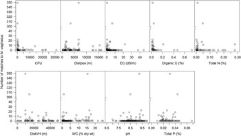
Plots of M. vaginatus counts against site characteristics are shown in Fig. 7. The high number of samples from which no counts were recorded creates difficulties with statistical modelling; however, negative relationships are suggested between M. vaginatus counts and CFU, EC, organic carbon and total nitrogen. A possible weak relationship was suggested with distance from the nearest positive site. Abundance of M. vaginatus was not influenced by distance from the valley head, water content of the soil, pH or total phosphorus.
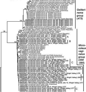
Neighbour-joining tree constructed from 16S rRNA gene sequences of Wright Valley cyanobacteria. Corrected distances were generated using the Kimura two-parameter model with gamma-distributed rate variation in MEGA version 6. Bootstrap support values are shown when >75%. Sequences contributed in this study are in bold. The lengths of 16S–23S internally transcribed spacer regions are shown in brackets. The tree is continued in Fig. 6.

Discussion
Most species are found rarely
Most species discovered in Wright Valley were rarely encountered; of the 19 taxa recorded, ten were found at only one site (Table I). It is not possible to draw meaningful conclusions regarding distribution of such species. Presumably, they occur in more sites, either those not sampled or at abundances too low to detect. The more common species showed no specialization by habitat. Microcoleus vaginatus was found in ten sites, including soils, ponds and streams. Chlorococcum sp. 1, found at six sites, was similarly found in all three habitats. However, ponds were clearly more diverse overall than soils or streams, with nearly double the species number recorded as soils for the same number of sites. Furthermore, 12 soil samples produced no result and are omitted from Table I; allowing for these, the species per site for soils would be 0.4. By contrast, all pond samples produced a result except one (site 31, which evaporation had reduced to mud). Ponds are much more reliably moist habitats than either soils or streams, and are less subject to freezing (depending on salt content). Therefore, a greater diversity of algae would be expected. The samples from Lake Canopus revealed the most species (seven) of any site.
Chlamydomonads are restricted to pond environments
Chlamydomonads (Chlamydomonas, Carteria and Brachiomonas, which have swimming vegetative cells) were restricted entirely to ponds. Interestingly, Chlamydomonas cf. gloeophila was found in three different ponds, unlike its co-inhabitant of the Labyrinth pond, C. cf. micropapillata. The former species produces akinetes, whereas these were not observed in the latter species; presumably these allow C. cf. gloeophila to more readily survive dispersal. Since no chlamydomonad taxa were found in soils, it appears that they must spread by aeolian dispersal between ponds. Chlamydomonads are commonly found in alpine soils, such as those in the Southern Alps of New Zealand, frequently appearing in cultures as palmelloid colonies (e.g. Novis & Visnovsky Reference Novis and Visnovsky2012), but they were not observed here suggesting that they cannot survive the harsher conditions in Wright Valley soils. Jungblut et al. (Reference Jungblut, Vincent and Lovejoy2012) noted that eukaryotes in cyanobacterial mats from ice shelf, pond and lake communities on the McMurdo Ice Shelf and in the High Arctic demonstrated habitat specificity; this was not always the case in Wright Valley but it was among chlamydomonads. Brachiomonas submarina Bohlin f. obtusa Hazen has previously been reported from warmer, saline conditions at 55–60 m depth in Lake Vanda (Seaburg et al. Reference Seaburg, Parker, Prescott and Whitford1979), but it is not known whether the clones from a pond in South Fork represent this taxon.
Life cycles involving zoospore production do not prevent colonization of soils
All eukaryotic genera reported here, apart from Chlorella, are known to produce zoospores and these were sometimes observed in culture (chlamydomonads, Chlorococcum sp. 1, Chlorellidium tetrabotrys, Heterococcus spp.). An opportunity to produce zoospores is typically regarded as necessary for life cycle completion in species able to produce them, and this requires sufficient liquid water. The capacity for zoospore production does not seem to impede some algal genera from occurring in Wright Valley soils, which are indisputably dry: Chlorococcum sp. 1, Heterococcus leptosiroides and Pseudopleurochloris sp. 1 were all recorded from soil samples. It is possible that zoospores are not produced in this habitat, growth of colonies proceeding by simple cell division, and zoospores are only seen in culture. However, this would be difficult to test in the field. Heterococcus is rarely seen in nature, and only as fragments of filaments; zoospores would be difficult to observe and difficult to link to the vegetative material.
Some Antarctic extremophiles appear to have lost the ability to form zoospores. In a study of Antarctic Heterococcus species, H. endolithicus Darling & Friedmann isolated from endolithic growths on Linnaeus Terrace failed to produce zoospores in culture, unlike species from other habitats. This species has since been re-determined by Rybalka et al. (Reference Rybalka, Wolf, Andersen and Friedl2013) as the green alga Desmococcus antarctica. Nonetheless, Desmococcus is also known to produce zoospores and their absence in this species may reflect an adaptation to an extremely water-limited environment. Other green algae known to grow cryptoendolithically in Beacon Sandstone lack flagellated stages, such as Hemichloris (Tschermak-Woess & Friedmann Reference Tschermak-Woess and Friedmann1984). There may be strong selection against zoospore production in this environment, with little recruitment from other biomes (Pointing et al. Reference Pointing, Chan, Lacap, Lau, Jurgens and Farrell2009). Conversely, the temperate near relatives of some strains (e.g. Heterococcus conicus, Chlorellidium tetrabotrys; Fig. 3) suggest that dispersal between Wright Valley and more tolerable environments probably occurs often enough to ensure against evolutionary loss of zoospore production. In the case of Chlorococcum sp. 1, exchange between soil and aquatic habitats in Wright Valley probably occurs (Table I).
On balance it seems probable that the zoosporic genera encountered in the Wright Valley soils do not produce zoospores in the field, since chlamydomonads (for which it is a prerequisite) are absent. Interestingly, chytridiomycete fungi, which form zoospores, have recently been reported to be widespread in Dry Valley soils, including high inland sites (Dreesens et al. Reference Dreesens, Lee and Cary2014); whether these organisms produce zoospores in situ is unknown.
Heterocytous cyanobacteria are infrequent in Wright Valley away from the coast
Nostoc commune Vaucher ex Bornet & Flahault has previously been cultured from a sand sample collected from the Wright Valley by Holm-Hansen (Reference Holm-Hansen1964) and from Lakes Brownworth, Vanda and Canopus by Seaburg et al. (Reference Seaburg, Parker, Prescott and Whitford1979). The latter also reported Calothrix gypsophila (Kützing) Thuret from these lakes, as well as Anabaena sp. and C. braunii Bornet & Flahault from the Onyx River. However, all cyanobacteria detected by us, whether by direct observation, culturing or molecular cloning, were oscillatorialean. The 454 Wright Valley soils dataset did not identify any OTUs belonging to Nostocales (data not shown). Lake Brownworth, an area from which nostocaleans are known, was not reached in this study and no samples were taken from Lake Vanda (no visible growths being present at the shoreline), and only three samples were collected from the Onyx, none of which contained nostocaleans. Seaburg et al. (Reference Seaburg, Parker, Prescott and Whitford1979) recorded six additional species of Nostoc from Taylor Valley and five additional nostocaleans from Miers Valley that were not observed in Wright Valley, suggesting that these organisms are indeed reduced in diversity in the latter valley. Nostocalean cyanobacteria are widespread and abundant in the Dry Valleys nearer the coast, such as the Garwood, Marshall and Miers valleys, lower Taylor Valley and Pyramid Trough (e.g. Niederberger et al. Reference Niederberger, Sohm, Tirindelli, Gunderson, Capone, Carpenter and Cary2012). Nostoc commune especially is often seen with the naked eye in these locations, forming large conspicuous colonies in areas receiving periodic irrigation or in ponds; none of these colonies were observed in Wright Valley in our study. Similarly, Fernandez-Carazo et al. (Reference Fernandez-Carazo, Hodgson, Convey and Wilmotte2011) reported that inland sites in the Shackleton Range contain a reduced diversity of cyanobacteria and an absence of nostocaleans compared with coastal sites. A reminiscent pattern of increased cyanobacterial diversity towards the coast in Taylor Valley was suggested to result from the prevailing down valley wind, rather than more stringent habitat conditions up valley (Michaud et al. Reference Michaud, Šabacká and Priscu2012).
The reduced diversity of nostocaleans in Wright Valley seems to confirm (as does the apparent absence of mosses) that the valley is a significantly harsher environment than many valleys nearby. The absence of Nostoc in the pond samples is interesting; these sites would not be expected to experience the severe water stress of soil sites that might exclude Nostoc. Water chemistry is known to affect the cyanobacterial composition of ponds, for example on the McMurdo Ice Shelf, where nostocalean cyanobacteria are absent from saline ponds and their abundance in others is inversely correlated with the concentration of dissolved nitrogen (Jungblut et al. Reference Jungblut, Hawes, Mountfort, Hitzfeld, Dietrich, Burns and Neilan2005). Oscillatorialean cyanobacteria do not show this pattern, being present in hypersaline ponds and without an apparent relationship between abundance and nitrogen levels. Therefore, it is possible that the reduced presence of Nostocales in Wright Valley results from a combination of low water availability and chemistry.
Influence of sampling time
Seasonal progression affects the detection of biota. Samples collected by Seaburg et al. (Reference Seaburg, Parker, Prescott and Whitford1979) were taken earlier in the summer season than ours; the Onyx River had not yet begun to flow from Lake Brownworth and ice still covered Lake Canopus where a water column between ice and benthos was lacking. The lower diversity recovered from the Onyx River is probably due to this difference, as the river was flowing rapidly and transporting substantial sediment during our collecting period. Under these conditions the area from which the samples of Seaburg et al. (Reference Seaburg, Parker, Prescott and Whitford1979) were collected (described as desiccated microbial mats in dry stream channels) would have been scoured. On the other hand, Lake Canopus was ice-free and highly productive during our sampling, with wind-blown algal detritus distributed for some metres from the open water onto the shoreline. Furthermore, some of the ponds sampled were in the process of evaporating completely, and in one case had been reduced to mud (site 31); neither culturing nor environmental cloning produced a result from this pond.
Distribution and dispersal as suggested by molecular data
Algae reported here that have very close relatives (sometimes identical according to molecular data) outside Antarctica include Chlorellidium tetrabotrys, Heterococcus conicus, Chlorella cf. mirabilis, Brachiomonas sp., M. vaginatus, L. cf. foveolarum, Unknown sp. 1 and 2, and Pseudanabaena cf. amphigranulata (Figs 3, 5 & 6). Many of these near relatives are alpine or polar, providing further evidence of a cold-adapted flora that is, nevertheless, widely dispersed. Jungblut et al. (Reference Jungblut, Lovejoy and Vincent2010) reached similar conclusions regarding cyanobacteria from cold environments. Michaud et al. (Reference Michaud, Šabacká and Priscu2012) found that 86% of cyanobacterial molecular OTUs from Taylor Valley were close relatives (≥98% identical) of sequences retrieved from polar or alpine environments; c. 16% of all their sequences were unique to the McMurdo Dry Valleys. They concluded that four of these OTUs may be endemic to this region and 22 to Continental Antarctica based on the content of databases, but acknowledged that sampling from temperate regions is incomplete. The problem of falsifiability is very significant in microbial biogeography, and in the case of eukaryota there is rightly no consensus as to the genetic distance separating species according to any gene. However, using the 98% delimiting criterion of Michaud et al. (Reference Michaud, Šabacká and Priscu2012) for the cyanobacteria recorded here indicates no ‘species’ endemic to the Dry Valleys or Antarctica as a whole. Interestingly, the M. vaginatus clade would comprise more than one species at a cut-off of 0.02 (Figs 5 & 6). However, this variation does not appear to be geographically correlated. De Wever et al. (Reference De Wever, Leliaert, Verleyen, Vanormelingen, van der Gucht, Hodgson, Sabbe and Vyverman2009) reported evidence for widespread endemism in lacustrine Antarctic microchlorophytes and Strunecký et al. (Reference Strunecký, Elster and Komárek2012) drew similar conclusions for some Antarctic isolates of M. vaginatus/Phormidium autumnale Gomont; although our results offer limited support for the view of De Wever et al. (Fig. 3), confirmation may be achieved through thorough sampling of nearby landmasses, particularly in alpine regions. If true, such endemism seems more likely to apply to lakes than to soils. No evidence for endemism of M. vaginatus strains was found in Wright Valley.
Neighbour-joining tree constructed from 16S rRNA gene sequences of Wright Valley cyanobacteria (continued from Fig. 5). Corrected distances were generated using the Kimura two-parameter model with gamma-distributed rate variation in MEGA version 6. Bootstrap support values are shown when >75%. Sequences contributed in this study are in bold. The lengths of 16S–23S internally transcribed spacer regions are shown in brackets. Asterisks indicate clones from Miers Valley reported by Wood et al. (Reference Wood, Rueckert, Cowan and Cary2008a). Arrows indicate the distribution of morphotypes named ‘Leptolyngbya foveolarum’.

Internally transcribed spacer length and species identification of cyanobacteria
The use of ITS fragment length as a proxy for species, for example in ARISA, is vulnerable to two types of error: overestimation of diversity when distinct species possess spacer regions of equal length and underestimation when one species possesses spacer regions of different lengths within the same genome (Kovacs et al. Reference Kovacs, Yacoby and Gophna2010). In our case, sequences consisting of a fragment of the 16S rRNA gene+ITS enabled comparison with previous studies from the McMurdo Dry Valleys that have used ARISA and 16S rRNA gene+ITS clone libraries to measure diversity in cyanobacteria. The data of Wood et al. (Reference Wood, Rueckert, Cowan and Cary2008a) contained 11 ITS lengths suggesting identity with some of the species reported by us; in fact, examination of 16S rRNA gene sequences showed that only two of these are positive according to the 16S rRNA gene cut-off used by Michaud et al. (Reference Michaud, Šabacká and Priscu2012): L. frigida and Unknown sp. 1. Although this is a small fraction of the bacterial diversity documented by ARISA in the Ross Sea region (e.g. Wood et al. Reference Wood, Mountfort, Selwood, Holland, Puddick and Cary2008b, Reference Wood, Rueckert, Cowan and Cary2008a, Smith et al. Reference Smith, Barrett, Tusnády, Rejtö and Cary2010), it suggests that ARISA could (perhaps greatly) underestimate diversity of Oscillatoriales in this environment and that attempts to relate ARISA fragment lengths to sequences from isolates or clone libraries are of dubious value in this group. Taxonomic utility is nonetheless retained when actual ITS sequences are available.
Abundance of M. vaginatus in Wright Valley soils displays negative relationships with colony forming units, electrical conductivity, organic carbon and total nitrogen
Interestingly, the abundance of M. vaginatus appeared to be random with respect to soil water content, pH and total phosphorus (Fig. 7), and appeared to be more influenced by EC (a proxy for salinity), organic C and total N. Records of both P. autumnale and M. vaginatus (the latter now being regarded as the accepted name) have been made from fresh, brackish and marine waters, but the more saline records have been suggested to relate to different species (Komárek & Anagnostidis Reference Komárek and Anagnostidis2005), which is supported by our results.
The other negative correlations are more counterintuitive. Literature has accumulated suggesting that carbon in Dry Valley soils may be supplemented by spatial subsidies of lacustrine origin, especially in smaller valleys where the lake-to-soil ratio is higher (e.g. Sparrow et al. Reference Sparrow, Gregorich, Hopkins, Novis, Elberling and Greenfield2011 and references therein). Since M. vaginatus is one of the most widespread algae in Wright Valley (Table I), a correlation between its abundance and soil organic C might be expected if such spatial subsidies are occurring. However, the opposite superficially appears to be the case (Fig. 7). Feng et al. (Reference Feng, Simpson, Gregorich, Elberling, Hopkins, Sparrow, Novis, Greenfield and Simpson2010) suggested that spatially redistributed algal biomass is turned over quickly by heterotrophs; hence our results do not necessarily contradict the spatial subsidy hypothesis. However, they do support the contention of Feng et al. (Reference Feng, Simpson, Gregorich, Elberling, Hopkins, Sparrow, Novis, Greenfield and Simpson2010) that the majority of Dry Valley soil organic matter is not contemporary lacustrine in provenance.
A corollary of the spatial subsidy model is that heterotrophs may utilize (or even depend upon) carbon from primary producers for their metabolism. However, the negative relationship between CFU and M. vaginatus abundance (Fig. 7) suggests otherwise (though relies on the assumption that CFUs generated in culture correlate with heterotrophs that are active in the field). A likely alternative is that heterotrophs that co-occur with M. vaginatus differ from those in more saline soils, where M. vaginatus seldom occurs (Fig. 7); variations in bacterial diversity according to soil characteristics in the McMurdo Dry Valleys is becoming well established (e.g. Lee et al. Reference Lee, Barbier, Bottos, McDonald and Cary2012). Further work is intended to investigate the relationship between cyanobacteria and heterotrophs using the Wright Valley 454 soil dataset.
Relationship between the occurrence of Microcoleus vaginatus in Wright Valley soils and the characteristics of the soils. Top row of plots are indicative of negative relationships; bottom row are not suggestive of any relationship. Abbreviations: CFU=colony forming units (heterotrophs), Distpos=distance from nearest site from which M. vaginatus was recorded during the algal survey (see Fig. 5), EC=electrical conductivity, DistVH=distance from valley head (i.e. distance down valley), WC=water content. Note the break in the dependent axis to accommodate the two outliers.

Conclusions
Apart from two widespread species, most species of algae found in the Wright Valley seem to occur rarely. Distribution patterns of these rare species in the valley are difficult to evaluate, but chlamydomonad taxa are probably restricted to ponds and are unable to colonize the soils investigated. However, other species that produce zoospores, but do not necessarily rely on them for life cycle completion, occur in both environments, raising questions around changes in life cycle in soils versus ponds and in the field versus cultures. Cyanobacteria in the valley are mostly Oscillatoriales, distinguishing Wright Valley from more coastal areas in which nostocaleans are frequent. This appears to be common to other areas of Antarctica. The dominant species of algae are mostly known from elsewhere, but are frequently obtained from similar environments (alpine or polar). The distribution of one of the most common species, M. vaginatus, in soils within Wright Valley appears to be related to salinity (with which several other variables are also correlated).
Acknowledgements
Logistic support in Antarctica was provided by Antarctica New Zealand Event K123. Funding was provided by the New Zealand Ministry of Business, Innovation and Employment (under contract CO9X1001). Two anonymous referees provided very helpful comments that strengthened the manuscript.
Author contributions
Planning and field sampling by PMN, JA, ST, MM; data collection and manuscript preparation by PMN, JA.
Supplemental material
Two tables and further supplemental material will be found at http://dx.doi.org/10.1017/S0954102015000164.