Introduction
The placenta is an organ that mediates communication between embryo and mother in eutherian mammals by producing and secreting hormones and supplying nutrients from the mother (Cross, Reference Cross2006). For normal embryo development, placental formation is critical, and many reports have shown that placental defects lead to embryonic lethality (Rossant and Cross, Reference Rossant and Cross2001; Rielland et al., Reference Rielland, Hue, Renard and Alice2008). However, the mechanism of placental differentiation is not fully understood.
In mouse, the placenta is composed of three layers: the maternal decidua, junctional zone, and labyrinthine layer (Watson and Cross, Reference Watson and Cross2005). These layers include various cell types including three cell types that have been well studied: the trophoblast giant cell (TGC) and spongiotrophoblast (SpT) in the junctional zone and the syncytiotrophoblast (SynT) in the labyrinth. TGC and SpT are derived from the ectoplacental cone and formed in the placenta at around E7.5 and E9.5, respectively (Hu and Cross, Reference Hu and Cross2010). Failure of their differentiation has repeatedly shown these cells to be essential for embryo development (Cross, Reference Cross2005; Watson and Cross, Reference Watson and Cross2005; Hu and Cross, Reference Hu and Cross2010).
We recently reported that mouse prolyl oligopeptidase (POP, E.C.3.4.21.26, also known as prolyl endopeptidase; gene symbol Prep), a multifunctional serine protease, contributes to TGC and SpT differentiation, using the trophoblast stem cell (TSC) system (Maruyama et al. Reference Maruyama, Matsubara and Kimura2017). TSCs are cells that can be easily differentiated into TGC, SpT, and SynT in vitro, and widely used in the study of placental differentiation (Tanaka et al. Reference Tanaka, Kunath, Hadjantonakis, Nagy and Rossant1998; Latos and Hemberger, Reference Latos and Hemberger2014). From these data, we suggested the importance of POP in placental differentiation, but the molecular mechanism was not clear.
In this study we attempted to elucidate the mechanism of TSC differentiation into SpT by focusing on three genes that showed abnormal SpT phenotypes in their knockout mice (Rossant and Cross, Reference Rossant and Cross2001; Cross, Reference Cross2005). A basic helix–loop–helix (bHLH) transcription factor, Achaete-scute family bHLH transcription factor 2 (Ascl2, also known as Mash2), is a famous regulator of SpT formation; its knockout mice completely lacked the SpT layer and the embryos die at around E10.5 (Guillemot et al. Reference Guillemot, Nagy, Auerbach, Rossant and Joyner1994, Reference Guillemot, Caspary, Tilghman, Copeland, Gilbert, Jenkins, Anderson, Joyner, Rossant and Nagy1995; Tanaka et al. Reference Tanaka, Gertsenstein, Rossant and Nagy1997). Aryl hydrocarbon receptor nuclear translocator (Arnt)-deficient mice show a reduction in SpT and labyrinthine layers, and the embryos die between E9.5 and E10.5 (Adelman et al. Reference Adelman, Gertsenstein, Nagy, Simon and Maltepe2000; Kozak et al. Reference Kozak, Abbott and Hankinson1997). Epidermal growth factor receptor (Egfr)-deficient mice also resulted in a reduced SpT layer and the embryonic lethality until E12.5, although milder phenotypes have been reported depending on the strain or genetic background (Miettinen et al. Reference Miettinen, Berger, Meneses, Phung, Pedersen, Werb and Derynck1995; Sibilia and Wagner, Reference Sibilia and Wagner1995; Threadgill et al. Reference Threadgill, Dlugosz, Hansen, Tennenbaum, Lichti, Yee, LaMantia, Mourton, Herrup and Harris1995). These findings indicate that these three genes may participate in SpT differentiation, which in turn suggests that POP regulates TSC differentiation by controlling one or more of these genes.
In this study, we tested the possibility that POP regulates TSC differentiation by controlling one or more of the Ascl2, Arnt, and Egfr genes using a POP-specific inhibitor, 3-({4-[2-(E)-styrylphenoxy]butanoyl}-L-4-hydroxyprolyl)-thiazolidine (SUAM-14746; Saito et al. Reference Saito, Hashimoto, Kawaguchi, Shibata, Fukami, Tanaka and Higuchi1991) in the TSC culture system.
Materials and methods
TSC culture, induction of differentiation, and treatment with inhibitors
TSCs were kindly provided by Dr Satoshi Tanaka (Tanaka et al. Reference Tanaka, Kunath, Hadjantonakis, Nagy and Rossant1998) and maintained as previously reported (Himeno et al. Reference Himeno, Tanaka and Kunath2008). TSC differentiation was performed as previously described (Maruyama et al. Reference Maruyama, Matsubara and Kimura2017). To induce differentiation, 2 × 10⁵ TSCs were seeded on a 35-mm dish and, on the next day, fibroblast growth factor 4 (FGF4) was removed to start the differentiation. A POP-specific inhibitor, SUAM-14746 (Peptide Institute, Osaka, Japan), or a PI3K-specific inhibitor, LY294002 (Wako Pure Chemical, Osaka, Japan) was added on the removal of FGF4. An equal volume of dimethyl sulfoxide (DMSO) was used as a control.
Quantitative reverse transcription-polymerase chain reaction (qRT-PCR) analysis
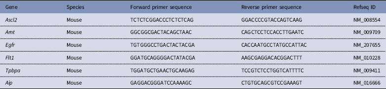
Total RNAs were prepared from the cultured cells at days 2 and 6, and from undifferentiated TSCs as the cells at day 0. After treatment with Turbo DNase (ThermoFisher Scientific, Walthum, USA), cDNA was synthesized by reverse transcription with oligo(dT) primer and 500 ng of each RNA, and quantitative PCR was performed using the ABI Prism 7300 real-time PCR system (Matsubara et al. Reference Matsubara, Takahashi and Kimura2010). All data were normalized to Aryl hydrocarbon receptor interacting protein (Aip) and the highest value was set to 1.0 in each experiment. Primer sequences are shown in Table 1.
Primers used in this study

Statistical analysis
Results were represented as the average ± standard deviation (SD), and their statistical significance was analyzed using Student’s t-test or one-way analysis of variance (ANOVA) followed by Tukey’s post hoc test or Dunnett’s test. P-values less than 0.05 were considered statistically significant.
Results
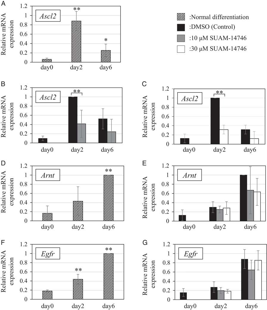
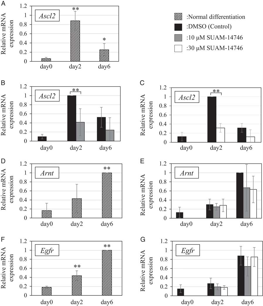
We first examined the expression patterns of Ascl2, Arnt, and Egfr during normal TSC differentiation. By qRT-PCR, Ascl2 mRNA was significantly increased at day 2 and decreased at day 6 (Fig. 1A ), while mRNA levels of the other two genes gradually increased to reach a peak at day 6 (Fig. 1D, F ). The Ascl2 gene pattern was in agreement with previous reports (Tanaka et al. Reference Tanaka, Kunath, Hadjantonakis, Nagy and Rossant1998; Hughes et al. Reference Hughes, Dobric, Scott, Su, Starovic, St-Pierre, Egan, Kingdom and Cross2004). Next, TSCs were treated with a POP-specific inhibitor, SUAM-14746, at 10 µM and 30 µM because these concentrations had been effective on TSC differentiation in our previous study (Maruyama et al. Reference Maruyama, Matsubara and Kimura2017). Compared with the control experiment with DMSO, Ascl2 expression was significantly reduced by both concentrations of SUAM-14746 at day 2 (Fig. 1B, C ). By contrast, the inhibitor did not change Arnt and Egfr expression (Fig. 1E, G ).
The effects of SUAM-14746 on expression patterns of Ascl2, Arnt, and Egfr genes. The relative expression of (A) Ascl2, (D) Arnt, and (F) Egfr during normal TSC differentiation is shown as mean ± SD, and the data were analyzed by one-way ANOVA followed by Tukey’s post hoc test. *P<0.05, **P<0.01 compared with day 0, n=3. To investigate the effects of a POP-specific inhibitor, we added 10 or 30 µM SUAM-14746 or DMSO (control) to TSCs. Total RNAs were isolated from the cells at days 0, 2, and 6, and the expression levels of (B, C) Ascl2, (E) Arnt and (G) Egfr were analyzed by qRT-PCR. These data are presented as mean ± SD, and were analyzed by Student’s t-test or one-way ANOVA followed by Dunnett’s test. **P<0.01 compared with the cells with DMSO, n=3.
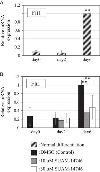
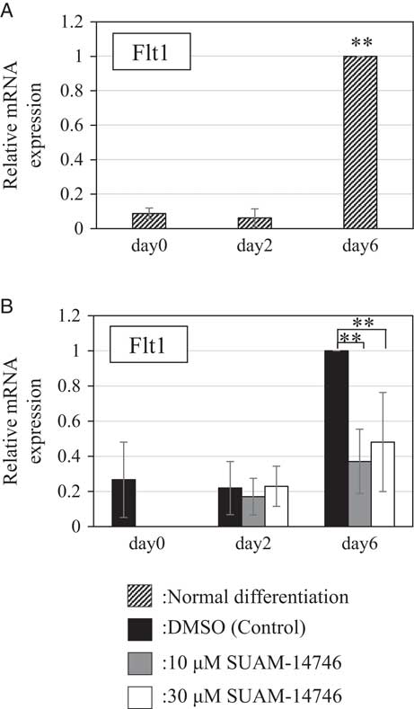
To further confirm the result, we checked the effect of SUAM-14746 on the expression of Flt1, a gene downstream of Ascl2 (Guillemot et al. Reference Guillemot, Nagy, Auerbach, Rossant and Joyner1994; El-Hashash et al. Reference El-Hashash, Warburton and Kimber2010). Flt1 expression was increased during normal differentiation and peaked at day 6. By addition of 10 µM and 30 µM SUAM-14746, it was significantly reduced at day 6 compared with the control (Fig. 2A, B ). These results indicated that POP is involved in regulation of the Ascl2 gene during mouse TSC differentiation into SpT.
The effect of SUAM-14746 on Flt1 expression. The relative expression of Flt1 during normal TSC differentiation (A) and in TSCs cultured with 10 or 30 µM SUAM-14746 or DMSO (control) (B) was analyzed by qRT-PCR. Both concentrations of SUAM-14746 significantly reduced the expression of Flt1 at day 6. The data are presented as mean ± SD, and were analyzed by one-way ANOVA followed by (A) Tukey’s post hoc test or (B) Dunnett’s test. **P<0.01 compared with (A) day 0 or (B) the cells with DMSO, n=3.
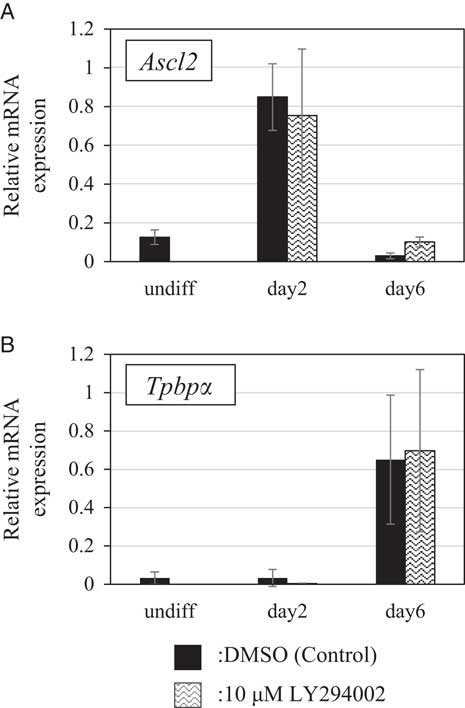
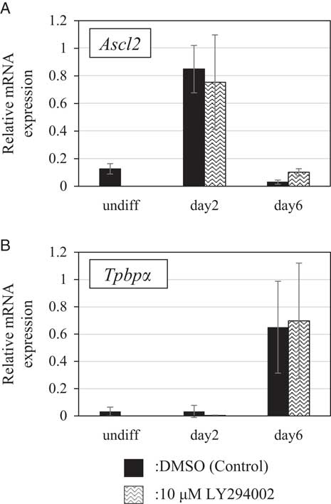
We next attempted to reveal the signalling pathway that POP regulated. Because POP is involved in the regulation of the PI3K-Akt signalling pathway, which controls the Ascl2 gene expression (Takao et al. Reference Takao, Asanoma, Tsunematsu, Kato and Wake2012; Duan et al. Reference Duan, Ying, Danzer, Perez, Shariat-Madar, Levenson and Maki2014), we investigated whether this pathway was related to SpT differentiation regulated by POP. We added a PI3K-specific inhibitor, LY294002, to the TSC culture system at 10 µM, and checked the expression of Ascl2 and Tpbpα. However, qRT-PCR showed that there was no significant difference in the expression of TSCs treated with DMSO or with LY294002 (Fig. 3A and B ). These results suggested that regulation of Ascl2 expression by POP was not through the PI3K-Akt pathway.
The effects of LY294002 on expression of SpT marker genes. (A) Ascl2 and (B) Tpbpα expression in TSCs supplemented with 10 µM LY294002 or DMSO (control) at days 2 and 6 and in undifferentiated cells (day 0) was analyzed by qRT-PCR; 10 µM LY294002 did not significantly affect the expression of SpT marker genes, n=5.
Discussion
Our present data clearly showed downregulation of Ascl2 after adding a POP-specific inhibitor. The Ascl2 gene encodes a transcription factor that is critical for SpT formation (Johnson et al. Reference Johnson, Birren, Saito and Anderson1992; Guillemot et al. Reference Guillemot, Nagy, Auerbach, Rossant and Joyner1994, Reference Guillemot, Caspary, Tilghman, Copeland, Gilbert, Jenkins, Anderson, Joyner, Rossant and Nagy1995). Flt1 is one of its downstream genes and was downregulated by the inhibitor, although this gene itself is dispensable for placental formation (Hirashima et al. Reference Hirashima, Lu, Byers and Rossant2003). Moreover, another downstream gene, Tpbpα, which is unrelated to Flt1 and essential for vascularization of the placenta (Hu and Cross, Reference Hu and Cross2011), was also significantly decreased by the same inhibitor (Maruyama et al. Reference Maruyama, Matsubara and Kimura2017). These results indicate that POP is directly or indirectly involved in regulation of the pathway originating from Ascl2 and leading to SpT differentiation in the placenta. However, it is also possible that POP controls glycogen trophoblast differentiation because Ascl2, Flt1, and Tpbpα genes are all expressed in both SpT and glycogen trophoblast. We can not completely rule out this possibility, but the low levels of POP expression found in glycogen trophoblasts were not necessarily consistent with our hypothesis (Matsubara et al. Reference Matsubara, Takahashi and Kimura2011). Therefore, we strongly suggest that POP regulates the Ascl2 gene pathway and therefore SpT differentiation.
POP was reported to be localized in the nuclei of some cell types (Ohtsuki et al. Reference Ohtsuki, Homma, Kurata, Komano and Natori1994; Ishino et al. Reference Ishino, Ohtsuki, Homma and Natori1998; Venditti and Minucci, Reference Venditti and Minucci2018), but cytoplasmic POP is more likely to be involved in the Ascl2 regulation because it is predominantly present in the cytoplasm of TSCs (Maruyama et al. Reference Maruyama, Matsubara and Kimura2017). Cytoplasmic POP is not only a protease that digests –Pro–X– peptide bonds in bioactive peptides, it also functions as an interacting protein regardless of its enzyme activity (García-Horsman et al. Reference García-Horsman, Männistö and Venäläinen2007; Myöhänen et al. Reference Myöhänen, García-Horsman, Tenorio-Laranga and Männistö2009). There are some possible pathways from POP to Ascl2. In cancerous cells, the Ascl2 gene is controlled by Wnt/β-catenin and PI3K-Akt pathways (Jubb et al. Reference Jubb, Chalasani, Frantz, Smits, Grabsch, Kavi, Maughan, Hillan, Quirke and Koeppen2006; Takao et al. Reference Takao, Asanoma, Tsunematsu, Kato and Wake2012; Zhou et al. Reference Zhou, Rao, Yang, Wu, Tan, Xu, Ding, Zhan, Hu, Cui, Zhang, Dong, Liu and Bian2015), and a relationship with the ERK pathway has also been shown (Sharma et al. Reference Sharma, Kubaczka, Kaiser, Nettersheim, Mughal, Riesenberg, Hölzel, Winterhager and Schorle2016). Here, we found that the PI3K-Akt pathway was unlikely to be a factor, so POP may be involved in other pathways. POP could control Ascl2 by changing the phosphorylation status of proteins in these pathways, as it has been reported to regulate protein phosphorylation (Williams et al. Reference Williams, Eames, Ryves, Viggars and Harwood1999; Duan et al. Reference Duan, Ying, Danzer, Perez, Shariat-Madar, Levenson and Maki2014). It is interesting that Ascl2 is activated by a placenta-specific transcription factor, AP-2γ (Sharma et al. Reference Sharma, Kubaczka, Kaiser, Nettersheim, Mughal, Riesenberg, Hölzel, Winterhager and Schorle2016), and POP is also suggested to be controlled by AP-2γ (Matsubara et al. Reference Matsubara, Maruyama and Kimura2013). SpT differentiation may be regulated by an AP-2γ–POP–Ascl2 pathway.
In conclusion, the addition of SUAM-14746 significantly decreased the expression of Ascl2 and its related genes in mouse TSCs. This suggests that POP plays a role in SpT differentiation by regulating the Ascl2 gene. Some strains of POP-deficient mice have shown embryonic lethality and growth retardation (IMPC, http://www.mousephenotype.org/; KOMP, https://www.komp.org/), possibly due to failure of SpT formation and abnormal communication between mother and embryo.
Financial support
This research received no specific grant from any funding agency, commercial or not-for-profit sectors.
Conflicts of interest
The authors certify that they have nothing to disclose.
Ethical standards
Not applicable.
Acknowledgements
We thank Dr Satoshi Tanaka at University of Tokyo, Japan for kindly gifting TSCs.