Introduction
The genus Oodinium Chatton, 1912 (Blastodiniales, Oodiniaceae) is a unique group of ectoparasitic dinoflagellates (Fensome et al., Reference Fensome, Taylor, Norris, Sarjeant, Wharton and Williams1993). Information about Oodinium species is scarce, which is probably explained by the rare findings of their trophonts on the host body that occur, in most cases, accidentally during routine examinations of plankton samples. Such samples are often fixed with formaldehyde, which makes further identification of the trophont species by electron microscopy methods challenging. In addition, the extent of the negative effect that Oodinium dinoflagellates exert on their hosts has not been fully elucidated. Therefore, every discovery of these ectoparasitic dinoflagellates on a host is of great interest, contributing to understanding their biodiversity and pathogenic effects. The life cycle of Oodinium consists of a free-living stage (dinospore, considered as the infective agent), a growth stage (trophont, which rarely resembles a dinoflagellate in gross appearance), and a sporogenic reproductive stage (generating large numbers of dinospores) (Chatton, Reference Chatton1920; Coats, Reference Coats1999; Gomez and Skovgaard, Reference Gomez and Skovgaard2015). The trophont (vegetative cell, or trophozoite), which is a feeding form of the organism, attaches to the surface of the host’s body by the adhesive disc of its peduncle (Horiguchi, Reference Horiguchi, Ohtsuka, Suzaki, Horiguchi, Suzuki and Not2015). To date, five species of Oodinium are known. Oodinium pouchetii (Lemmerman, 1899) Chatton, 1912, the type species of the genus, is an ectoparasite of the appendicularian Oikopleura (Vexillaria) dioica Fol, 1872 (Oikopleuridae) (Chatton, Reference Chatton1912). Trophonts of O. fritillaria Chatton, 1912, are also found on other appendicularians: Fritillaria pellucida Busch, 1851; F. formica Fol, 1872; and F. megachile Fol, 1872 (Fritillariidae) (Chatton, Reference Chatton1920; Cachon and Cachon, Reference Cachon and Cachon1971). Trophonts of O. dogieli (Dogiel, Reference Dogiel1910) Cachon and Cachon, 1971 have been described from annelids (Dogiel, Reference Dogiel1910; Cachon and Cachon, Reference Cachon and Cachon1971). Oodinium jordani McLean and Nielsen, 1989 and O. inlandicum Horiguchi and Ohtsuka, 2001 are ectoparasites of the chaetognaths Sagitta elegans Verrill, 1873 and Aidanosagitta crassa (S. crassa) Tokioka, 1938, respectively (McLean and Nielsen, Reference McLean and Nielsen1989; Horiguchi and Ohtsuka, Reference Horiguchi and Ohtsuka2001). Oodinium acanthometrae Cachon, 1964 is a parasite of acantharians from the Mediterranean Sea, whose morphology is poorly known (Cachon, Reference Cachon1964; Cachon et al., Reference Cachon, Cachon and Greuet1970). In addition, an unknown species of Oodinium, closely resembling O. jordani McLean and Nielsen, 1989, has been reported as a parasite of ctenophores (Ctenophora) and a hydromedusa (Cnidaria) (Mills and McLean, Reference Mills and McLean1991). Oodinium fritillaria is the only species in which a single trophont (rarely, two) parasitises the host. Trophonts of all other Oodinium species infect the host’s body in large numbers.
It should also be noted that Oodinium species have been mainly found in the Mediterranean Sea, with the exception of O. jordani described from the north-western USA coast, Washington (McLean and Nielsen, Reference McLean and Nielsen1989) and O. inlandicum from the Seto Inland Sea, Japan, and waters along the Korean coasts (Horiguchi and Ohtsuka, Reference Horiguchi and Ohtsuka2001; Horiguchi et al., Reference Horiguchi, Harada, Ohtsuka, Soh and Yoon2004). The species composition of parasitic dinoflagellates in the Russian waters of the Sea of Japan, in contrast to free-living ones, has been studied very poorly to date. Five species of parasitic dinoflagellates from the genera Protoodinium, Paulsenella, Duboscquella, Syndinium, and Dissodinium and two species of ellobiopsids of the genus Ellobiopsis are known (Konovalova, Reference Konovalova2008). Parasitic dinoflagellates from the genus Oodinium have never been reported before for the Russian waters of the Sea of Japan. In Vostok Bay, fritillariid appendicularians appear in plankton from mid-September to mid-October. They also sometimes occur in June and August, when the water temperature is below 22°C. Fritillariids infected by Oodinium are rarely found in plankton samples.
This study aimed to describe the morphology and ultrastructure of an Oodinium species whose trophonts were found on the fritillarian, F. borealis, from Vostok Bay, Peter the Great Bay, Sea of Japan. A number of ultrastructural features of the trophonts, the host specificity, and the geographic distribution allowed us to identify it as a new species of the genus Oodinium, O. vostokense sp. nov. Savelieva.
Materials and methods
Specimens of Fritillaria borealis were collected in Vostok Bay, Peter the Great Bay, Sea of Japan, by vertical towing of a Juday plankton net (mesh size 112 μm) from a depth of 10 m on 10 June 2018 and 14 October 2019. Samples were examined in a Bogorov chamber under an MBS-10 stereo microscope (Russian Federation). The F. borealis specimens were pipetted onto a glass slide and examined for ectoparasites under a Zeiss Primo Star light microscope. One more trophont of Oodinium vostokense sp. nov., goldish in colour, which did not belong to the described holotype and isotype, was also found and examined (Figure 1A–C).
Oodinium vostokense sp. nov. attached to appendicularian, Fritillaria borealis. (A–C) Optical sections through trophont attached to host’s pharyngeal epidermis. (D, E) Semithin sagittal sections through trophont attached to host’s pharyngeal epidermis. (Inset D) Photo of F. borealis with attached trophont, embedded in Epon–Araldite resin. (F, G, inset G) Optical sections through trophont attached to host’s oesophageal epidermis closer to pharynx. (H, I, inset I) Semithin longitudinal sections through trophont attached to host’s oesophageal epidermis closer to pharynx. Letter designations: ad, adhesive disc; en, endostyle; ep, epidermis; eso, oesophagus; esep, oesophageal epidermis; g, gonads; m, mouth; oik, oikoplast; ov, ovary; ph, pharynx; phep, pharyngeal epithelium; sg, stigma; st, stomach; t, testis; tl, tail; tr, trophont.

For transmission electron microscopy (TEM), the F. borealis specimens with attached trophonts were fixed with 2.5% glutaraldehyde (EMS) solution in 0.1 M cacodylate buffer (pH 7.4) supplemented with NaCl (21 mg/mL) for 8 hr at 4℃, and then rinsed three times in the buffer (pH 7.4). The specimens were post-fixed for 2 hr with a 1% osmium tetroxide solution, prepared in the same buffer, dehydrated in a graded ethanol series (30, 50, 70, 80, 90, and 96% for 15 min each), in ethanol: acetone mixtures (3:1, 1:1, and 1:3, v/v), and in pure acetone. The dehydrated specimens were embedded in Epon–Araldite resin (EMS). Semithin sections (0.9 μm) were cut on a Reichert Ultracut E ultramicrotome, stained with methylene blue, and photographed through a Keyence BZ-9000 BIOREVO fluorescent microscope. Ultrathin sections (60 nm) were stained with an aqueous uranyl acetate solution or UranyLess EM Stain (EMS) and with a lead citrate solution and then examined under a Zeiss Sigma 300 VP scanning electron microscope operated at 25 kV in STEM detection mode. The microscopes were provided by the Far Eastern Center for Electron Microscopy, A.V. Zhirmunsky National Scientific Center of Marine Biology, Far Eastern Branch, Russian Academy of Sciences (NSCMB FEB RAS).
The slides with semithin sections and cuprum grids A1, A3, A5, A7, A9 with ultrathin sections in collected box grids # 71140 for illustration of the holotype were deposited at MIMB (The Museum of the A.V. Zhirmunsky National Scientific Center of Marine Biology), NSCMB FEB RAS, Vladivostok, Russia (accession no. MIMB 53540).
Unfortunately, due to the rarity of the findings and the small number and size of the specimens, we could not obtain gene marker sequences from the ectoparasitic dinoflagellate identified in the present study.
Results
On the basis of detailed TEM examination, we assigned this ectoparasitic dinoflagellate to the genus Oodinium. The clear ultrastructural differences from other species of the genus allowed us to identify it as a new species, Oodinium vostokense sp. nov.
Class Dinophyceae West et Fritch (1927)
Order Blastodiniales Chatton (1906)
Family Oodiniaceae Chatton (Reference Chatton1920)
Genus Oodinium Chatton (Reference Chatton1912)
Oodinium vostokense Savelieva sp. nov. (Figures 1–4)
Material examined
Russia, Sea of Japan, Peter the Great Bay, Vostok Bay, Vostok Marine Biological Station, NSCMB FEB RAS. Holotype (MIMB 53540), collected on 14 October 2019. Slides with semithin sections and cuprum grids (A1, A3, A5, A7, and A9) with ultrathin sections in collected grids box # 71140. One isotype (MIMB 53541, collected on 10 June 2018). Slide with semithin sections and cuprum grids (B2, B4, B6) with ultrathin sections in collected grids box # 71140.
Description
Localisation of O. vostokense sp. nov. on the host
Two specimens of F. borealis (ca. 400 µm in trunk length and ca. 450 µm in tail length) bear two and five trophonts, respectively (four of which are ca. 25 µm, one is ca. 30 µm, and two are ca. 50 µm in length). The trophonts are attached to different sites on the epidermis of the trunk, including the gonad epidermis, and the epidermis of the tail (Figure 1D, inset D). In the cases of infection of F. borealis by a single trophont, the attachment site is the pharyngeal epidermis of the host (ca. 400 µm in trunk length and ca. 450 µm in tail length) closer to the oesophagus (holotype) (Figure 1D, E), or oesophageal epidermis closer to the pharynx (isotype) (Figure 1F–I), or on the tail epidermis. Most trophonts are transparent (Figure 1F and G, inset G). Only one F. borealis specimen (ca. 450 µm in trunk length and ca. 500 µm in tail length) is infected by a single trophont (with a length of ca. 50 µm), goldish in colour (Figure 1B and C).
Holotype
The trophont of O. vostokense sp. nov. attached to the pharyngeal epidermis closer to the oesophagus of F. borealis is transparent, balloon-shaped, 55 µm in length (Figure 1D, inset D and E). Thecal plates of the trophonts are smooth, with amorphous material on the surface (Figure 2A–C). The areas where one plate overlaps another are visualised as electron-dense thickening (Figure 2C). The cytoplasm contains numerous elongated or roundish mitochondria, inclusions with electron-dense granules, electron-lucent vacuoles, and many large, electron-dense, roundish lipid droplets reaching 2 µm in diameter. Clusters of up to 11 droplets are located in the cortical cytoplasm (Figure 2A–D). A large, spherical nucleus, approximately 20 µm in diameter, is located in the upper portion of the cytoplasm. The nucleoplasm contains condensed chromatin in the form of large irregular clumps (Figures 2B and 3F). The peduncle of the trophont consists of an external adhesive disc, approximately 25 µm in diameter, and a 15-µm long peduncle stalk partly extending from the trophont (Figures 2A, F, G and 3A–C). The adhesive disk is attached to the flattened pharyngeal epidermis of F. borealis via numerous double cytoplasmic invaginations that form channels of the peduncle stalk (Figures 2A, F, G and 3A, C). The adhesive disc and the proximal portion of the peduncle stalk below the osmiophilic ring have two zones: a peripheral cortex, represented by a moderately dense granulated layer, and a medulla, containing membranous channels packed in the peduncle stalk (Figures 2G and 3A, C). In the attachment (infection) area, the host’s epidermis is stretched towards the adhesive disc like a suction cup (Figure 2A, F), remaining, however, not damaged. The hemocoel beneath the epidermis contains fibrous-like material that is also found in the adhesive disc channels. The cytoplasmic invaginations extend into the trophont’s body where the channels are tightly packed, forming an electron-dense peduncle stalk (Figure 3C). The peduncular bulb is constricted centrally by an osmiophilic ring, from which a double membrane extends, giving rise to the osmiophilic ring of the pusule, with both osmiophilic rings located on the same level (Figures 2A and 3A–D). The pusule is represented by a roundish, electron-lucent sac from which electron-lucent membrane channels extend into the cytoplasm (Figure 3A, D, E, and G). The pusular sac opens outside through a pore represented by a long canal (Figure 3A and C). Proximally, above the peduncular bulb, the stalk extends into the cytoplasm (Figure 3C and E). In a deeper layer of the cytoplasm, beneath the nucleus, the membranous channels of the stalk expand greatly. In longitudinal sections, this expansion appears as an electron-lucent area of the cytoplasm where the double-membrane channels are irregularly intertwined (Figure 3F). Through the proximal, narrowed part of this conglomerate, the membrane channels enter the cytoplasm and are distributed throughout its volume.
Ultrastructure of Oodinium vostokense sp. nov. attached to host’s pharynx. (A) Longitudinal section through the major portion of trophont attached to pharyngeal epidermis of Fritillaria borealis. (B) Nuclear portion of trophont. (C) Thecal plates (tcp) with amorphous material (am) on surface. (D, E) Portion of cytoplasm with trichocyst (tr), lipid droplets (ld), various-sized vesicles (v), and electron-dense granules (gr). (F) Part of adhesive disc. (G) Two zones of proximal part of peduncle stalk. Letter designations: ad, adhesive disc; c, cortex; chc, chromatin clump; ep, host’s epidermis; m, medulla; N, nucleus; pst, peduncle stalk; tc, theca; *, host’s hemocoel; **, environment.

Ultrastructure of peduncle and pusular system of Oodinium vostokense sp. nov. attached to host’s pharynx. (A, B) Longitudinal section showing the proximal portion of peduncle stalk below osmiophilic ring and pusule. (C) Longitudinal section through peduncle stalk (pst), adhesive disc (ad), and pusule (p). (D) Pusular osmiophilic ring (osp). (E) Pusule and cytoplasmic portion of peduncle stalk (cps). (F) Conglomerate of double-membrane channels of peduncle stalk in cytoplasm (cmp). (G) Ending of pusule with its osmiophilic ring. Letter designations: c, cortex; ep, oesophageal epidermis of the host; m, medulla; N, nucleus; osb, osmiophilic ring of peduncle bulb; pb, pusular bulb; pc, pusular channel extending into cytoplasm; pp, proximal part of peduncle stalk; ppr, pusular pore; ps, pusular sac; v, vesicle; *, host’s hemocoel; **, environment.

Isotype
The trophont of O. vostokense sp. nov. from another individual of F. borealis, attached to the oesophageal epidermis close to the pharynx, has two ultrastructural differences from the trophont that represents the holotype (Figures 1f–I and 4). Its cortical cytoplasm contains large, oval, moderately electron-dense lipid droplets, reaching 5 µm in size and arranged into a single layer (Figures 4A, C and 5B). The cytoplasmic portion of the peduncle stalk, located under the nucleus, forms an oval, more regularly arranged conglomerate of anastomosing double-membrane channels (Figure 4C–E). Differences between these trophonts are presented in Figure 5 and Table 1.
Ultrastructure of Oodinium vostokense sp. nov. attached to host’s oesophagus. (A, C) Longitudinal sections through trophont. (B) Longitudinal section through adhesive disc (ad) and lower part of trophont. (D) Proximal portion of peduncle stalk forming an ovoid conglomerate of double-membrane channels (cmp). (E) Double-membrane channels of peduncle stalk extending from conglomerate into cytoplasm (arrowheads). Letter designations: c, cortex; ep, host’s epidermis; ld, lipid droplets; m, medulla; N, nucleus; osb, osmiophilic ring of peduncle bulb; pb, peduncle bulb; pc, pusular channels; pp, proximal portion of peduncle; ppr, pusular pore; ps, pusular sac; pst, peduncle stalk; tc, theca; *, host’s hemocoel; **, environment.

Drawing of Oodinium vostokense sp. nov. trophont. (A) General view of trophont attached to host’s pharyngeal epidermis (holotype). (B) Lower portion of trophont attached to host’s oesophageal epidermis via peduncle stalk (pst), pusule, and adhesive disc (ad) (isotype). Letter designations: c, cortex; ep, host’s epidermis; ld, lipid droplets; m, medulla; N, nucleus; osb, osmiophilic ring of peduncle bulb; osp, pusular osmiophilic ring; pc, pusular channels; ppr, pusular pore; ps, pusular sac; tc, theca; v, vesicle; *, host’s hemocoel; **, environment.

Variability of trophonts in O. vostokense sp. nov

Table 1 Long description
Measurements are given in micrometers for trophont length and width and for peduncle stalk length and diameter, for two specimens labeled holotype and isotype. For the holotype, trophont length is 55 and width is 37, and peduncle stalk length is 15 with diameter 25. For the isotype, trophont length is 50 and width is 35, and peduncle stalk length is 15 with diameter 35.
Diagnosis
Vegetative cell (trophont or trophozoite) balloon-shaped, 55 µm in length; thecal plates present; nucleus spherical, dinokaryotic, localised closer to upper portion of trophont; cytoplasm granulated, containing numerous large lipid droplets lacking chloroplasts; trychosysts present; attachment (adhesive) disc of peduncle stalk 25 µm in diameter, external, with cortex and medulla zones; peduncle stalk completely internal, approximately 15 µm in length; peduncular bulb constricted centrally by osmiophilic ring; pusular sac constricted by osmiophilic ring and opening outside through pore represented by long canal. Host: appendicularian, Fritillaria borealis.
Distribution
Only known from Vostok Bay in Peter the Great Bay, Sea of Japan.
Type locality
One transparent trophont on appendicularian, F. borealis; Sea of Japan, Peter the Great Bay, Vostok Bay, Vostok Marine Biological Station, NSCMB FEB RAS, 42°54ʹ07.7ʺ N, 132°44ʹ20.5ʺ E; depth: 10 m, plankton sample; holotype, collected on 14 October 2019; isotype, collected on 10 June 2018.
Etymology
The specific epithet refers to the type locality, Vostok Marine Biological Station NSCMB FEB RAS, situated in Vostok Bay, Peter the Great Bay, Sea of Japan.
Conclusion remarks
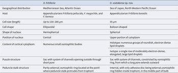
The presence of both the thecal plates and the peduncle indicates that this ectoparasitic dinoflagellate belongs to the family Oodiniaceae from the order Blastodiniales (Fensome et al., Reference Fensome, Taylor, Norris, Sarjeant, Wharton and Williams1993). The above-described ultrastructure of trophonts of Oodinium vostokense sp. nov. shows similarity with that of Oodinium fritillaria Chatton, 1912, another species of the genus Oodinium parasitising fritillariids (Fritillaria pellucida) from the Mediterranean Sea. The morphology and ultrastructure of the known Oodinium species have been investigated to varying degrees of detail (Cachon et al., Reference Cachon, Cachon and Greuet1970; Cachon and Cachon, Reference Cachon and Cachon1971, Reference Cachon and Cachon1977; McLean and Nielsen, Reference McLean and Nielsen1989; Horiguchi and Ohtsuka, Reference Horiguchi and Ohtsuka2001). The ultrastructure of trophonts in O. jordani, O. inlandicum, and O. fritillaria is described in more detail than that in O. dogieli and O. pouchetii. Oodinium acanthometrae is poorly studied (Cachon, Reference Cachon1964; Cachon et al., Reference Cachon, Cachon and Greuet1970). Trophonts of O. vostokense sp. nov. are quite similar to those of O. fritillaria in terms of both general morphology and their hosts, which are various fritillariid species. However, the trophonts of both species differ in their geographic locality, the number of trophonts on a single host, the attachment site on the host, and several ultrastructural features. Oodinium fritillaria is attached to the stomach region in Fritillaria pellucida, F. formica, and F. megachile (Chatton, Reference Chatton1920; Cachon and Cachon, Reference Cachon and Cachon1971). A single trophont (or, in rare cases, two trophonts) of O. fritillaria is attached to the stomach region in F. pellucida, F. formica, and F. megachile (Chatton, Reference Chatton1920; Cachon and Cachon, Reference Cachon and Cachon1971). From one to five trophonts of Oodinium sp. have been found on a single F. borealis, with different attachment sites on the trunk and tail. In cases of infection by a single O. vostokense sp. nov. trophont, it is localised in the pharyngeal or oesophageal region of the host’s body. The ultrastructural features include the shape of the trophont and nucleus, the cytoplasmic content, and the structure of the stalk and pusule. Table 2 summarises the morphological characteristics of O. vostokense sp. nov. and O. fritillaria (Cachon and Cachon, Reference Cachon and Cachon1971), highlighting the differences between these species. One of the important ultrastructural features of the trophont of O. vostokense sp. nov. is the osmiophilic ring of the pusule from which the pusular canal extends outward along the peduncle pore. In the O. fritillaria trophonts, as well as in O. dogieli, O. pouchetii, O. inlandicum, and O. jordani, the pusule lacks the transverse construction in the form of an osmiophilic ring. Additionally, the pusule in O. fritillaria is located lower than in O. vostokense sp. nov. and opens directly outside through a short peduncle pore (Cachon and Cachon, Reference Cachon and Cachon1971). The function of the pusule in Oodinium trophonts remains unclear. This structure has been suggested to perform osmoregulation, absorption, and secretion of macromolecules (Dodge, Reference Dodge1972; Klut et al., Reference Klut, Bisalputra and Antia1987; Kalinina et al., Reference Kalinina, Matantseva, Berdieva and Skarlato2018). The presence of an osmiophilic ring in the pusule of O. vostokense sp. nov. indicates a more complex structure compared to that of O. fritillaria and other Oodinium species. However, this finding does not clarify the pusule function in dinoflagellates; it only highlights differences in its ultrastructure between Oodinium species. Another ultrastructural feature of O. vostokense sp. nov. trophonts is the presence of two layers (cortex and medulla) in the adhesive disc and the proximal portion of the peduncle stalk that have not been found in the peduncle of O. fritillaria. Among Oodinium species, only the peduncle of O. jordani also possesses the cortex and medulla layers (McLean and Nielsen, Reference McLean and Nielsen1989). The peduncle stalk of O. vostokense sp. nov. is internal, with only the adhesive disc protruding outside. In contrast, the peduncle stalk of O. fritillaria trophonts is partly external, and its osmiophilic ring is located at the point where the peduncle stalk protrudes from the trophont. In O. vostokense sp. nov., the osmiophilic ring of the stalk is hidden inside the trophont, in the middle part of the bulb. Furthermore, the cortical cytoplasm in O. vostokense sp. nov. lacks osmiophilic bodies, unlike that in O. fritillaria. Instead, it contains groups of roundish, electron-dense or single oval, moderately electron-dense lipid droplets arranged in a single row.
Comparison of morphological characteristics of trophonts between Oodinium fritillaria and Oodinium vostokense sp. nov

Table 2 Long description
Morphological characters are compared between O. fritillaria and O. vostokense sp. nov., covering distribution, host, cell size and shape, nucleus features, cortical cytoplasm content, pusule structure, and peduncle stalk structure. Geographical distribution lists Mediterranean Sea and Atlantic Ocean for O. fritillaria, and Sea of Japan and North Western Pacific Ocean for O. vostokense sp. nov. Host lists appendicularians Fritillaria pellucida, F. megachile, and F. formica for O. fritillaria, and appendicularian Fritillaria borealis for O. vostokense sp. nov. Cell size length is up to 130 to 285 micrometers for O. fritillaria and 55 micrometers for O. vostokense sp. nov. Nucleus shape is hemispherical with a central position for O. fritillaria, and spherical in the upper portion of cytoplasm for O. vostokense sp. nov. Cortical cytoplasm content is numerous small osmiophilic bodies for O. fritillaria; for O. vostokense sp. nov., holotype has numerous groups of roundish electron dense lipid droplets, and isotype has a single row of moderately electron dense elongated large lipid droplets. Pusule structure is a sac with a system of channels opening outside through a short pore for O. fritillaria, and a sac with a system of channels constricted by an osmiophilic ring with a long pore extending outward for O. vostokense sp. nov. Peduncle stalk structure is partly external with an osmiophilic ring at the point where the stalk protrudes from the trophont for O. fritillaria, and internal with only the adhesive disc external and an osmiophilic ring hidden inside the trophont in the middle part of the bulb for O. vostokense sp. nov.
Discussion
Summarising the findings above, we can conclude that the new parasitic dinoflagellate species from the genus Oodinium is clearly distinguishable from the five known species and, therefore, it is relevant to establish a new species, O. vostokense sp. nov. Unfortunately, the ontogeny of dinospores remains undescribed for O. vostokense sp. nov., as for most other species. The life cycle, from young trophonts to free-living dinospores, has been studied in detail in O. pouchetii. Mature O. pouchetii trophonts become dark in colour, and the nucleus undergoes division to form free-living dinospores (Gomez and Skovgaard, Reference Gomez and Skovgaard2015). Thus, the ultrastructural differences between young and mature Oodinium trophonts mainly consist in their size, colour, and the nuclear structure that becomes non-dinokaryotic (Horiguchi and Ohtsuka, Reference Horiguchi and Ohtsuka2001).
Elucidation of phylogenetic relationships between trophonts of O. fritillaria and O. vostokense sp. nov., which infect fritillariids, is challenging because they are extremely rarely found. Additionally, the small size of both trophonts and their hosts makes such studies difficult. Currently, only O. pouchetii and O. inlandicum have been subject to phylogenetic analysis (Gomez and Skovgaard, Reference Gomez and Skovgaard2015; Horiguchi, Reference Horiguchi, Ohtsuka, Suzaki, Horiguchi, Suzuki and Not2015). However, no clear relationships with other dinoflagellates have been reported, and the taxonomic affinity of the family Oodiniaceae remains unresolved (Gomez and Skovgaard, Reference Gomez and Skovgaard2015; Horiguchi, Reference Horiguchi, Ohtsuka, Suzaki, Horiguchi, Suzuki and Not2015).
Fritillarians, like other appendicularians, are an important link in marine food chains, providing food supply for other invertebrates (e.g., chaetognaths) and fish (Kodama et al., Reference Kodama, Iguchi, Tomita, Morimoto, Ota and Ohshimo2018). Therefore, studying the degree of the pathogenic effect that ectoparasitic dinoflagellates, in particular Oodinium, exert on these hosts may potentially contribute to biodiversity conservation. Except O. fritillaria, other Oodinium species may abundantly parasitise the host’s body. When infected, a fritillariid individual usually bears a single trophont of O. fritillaria. In rare cases, two trophonts per host have been recorded (Chatton, Reference Chatton1920). Pathological effects of O. vostokense sp. nov. on F. borealis are still unclear, but the present observation suggests that they can be severe for fritillariids if a host is infected by several trophonts.
The finding of a new species of Oodinium is a contribution to the knowledge of this enigmatic group of parasitic dinoflagellates. However, further investigations into their life cycle and spore morphology and genetic analyses of O. vostokense sp. nov. are required to address a number of phylogenetic, ecological, and other issues for this genus that still remain to be resolved.
Acknowledgements
The author is grateful to D.V. Fomin and K.A. Sheffer, engineers of the Electron Microscopy Center, A.V. Zhirmunsky National Scientific Center of Marine Biology, Far Eastern Branch, Russian Academy of Sciences, for their technical assistance with electron microscopy studies. The author thanks Evgeny Shvetsov (NSCMB FEB RAS) for proofreading the English of the manuscript.
Declaration of interest
The author declares no competing interests.
Data availability statement
The datasets generated or analysed during the current study are available from the corresponding author upon request.
Ethics and approach
All applicable international, national, and/or institutional guidelines for the care and use of animals were followed by the author.
Funding statement
This research received no specific grant from any funding agency, commercial, or not-for-profit sectors.