I. INTRODUCTION
Osimertinib mesylate is the methanesulfonate salt of osimertinib. Osimertinib mesylate (brand name Tagrisso) is used for treatment of non-small cell lung cancer (NSCLC). It has a role as an antineoplastic agent and as an epidermal growth factor receptor antagonist (PubChem; Kim et al., Reference Kim, Chen, Cheng, Gindulyte, He, He, Li, Shoemaker, Thiessen, Yu, Zaslavsky, Zhang and Bolton2019). Osimertinib mesylate can be prescribed for prevention of some NSCLC from returning after surgical removal of tumors and can be used as an intial treatment for certain NSCLC that have metastasized. The systematic name (CAS Registry Number 1421373-66-1) is N-[2-[2-(dimethylamino)ethyl-methylamino]-4-methoxy-5-[[4-(1-methylindol-3-yl)pyrimidin-2-yl]amino]phenyl]prop-2-enamide methanesulfonate. A two-dimensional (2D) molecular diagram is shown in Figure 1.
The 2D molecular structure of osimertinib mesylate.

X-ray powder diffraction data are reported for a named Form A and Form B of osimertinib mesylate in US Patent 8,946,235 B2 (Butterworth et al., Reference Butterworth, Finlay, Ward and Redfern2015; AstraZeneca). Powder data for another named Form A has been reported in US Patent Application 2019/0194199 A1 (Zhou et al., Reference Zhou, Wu, Jin and Li2019; Wuxi Shuangliang Biotechnology), as well as data for other salts of osimertinib. The diffraction pattern for osimertinib mesylate “Form A” in US Patent 8,946,235 B2 does not agree with the osimertinib mesylate “Form A” in US Patent Application 2019/0194199 A1. Neither patent reports crystal structure data for any osimertinib mesylate phase.
This work was carried out as part of a project (Kaduk et al., Reference Kaduk, Crowder, Zhong, Fawcett and Suchomel2014) to determine the crystal structures of large-volume commercial pharmaceuticals, and include high-quality powder diffraction data for them in the Powder Diffraction File (Gates-Rector and Blanton, Reference Gates-Rector and Blanton2019).
II. EXPERIMENTAL
Osimertinib mesylate was a commercial reagent, purchased from Sigma (Lot # A188322-006), and was used as-received. The pale yellow powder was packed into a 1.5 mm diameter Kapton capillary and rotated during the measurement at ~50 Hz. The powder pattern was measured at 295 K at beamline 11-BM (Antao et al., Reference Antao, Hassan, Wang, Lee and Toby2008; Lee et al., Reference Lee, Shu, Ramanathan, Preissner, Wang, Beno, Von Dreele, Ribaud, Kurtz, Antao, Jiao and Toby2008; Wang et al., Reference Wang, Toby, Lee, Ribaud, Antao, Kurtz, Ramanathan, Von Dreele and Beno2008) of the Advanced Photon Source at Argonne National Laboratory using a wavelength of 0.457912(2) Å from 0.5 to 50° 2θ with a step size of 0.001° and a counting time of 0.1 s per step. The high-resolution powder diffraction data were collected using 12 silicon crystal analyzers that allow for high angular resolution, high precision, and accurate peak positions. A silicon (NIST SRM 640c) and alumina (SRM 676a) standard (ratio Al2O3:Si = 2:1 by weight) was used to calibrate the instrument and refine the monochromatic wavelength used in the experiment.
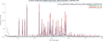
The pattern was difficult to index initially, until it was realized that the sample was not a single phase. The sample consists mainly of AstraZeneca Form B, with a small amount of Form A impurity (Figure 2). After removing the Form A peaks (especially the two lowest-angle peaks), Jade Pro 8.1 (MDI, 2021) yielded a primitive triclinic unit cell with a = 11.43332, b = 11.72806, c = 13.32765 Å, α = 69.02, β = 74.59, γ = 66.38°, V = 1513.23 Å3, and Z = 2. The space group was assumed to be P-1, which was confirmed by successful solution and refinement of the structure. A reduced cell search in the Cambridge Structural Database (Groom et al., Reference Groom, Bruno, Lightfoot and Ward2016) yielded two hits, but no structures of osimertinib derivatives.
Comparison of the synchrotron pattern (black) of osimertinib mesylate to the patterns of Form A (Green) and Form B (red) reported by Butterworth et al. (Reference Butterworth, Finlay, Ward and Redfern2015). The published patterns were digitized using UN-SCAN-IT (Silk Scientific, 2013) and scaled to the synchrotron wavelength of 0.457912 Å using MDI JADE Pro (MDI, 2021).

An osimertinib molecule was downloaded from PubChem (Kim et al., Reference Kim, Chen, Cheng, Gindulyte, He, He, Li, Shoemaker, Thiessen, Yu, Zaslavsky, Zhang and Bolton2019) as Conformer3D_CID71496458.sdf. It was converted into a .mol2 file using Mercury (Macrae et al., Reference Macrae, Sovago, Cottrell, Galek, McCabe, Pidcock, Platings, Shields, Stevens, Towler and Wood2020), and into a Fenske-Hall Z-matrix file using OpenBabel (O'Boyle et al., Reference O'Boyle, Banck, James, Morley, Vandermeersch and Hutchison2011). A mesylate anion was built using Spartan ‘18 (Wavefunction, 2020) and converted into a Fenske-Hall Z-Matrix file using the same tools. The structure was solved using FOX (Favre-Nicolin and Černý, Reference Favre-Nicolin and Černý2002) using sinθ/λ max = 0.4 Å−1. One of the 26 runs yielded a cost factor much lower than the others (Figure 3). The Form A peaks from the pattern of US Patent 8 946 235 B2 (Figure 18) were indexed on a primitive monoclinic unit cell with a = 6.9261, b = 32.0935, c = 15.7046 Å, β = 105.09°, V = 3370.48 Å3, and Z = 4 using DICVOL14 (Louër and Boultif, Reference Louër and Boultif2014). The space group was tentatively assigned as P21/c, and an approximate/tentative structure was obtained using FOX. This model was used to include Form A in the Rietveld refinement.
Distribution of the cost factors (×10−3) from the 26 FOX structure solutions.

NMR spectra were obtained using a 400 MHz Bruker Avance Spectrometer equipped with a multinuclear probe. Spectra of osimertinib mesylate were measured in a solution of dry d6-DMSO (Sigma-Aldrich, stored over flame dried 3-Å molecular sieves). The 1H and 13C NMR spectra matched previously reported spectra (Supplementary Figures S1 and S2). In their synthesis of osimertinib mesylate, Finlay et al. (Reference Finlay, Anderton, Ashton, Ballard, Bethel, Box, Bradbury, Brown, Butterworth, Campbell, Chorley, Colclough, Cross, Currie, Grist, Hassall, Hill, James, James, Kemmitt, Klinowska, Lamont, Lamont, Martin, McFarland, Mellor, Orme, Perkins, Perkins, Richmond, Smith, Wards, Waring, Whittaker, Wells and Wrigley2014) reported that the proton from methane sulfonic acid protonates the dimethyl amine moiety on the pharmaceutical to yield the product salt, as shown in Figure 4, however they do not indicate that this was verified spectroscopically. To ensure that the regiochemistry of the protonation of osimertinib mesylate in solution is properly elucidated, 2D NMR analysis was utilized. A 2D HSQC (1H–13C) spectrum of osimertinib mesylate indicated that three 1H NMR signals in the pharmaceutical salt integrating to a single proton (δ = 9.57, 9.17, and 7.97 ppm) did not have a corresponding 13C signal and were assigned to the heteroatom bound protons on the molecule (Supplementary Figure S3). 2D COSY and TOCSY (both 1H–1H) indicated that only the resonance at 7.97 ppm showed coupling with other 1H resonances on the pharmaceutical (Supplementary Figures S4 and S5). This allowed the resonances at δ = 9.57 and 9.17 ppm to be assigned as the amide N–H and aminopyrimidine N–H, labeled as protons A and B, respectively, which do not have nearest neighbors with protons capable of J-coupling. The heteroatom bound proton resonance at 7.97 ppm indicated coupling to resonances assigned to the methylene and geminal dimethyl groups bound to the terminal amine nitrogen. This coupling allowed for the location of protonation to be spectroscopically determined as the terminal amine labeled as N–H C in Figure 4.
Proposed solution structure of osimertinib mesylate in d6-DMSO. COSY and TOCSY spectra indicate that the signal assigned to the N–H signal assigned as C shows J-coupling with both the geminal dimethyl groups and methylene group attached to the ammonium moiety.

Analysis of potential hydrogen bonding interactions in Form B using Mercury (Macrae et al., Reference Macrae, Sovago, Cottrell, Galek, McCabe, Pidcock, Platings, Shields, Stevens, Towler and Wood2020) indicated that N6 was protonated, so H79 was added to it in an approximate position. This model refined to R wp = 0.1062, but was unsatisfactory in several ways. There were close methyl–methyl contacts between the mesylate anions. The agreement of the refined and DFT-optimized structures (rms Cartesian displacements >0.4 Å) was outside the normal range for correct structures. Both “normal” DFT optimizations using CRYSTAL14 and VASP, as well as “higher-level” calculations (more k-points, higher energy cutoffs, better basis functions) moved one of the hydrogen atoms from N6 to the mesylate anion – a chemically unreasonable result. Apparently the model from FOX, despite its superior cost factor, represented a local (and not global) minimum.
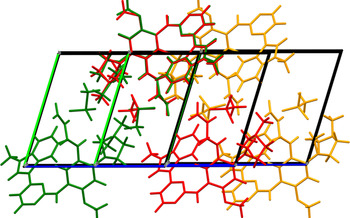
Accordingly, an alternate approach was attempted, using DASH (David et al., Reference David, Shankland, van de Streek, Pidcock, Motherwell and Cole2006). The same fragments were used, but no Mogul Distribution Bias was applied. Six of the 100 runs yielded low values for profile χ 2 (Table I). The next “best” solutions had values > 58 and up to >100. Analysis of potential N–H⋅⋅⋅O hydrogen bonds between the cation and the anion suggested that three of these models were protonated at N5, as indicated by the NMR results. These three models were subjected to Rietveld refinement. The best structure yielded by DASH yielded R wp = 0.1002, while the other two structures refined to lower residuals. The two better refinements yielded essentially identical structures for the cations, but different orientations of the anions (Figure 5); the positions of S1 were the same. The “worst” refinement has a somewhat different cation conformation, but the same anion location and orientation as in the middle refinement (Figure 6). The final refinement and optimization were carried out on the best of these three structures.
Overlay of the two better refinements. The best refinement is in green, and the second-best is illustrated in orange.

Addition of the best DASH solution (red) to the two structures of Figure 5.

Best structure models of osimertinib mesylate from DASH

Rietveld refinement was carried out using GSAS-II (Toby and Von Dreele, Reference Toby and Von Dreele2013). Only the 1.5–25.0° portion of the pattern was included in the refinement (d min = 1.058 Å). All non-H bond distances and angles were subjected to restraints, based on a Mercury/Mogul Geometry Check (Bruno et al., Reference Bruno, Cole, Kessler, Luo, Motherwell, Purkis, Smith, Taylor, Cooper, Harris and Orpen2004; Sykes et al., Reference Sykes, McCabe, Allen, Battle, Bruno and Wood2011) of the cation and the anion. The Mogul average and standard deviation for each quantity were used as the restraint parameters. The restraints contributed 2.5% to the final χ 2. The hydrogen atoms were included in calculated positions, which were recalculated during the refinement using Materials Studio (Dassault, 2020). The U iso were grouped by chemical similarity; the U iso for the H atoms were fixed at 1.3 × the U iso of the heavy atoms to which they are attached. The peak profiles were described using the generalized microstrain model. The background was modeled using a 6-term shifted Chebyshev polynomial, along with a peak at 5.90° to model the scattering from the Kapton capillary and any amorphous component of the sample.
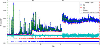
The final refinement of 169 variables using 23 538 observations and 106 restraints yielded the residuals R wp = 0.0665 and GOF = 1.38. The largest peak (1.84 Å from O1) and hole (2.05 Å from C15) in the difference Fourier map were 0.27 and −0.21(4) eÅ−3, respectively. The largest errors in the difference plot (Figure 7) are in the shapes and/or intensities of some of the strong low-angle Form B peaks and Form A peaks, and may reflect changes in the specimen during the measurement.
The Rietveld plot for the refinement of osimertinib mesylate Form B. The blue crosses represent the observed data points, and the green line is the calculated pattern. The cyan curve is the normalized error plot. The vertical scale has been multiplied by a factor of 10× for 2θ > 7.85°, and by a factor of 40× for 2θ > 15.45°. The row of blue tick marks indicates the calculated reflection positions for osimertinib mesylate Form B, and the row of red tick marks indicates the Form A peak positions.

A density functional geometry optimization (fixed experimental cell) was carried out using CRYSTAL14 (Dovesi et al., Reference Dovesi, Orlando, Erba, Zicovich-Wilson, Civalleri, Casassa, Maschio, Ferrabone, De La Pierre, D-Arco, Noël, Causà and Kirtman2014). The basis sets for the H, C, N, and O atoms in the CRYSTAL14 calculations were those of Gatti et al. (Reference Gatti, Saunders and Roetti1994), and the basis set for S was that of Peintinger et al. (Reference Peintinger, Vilela Oliveira and Bredow2013). The calculations were run on eight 2.1 GHz Xeon cores (each with 6 GB RAM) of a 304-core Dell Linux cluster at IIT, using 8 k-points and the B3LYP functional. A Mulliken population analysis was carried out after geometry optimization.
III. RESULTS AND DISCUSSION
The synchrotron powder pattern of this study matches that of Butterworth et al. (Reference Butterworth, Finlay, Ward and Redfern2015) well enough (Figure 2) to conclude that our material is AstraZeneca's Form B, with a Form A impurity. The concentration of Form A was ~0.8 wt%. The refined atom coordinates of osimertinib mesylate Form B and the coordinates from the DFT optimization are reported in the CIFs which have been deposited with ICDD.
The rms Cartesian displacement of the non-hydrogen atoms in the Rietveld-refined and DFT-optimized structures is 0.279 Å (Figure 8). The maximum displacement is 0.750 Å, at the methyl group C70. The good agreement is evidence that the structure is correct (van de Streek and Neumann, Reference van de Streek and Neumann2014). This discussion concentrates on the DFT-optimized structure. The asymmetric unit (with atom numbering) is illustrated in Figure 9, and the crystal structure is presented in Figure 10.
Comparison of the Rietveld-refined (red) and VASP-optimized (blue) structures of the cation in osimertinib mesylate Form B. The rms Cartesian displacement is 0.279 Å.

The asymmetric unit of osimertinib mesylate Form B, with the atom numbering. The atoms are represented by 50% probability spheroids.

The crystal structure of osimertinib mesylate Form B, viewed down the b-axis.

The crystal structure (Figure 10) is characterized by alternating layers of cation–anion and parallel stacking interactions parallel to the ab-planes. Almost all of the bond distances, angles, and torsion angles fall within the normal ranges indicated by a Mercury/Mogul Geometry check (Macrae et al., Reference Macrae, Sovago, Cottrell, Galek, McCabe, Pidcock, Platings, Shields, Stevens, Towler and Wood2020). The S1–O7 distance of 1.549 Å (average = 1.456(31) Å, Z-score = 3.03) is flagged as unusual. This is the oxygen atom involved in two cation–anion hydrogen bonds, so we might expect the covalent bond to be longer than average.
Quantum chemical geometry optimization of the osimertinib cation (DFT/B3LYP/6-31G*/water) using Spartan ‘18 (Wavefunction, 2020) indicated that the observed conformation is 10.1 kcal mol−1 higher in energy than the local minimum. The major differences are in the conformations of the vinyl and dimethylamino groups. The minimum-energy conformation is 9.6 kcal mol−1 lower in energy, and contains an intramolecular N5⋅⋅⋅O2 hydrogen bond. The cation⋅⋅⋅anion hydrogen bonds are apparently important for determining the solid-state conformation.
Analysis of the contributions to the total crystal energy using the Forcite module of Materials Studio (Dassault, 2021) suggests that angle distortion terms dominate the intramolecular deformation energy. The intermolecular energy is dominated by electrostatic attractions, which in this force-field-based analysis include cation coordination and hydrogen bonds. The hydrogen bonds are better analyzed using the results of the DFT calculation.
Hydrogen bonds are significant in the crystal structure (Table II). There is a strong N5–H79⋅⋅⋅O7 hydrogen bond between the cation and the anion. H79 also participates in a weaker intramolecular hydrogen bond to the amino nitrogen N3. There are two additional N–H⋅⋅⋅O hydrogen bonds between the cation and the anion, from the amino nitrogens N6 and N7. The most important hydrogen bonds are illustrated in Figure 11. Several C–H⋅⋅⋅O hydrogen bonds also link the cations and anions. The carbonyl O2 acts as an acceptor in three additional C–H⋅⋅⋅O hydrogen bonds. C–H⋅⋅⋅N and C–H⋅⋅⋅C hydrogen bonds also contribute to the crystal energy.
The principal hydrogen bonds in osimertinib mesylate Form B.

Hydrogen bonds (CRYSTAL14) in osimertinib mesylate Form B

a Intramolecular.
The volume enclosed by the Hirshfeld surface (Figure 12; Hirshfeld, Reference Hirshfeld1977; Turner et al., Reference Turner, Mckinnon, Wolff, Grimwood, Spackman, Jayatilaka and Spackman2017) is 746.68 Å3, 98.80% of half the unit cell volume. The packing density is, thus, fairly typical. All of the significant-close contacts (red in Figure 12) involve the hydrogen bonds. The volume/non-hydrogen atom is 18.0 Å3.
The Hirshfeld surface of osimertinib mesylate Form B. Intermolecular contacts longer than the sums of the van der Waals radii are colored blue, and contacts shorter than the sums of the radii are colored red. Contacts equal to the sums of radii are white.

The Bravais–Friedel–Donnay–Harker (Bravais, Reference Bravais1866; Friedel, Reference Friedel1907; Donnay and Harker, Reference Donnay and Harker1937) morphology suggests that we might expect blocky morphology for osimertinib mesylate Form B. A second-order spherical harmonic model for preferred orientation was incorporated into the refinement. The texture index was 1.001(0), indicating that preferred orientation was not significant for this rotated capillary specimen. The powder pattern of osimertinib mesylate Form B from this synchrotron data set has been submitted to ICDD for inclusion in the Powder Diffraction File.
IV. DEPOSITED DATA
The Crystallographic Information Framework (CIF) files containing the results of the Rietveld refinement (including the raw data) and the DFT geometry optimization were deposited with the ICDD. The data can be requested at info@icdd.com.
SUPPLEMENTARY MATERIAL
The supplementary material for this article can be found at https://doi.org/10.1017/S0885715621000555.
ACKNOWLEDGEMENTS
The use of the Advanced Photon Source at Argonne National Laboratory was supported by the U.S. Department of Energy, Office of Science, Office of Basic Energy Sciences, under Contract No. DE-AC02-06CH11357. This work was partially supported by the International Centre for Diffraction Data. We thank Lynn Ribaud and Saul Lapidus for their assistance in the data collection, and Andrey Rogachev for the use of computing resources at IIT.
CONFLICTS OF INTEREST
The authors have no conflicts of interest to declare.