Introduction
Glass-ceramics are partially crystallized inorganic materials obtained by controlled heat treatment of the parent glass above its crystallization temperature. Reference Serbena, Mathias, Foerster and Zanotto1–Reference Gerhardt and Boccaccini3 The resulting glass-ceramics contain one or more crystalline phases embedded in a residual glassy phase. Reference Höland and Beall4,Reference Montazerian and Zanotto5 In the late 1960s, Hench Reference Hench, Splinter, Allen and Greenlee6 discovered the first bioactive glass with composition (wt%) 45SiO2-24.5CaO-24.5Na2O-6P2O5, termed 45S5 bioactive glass. This was the first man-made material, which was shown to develop strong bonding to bone upon implantation, providing an alternative to inert materials in orthopedic and bone-replacement applications. Developing bioactive glass-ceramics has been a natural extension of the field of bioactive glasses in order to design bioactive materials with higher mechanical strength, but similar bioactivity to bioactive glass. Bioactive glass-ceramics are usually characterized by superior mechanical properties, including higher elastic modulus, failure strength, and hardness, than bioactive glasses. Reference Gerhardt and Boccaccini3,Reference Baino, Novajra and Vitale-Brovarone7 However, the brittleness and low fracture toughness of bioactive glass-ceramics have remained major obstacles for their applications in load-bearing sites. Reference Höland and Beall4,Reference Montazerian and Zanotto5
Bioactive glasses and bioactive glass-ceramics elicit specific biological reactions on their surfaces when in contact with the biological environment, which can stimulate cell attachment, proliferation, and differentiation. Reference Jones8,Reference Hench9 In particular, once in contact with biological fluids, bioactive glasses and bioactive glass-ceramics develop a biological active hydrocarbonate apatite (HCA) layer, which is equivalent to the mineral phase of bone. This layer is essential for the binding of the material to bone. Reference Ducheyne and Qiu10,Reference Sanz-Herrera and Boccaccini11 Bioactive glasses and bioactive glass-ceramics can also degrade over time, releasing biologically active ions that have positive specific effects on cells (e.g., increase proliferation of human osteoblasts (cells that are able to form new bone matrix), as well as angiogenesis (induction of new blood vessel formation), and anti-microbial and anti-inflammatory effects in vitro and in vivo). Reference Hoppe, Güldal and Boccaccini12 Investigations have started to emerge on the potential of bioactive glasses for the regeneration and repair of soft tissues. Reference Miguez-Pacheco, Hench and Boccaccini13
Several review papers are available covering the general field of bioactive glasses and bioactive glass-ceramics. Reference Gerhardt and Boccaccini3,Reference Montazerian and Zanotto5,Reference Jones8,Reference Hench9,Reference Rahaman, Day, Bal, Fu, Jung, Bonewald and Tomsia14 An extensive review on bioactive glass-ceramics in monolithic form has recently been published. Reference Montazerian and Zanotto5 This article was designed to fill the specific lack of recent review articles concerning porous bioactive glass-ceramics intended for applications in bone-tissue engineering (i.e., scaffolds), discussing the latest achievements in processing methods, microstructure, and properties of such systems.
Current developments in porous bioactive glass-ceramic scaffolds
Tissue-engineering strategies involve the development of biological substitutes capable of inducing the growth of new tissues. Reference Amini, Laurencin and Nukavarapu15 This can be achieved by combining three-dimensional (3D) porous substrates, termed scaffolds, with cells and biological signals (e.g., growth factors). Reference Gerhardt and Boccaccini3,Reference Amini, Laurencin and Nukavarapu15 The porous 3D scaffold should provide a mechanically stable environment (e.g., compressive strength comparable with that of trabecular bone), which is one of the two bone structures characterized by interconnected porosity and high vascularization, (2–12 MPa); adequate interconnected porosity (>90%); and pore size (150–500 µm) to enable cell adhesion, migration, and proliferation supporting the growth of new tissue. Reference Gerhardt and Boccaccini3 A major current limitation in the field is that most of the available biomaterials are not simultaneously biologically active, mechanically competent, and bioresorbable. Bioactive glass-ceramics are a class of biomaterials that can have all of these attributes. Different bioactive glass-ceramic compositions and fabrication techniques for bioactive glass-ceramic scaffolds have been reported in the literature and these are discussed in this section with a focus on the most recent achievements.
Foam replica method
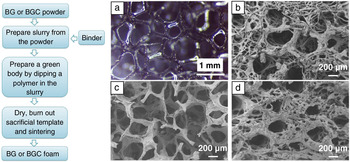
The foam replica method (FRM) for the fabrication of ceramic foams was patented in 1963. Reference Schwartzwalder and Somers16 In 2006, Reference Chen, Thompson and Boccaccini17 the technique was developed for the first time for the fabrication of bioactive glass-ceramic scaffolds based on 45S5 bioactive glass. Scaffolds produced by this technique are a positive replica of an open-cell porous template, which is usually a polyurethane foam ( Figure 1a). After coating with a bioactive glass or bioactive glass-ceramic slurry, the template is burned out and the bioactive glass is sintered at high temperature, which, in the case of 45S5 bioactive glass, leads to crystallization. Reference Chen, Thompson and Boccaccini17,Reference Lefebvre, Gremillard, Chevalier, Zenati and Bernache-Assolant18 The resulting 3D structures are characterized by an open and interconnected porous network that mimics the architecture of trabecular bone, Reference Chen, Thompson and Boccaccini17,Reference Bretcanu, Samaille and Boccaccini19,Reference Vitale-Brovarone, Baino and Verne20 however, it is at the expense of a relatively low compressive strength of the structures. Reference Gerhardt and Boccaccini3 Using this technique, foams with graded porosity can also be obtained. Reference Bretcanu, Samaille and Boccaccini19
Flowchart for the production of bioactive glass (BG) and bioactive glass-ceramic (BGC) scaffolds via foam replica method. Reference Schwartzwalder and Somers16,Reference Chen, Thompson and Boccaccini17 (a) Polyurethane foam (45 ppi, [pores per inch]) and (b) natural marine sponge used to produce (c–d) bioactive glass-ceramic scaffolds of crystallized 45S5 (45SiO2-24.5CaO-24.5Na2O-6P2O5, wt%) bioactive glass composition. Micrographs courtesy of Friedrich-Alexander-Universität Erlangen-Nürnberg, Institute of Biomaterials.

Baino et al. Reference Baino, Minguella, Kirk, Montealegre, Fiaschi, Korkusuz, Orlygsson and Vitale-Brovarone21,Reference Baino, Marshall, Kirk and Vitale-Brovarone22 recently developed a hemispheric highly porous bioactive glass-ceramic shell as a trabecular-like coating for the bioceramic acetabular cup (the semispherical component of a hip implant that allows movement inside the joint) in hip-replacement devices that demonstrates the versatility of the FRM. Moreover, the use of different templates, including marine sponges, Reference Boccardi, Philippart, Juhasz-Bortuzzo, Novajra, Vitale-Brovarone and Boccaccini23 has been shown to lead to increased mechanical properties, especially the compressive strength, by a reduction of the total porosity of the scaffolds, without affecting the pore interconnectivity (Figure 1b,d).
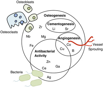
The bioactivity of crystallized 45S5-based bioactive glass-ceramic scaffolds has been proven, Reference Chen, Thompson and Boccaccini17 confirming earlier studies Reference Peitl, Zanotto and Hench24 that had shown that crystallization of bioactive glass reduces, but does not eliminate, the bioactive character of the 45S5 bioactive glass composition. It is also well known that bioactive glasses and bioactive glass-ceramics can be doped with a variety of metallic ions to enhance their biological activity Reference Hoppe, Güldal and Boccaccini12,Reference Salinas and Vallet-Regí25 ( Figure 2 Footnote *).
Selected examples of the biologically active ions being investigated to enhance the performance of bioactive glasses and bioactive glass-ceramics. Reference Hoppe, Güldal and Boccaccini12,Reference Salinas and Vallet-Regí25

FRM is also versatile in terms of the type of starting slurry suitable for producing foams. Zhu et al. Reference Zhu, Wu, Ramaswamy, Kockrick, Simon, Kaskel and Zreiqat26 proposed using a sol-gel synthesis solution to coat the sacrificial template. Cabañas-Polo et al. Reference Cabañas-Polo, Philippart, Boccardi, Hazur and Boccaccini27 applied electrophoretic deposition of a sol-gel solution in combination with FRM, accelerating the scaffold production time. Bioactive glass-ceramic scaffolds made by FRM have also been coated with ordered mesoporous silica particles, Reference Boccardi, Philippart, Juhasz-Bortuzzo, Beltrán, Novajra, Vitale-Brovarone, Spiecker and Boccaccini28 biodegradable polymers, Reference Desimone, Li, Roether, Schubert, Crovace, Rodrigues, Zanotto and Boccaccini29 and polymer microspheres, Reference Li, Nooeaid, Roether, Schubert and Boccaccini30 transforming them in local drug delivery systems. In addition, polymer coatings on bioactive glass-ceramic foams usually lead to enhanced mechanical properties. Reference Desimone, Li, Roether, Schubert, Crovace, Rodrigues, Zanotto and Boccaccini29,Reference Philippart, Boccaccini, Fleck, Schubert and Roether31,Reference Yunos, Bretcanu and Boccaccini32
The versatility and simplicity of FRM as well as the high reproducibility of the microstructure of the scaffolds produced explain the popularity of this method. A critical FRM challenge is to produce a uniform coating of the sacrificial template, which can be achieved by using slurries of suitable viscosity; the incomplete removal of excess slurry could lead to the presence of closed pores in the final bioactive glass-ceramic scaffolds, decreasing pore interconnectivity. Reference Colombo33
Foaming techniques
Highly porous bioactive glass and bioactive glass-ceramic scaffolds can be produced by directly foaming a colloidal sol or a powder suspension, followed by a solidification treatment. The resulting foams exhibit a hierarchical structure with interconnected macropores (ranging from ∼20 µm to 1–2 mm). Reference Jones and Hench34 The main challenge is to prevent the foam struts from collapsing, which may result from the loss of the cellular shape of the bubbles. For this reason, a surfactant is used to stabilize the bubbles.
One of the most successful foaming methods is gel casting, Reference Sepulveda, Jones and Hench35 in which in situ polymerization of monomers can be initiated, forming a 3D polymeric network, a gel, which produces mechanically stable structures. Reference Jones and Hench34 The samples are then sintered to provide mechanical strength. The total porosity and the pore size can be controlled by the surfactant concentration. A further development of this technique is sol-gel foaming. Reference Sepulveda, Jones and Hench35 In this method, a sol-gel glass synthesis solution is prepared and a surfactant is added, the foam is generated by vigorous agitation, the sol is transferred to a mold for aging, and finally, the porous body is heat-treated. Reference Sepulveda, Jones and Hench35
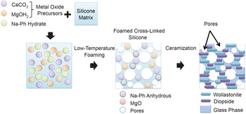
A more recent technology developed for bioactive glass-ceramic foams involves the use of polymer-derived ceramics. Reference Elsayed, Zocca, Franchin, Bernardo and Colombo36 In this approach, metal oxide precursors in the form of micro- or nano-sized particles are added to a polymeric precursor (e.g., a silicone resin), allowing the production of silicate bioceramics ( Figure 3 ). The foaming is obtained by water release from specific hydrated fillers. The foams are then sintered. Fiocco et al. Reference Fiocco, Elsayed, Daguano, Soares and Bernardo37,Reference Fiocco, Elsayed, Ferroni, Gardin, Zavan and Bernardo38 showed the possibility of obtaining wollastonite-diopside (CaSiO3-CaMgSi2O6) foams with 77% porosity and compressive strength of 1.8 ± 0.3 MPa.
Schematic diagram of the production of wollastonite-diopside (CaSiO3-CaMgSi2O6) polymer derived glass-ceramic foams, using sodium hydrate phosphate filler (Na-Ph hydrate). The process leads to the fabrication of highly porous glass-ceramic foams by mixing preceramic polymers (e.g., silicone resins that act as a silica precursor) and powdered metal oxide precursors. Reference Fiocco, Elsayed, Ferroni, Gardin, Zavan and Bernardo38

The quality of the obtained porous scaffolds can be improved by selecting different silicon precursors (e.g., polysiloxane) and by using CO2-assisted extrusion. Another approach to produce porous bioactive glass-ceramics involves a powder-metallurgy technique. Reference Auguilar-Reyes, León-Patiño, Jacinto-Díaz, Perdomo-Cervantes and Lefebvre39 In this method, bioactive glass powder is dry mixed with a solid polymeric binder and a foaming agent. The mixture is then molded and heat-treated. Reference Aguilar-Reyes, León-Patiño, Jacinto-Diaz and Lefebvre40 The resulting scaffolds showed compressive strength values in the range of 5–40 MPa (i.e., in the upper range of values reported so far for this type of porous materials). The scaffolds exhibited highly interconnected pore structure and tunable porosity (55–77%), and they retained satisfactory compressive strength after immersion for one month in simulated body fluid (6 MPa). Reference Boccardi, Melli, Catignoli, Altomare, Jahromi, Cerruti, Lefebvre and De Nardo41
The major drawback of this method is that the process could yield a structure with unconnected pores that are essential for bone integration and vascularization. Reference Zhu, Chen and Pignatello42 Interconnected porosity is a key property required in bone-tissue engineering scaffolds as it is essential to enhance cell invasion and new tissue growth. In addition, the ability of these scaffolds to promote vascularization has not been yet investigated, as opposed to bioactive glass-ceramics produced by FRM. Reference Arkudas, Balzer, Buehrer, Arnold, Hoppe, Detsch, Newby, Fey, Greil, Horch, Boccaccini and Kneser43
Additive manufacturing technologies
Additive manufacturing technology (AMT) is the usual term given to fabrication processes where 3D structures are fabricated layer by layer without any specific tooling to obtain the targeted geometry. Reference Bose, Vahabzadeh and Bandyopadhyay44,Reference Gmeiner, Deisinger, Schönherr, Lechner, Detsch, Boccaccini and Stampfl45 Of relevance for bone-tissue engineering, these methods allow for the customization the final cellular scaffolds from the patient-specific (bone) defect using a computer-aided design file. Reference Gmeiner, Deisinger, Schönherr, Lechner, Detsch, Boccaccini and Stampfl45
Tesavibul et al. Reference Tesavibul, Felzmann, Gruber, Liska, Thompson, Boccaccini and Stampfl46 were the first to process 45S5 bioactive glass powder, obtaining porous bioactive glass-ceramic scaffolds with 50% porosity through stereolithography ceramic manufacturing (SLCM). SLCM starts with a layer-by-layer buildup of a photoreactive polymer filled with bioactive glass or bioactive glass-ceramic powders. Reference Gmeiner, Deisinger, Schönherr, Lechner, Detsch, Boccaccini and Stampfl45 Gmeiner et al. Reference Gmeiner, Mitteramskogler, Stampfl and Boccaccini47 recently demonstrated the possibility of processing different bioactive glasses and bioactive glass-ceramics by SLCM using raw materials with specific particle sizes (0.16–4.3 µm). Reference Gmeiner, Deisinger, Schönherr, Lechner, Detsch, Boccaccini and Stampfl45 They succeeded in reproducing the microstructure of a human femoral bone with a precision of micrometers. The authors also reported the possibility to increase the structural strength Reference Gmeiner, Deisinger, Schönherr, Lechner, Detsch, Boccaccini and Stampfl45 of these scaffolds by adding artificial support structures (e.g., barrel rings, shifted honeycomb structure). The strength values were increased by a factor of three. Reference Gmeiner, Deisinger, Schönherr, Lechner, Detsch, Boccaccini and Stampfl45,Reference Gmeiner, Mitteramskogler, Stampfl and Boccaccini47
Three-dimensional printing has mainly been used for the production of bioactive glass-ceramic composite scaffolds. Reference Suwanprateeb, Sanngam, Suvannapruk and Panyathanmaporn48,Reference Shao, He, Fu, He, Yang, Xie, Yao, Ye, Xu and Gou49 A liquid binder is printed onto a powder bed layer by layer, gluing the powder together in the desired areas until the 3D structure is completely printed. Reference Gmeiner, Deisinger, Schönherr, Lechner, Detsch, Boccaccini and Stampfl45 Zocca et al. Reference Zocca, Gomes, Bernardo, Müller, Günster and Colombo50 developed 3D printed Li-aluminosilicate crystallized scaffolds characterized by 60% porosity and 15 MPa maximum compressive strength. More recently, a nonsacrificial preceramic binder (e.g., silicon resin), combined with reactive fillers (e.g., calcium carbonate, CaCO3, and bioactive glasses) was used to produce complex porous wollastonite 3D printed structures. Reference Zocca, Elsayed, Bernardo, Gomes, Lopez-Heredia, Knabe, Colombo and Günster51 The resin plays a dual role: first, as a binder during the printing process, and second, as a reactive phase to form the desired bioceramic during the thermal treatment. Highly porous (80%) structures exhibiting a compressive strength of 1.0 ± 0.3 MPa were produced.
Lee et al. Reference Lee, Barlow, Marcus, Beaman, Barlow, Bourell and Crawford52 proposed in early investigations the selective laser sintering (SLS) technique for the development of ceramic porous structures. This technique is analogous to 3D printing, consisting of a free-flowing powder fused by a laser in defined areas. Reference Gmeiner, Deisinger, Schönherr, Lechner, Detsch, Boccaccini and Stampfl45,Reference Lusquiños, del Val, Arias-González, Comesaña, Quintero, Riveiro, Boutinguiza, Jones, Hill and Pou53 Liu et al. Reference Liu, Hu, Li, Shuai and Peng54 showed that the processing of 45S5 bioactive glass by SLS led to the same crystalline phases obtained in conventionally sintered 45S5 bioactive glass, namely combeite (Na2Ca2Si3O9) as the main phase and rhenanite (NaCaPO4) as the secondary phase, which confirms SLS as a suitable alternative method to fabricate this type of scaffold.
The dispense plotting technique, also known as direct ink writing or robocasting, is a method in which a paste-like material is extruded through a nozzle onto a building platform. Reference Fu, Saiz and Tomsia55 Eqtesadi et al. Reference Eqtesadi, Motealleh, Miranda, Pajares, Lemos and Ferreira56 fabricated 45S5 bioactive glass-based scaffolds with 60–80% porosity and compressive strength of 2–13 MPa by this technique. Shao et al. Reference Shao, Yang, He, Fu, Liu, Ma, Zhang, Yang, Gao and Gou57 reported a study on low-melting-point bioactive glass evaluating the effect of pore morphology on the mechanical properties of the scaffolds (compressive strength of 48 MPa and porosity of 60%). Pierin et al. Reference Pierin, Grotta, Colombo and Mattevi58 used the same technique and produced for the first time a porous bioactive glass-ceramic scaffold using an ink made of a pure preceramic polymer ([CH3]0.96 [OR]0.04 SiO1.5)n (where the cross-linking groups [OR] are –OH and –OC2H5) with 64% porosity and compressive strength of 2.5 MPa, which increased to 3.1 MPa with the addition of 0.1 wt% graphene oxide.
While AMTs for bioactive glass-ceramics continue to be developed, novel combinations of bioactive amorphous and crystalline phases are expected, which should enhance the biological activity of the scaffolds without compromising their mechanical stability. However, the main disadvantage of the techniques involving organic inks and resins is the difficulty in removing unprocessed powders trapped in the pores of the scaffolds, which requires optimized heat treatment in each case. Reference Thavornyutikarn, Chantarapanick, Sitthiseripratip, Thouas and Chen59
Freeze casting
Freeze casting generally involves freezing a ceramic slurry, inducing the formation of ice crystals along the freezing direction and agglomeration of bioactive glass or bioactive glass-ceramic particles between the crystals. Reference Deville, Saiz and Tomsia60 The obtained structure undergoes sublimation to remove the ice. The obtained green body is then thermally treated to consolidate the structure. Reference Gaudillere and Serra61 Similar methods have been used for the production of porous polymeric composite scaffolds containing bioactive glass and bioactive glass-ceramic as the filler. Reference Roether, Gough, Boccaccini, Hench, Maquet and Jérôme62,Reference Pourhaghgouy, Zamanian, Shahrezaee and Masouleh63 Limited work has been reported in the literature on bioactive glass-ceramic scaffolds developed by this method, and the resulting foams are mainly characterized by relatively small pores (≤100 µm). Reference Mallick64,Reference Song, Koh, Kim, Li and Bahn65 Thus, a drawback of this technique is the presence of small pores in the final structure and the long processing time required to produce scaffolds of relevant dimensions. Reference Subia, Kundu, Kundu and Eberli66
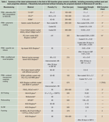
Table I Reference Kokubo and Takadama67–Reference Wu, Hill, Yue, Nightingale, Lee and Jones80 lists a summary of the latest achievements in the field of 3D bioactive glass-ceramic scaffolds, reporting the fabrication techniques used and the main properties of the scaffolds.
Summary of the latest achievements in the field of 3D bioactive glass-ceramic scaffolds, including processing method used and main properties obtained. (*Bioactivity test performed without following the proposed standard in References Reference Kokubo and Takadama67 and Reference Cerruti, Greenspan and Powers68.)

Note: HCA, hydrocarbonate apatite; FRM, foam replica method; BG, bioactive glass; BGC, bioactive glass-ceramic; CEL2, 45SiO2-26CaO-15Na2O-3P2O5-4K2O-7MgO mol%; SCNA, 57SiO2-34CaO-6Na2O-3Al2O3 mol%; PCL, polycaprolactone; MBG, mesoporous bioactive glass; MCM, mobil composition of matter; HA/A-W hydroxyapatite/apatite-wollastonite; 6P53B, 52.7SiO2, 10.3Na2O, 2.8K2O, 10.2MgO, 18.0CaO, 6.0P2O5 (wt%); SLCM, stereolithography ceramic manufacturing; SBF, simulated body fluid; TRIS, tris-(hydroxymethyl) aminomethane; T sint, sintering temperature; 45S5, 45SiO2–24.5CaO-24.5Na2O-6P2O5, in wt%.
Discussion
Bioactive glass-ceramics are partially crystallized inorganic materials obtained by heat treatment of parent bioactive glasses above the crystallization temperature (i.e., the temperature corresponding to the structural change of the glass into a crystalline solid). Reference Serbena, Mathias, Foerster and Zanotto1–Reference Gerhardt and Boccaccini3,Reference Rawlings, Wu and Boccaccini81
Since the first series of scaffolds based on 45S5 bioactive glass was developed 10 years ago, Reference Chen, Thompson and Boccaccini17 the high potential for applications of bioactive glasses and bioactive glass-ceramics as 3D scaffolds for bone-tissue engineering has prompted substantial research in the field using melt-derived bioactive glass powders. However, due to their relatively low compressive strength, no clinical applications have been reported for these scaffolds, despite their notable characteristics in terms of osteogenic (formation of boney tissue) and angiogenic (formation of new blood vessels) responses. Reference Gerhardt and Boccaccini3,Reference Arkudas, Balzer, Buehrer, Arnold, Hoppe, Detsch, Newby, Fey, Greil, Horch, Boccaccini and Kneser43,Reference Newby, El-Gendy, Kirkham, Yang, Thompson and Boccaccini74 FRM is still the most widely used technique for the production of porous bioactive glass-ceramic scaffolds. By modifying the glass composition or choosing alternative sacrificial templates, it is possible to obtain bioactive glass-ceramic scaffolds with compressive strength values comparable to those of human trabecular bone (2–12 MPa). Coating and infiltrating bioactive glass-ceramic scaffolds with biodegradable polymers is also being explored to enhance the resulting mechanical properties. Reference Desimone, Li, Roether, Schubert, Crovace, Rodrigues, Zanotto and Boccaccini29,Reference Philippart, Boccaccini, Fleck, Schubert and Roether31,Reference Yunos, Bretcanu and Boccaccini32
Improving foaming techniques is an alternative approach to develop robust bioactive glass-ceramic scaffolds, for example, by using polymeric precursors or by developing powder-metallurgy methods for the production of 45S5-based bioactive glass-ceramic scaffolds with high pore interconnectivity and increased mechanical properties, opening the possibility to machine the obtained foams to required shapes for the intended applications. Reference Boccardi, Melli, Catignoli, Altomare, Jahromi, Cerruti, Lefebvre and De Nardo41
More recently, research efforts have increased in the use of AMTs, especially the robocasting method, to produce mechanically competent bioactive glass-ceramic scaffolds. Reference Gmeiner, Deisinger, Schönherr, Lechner, Detsch, Boccaccini and Stampfl45,Reference Shao, He, Fu, He, Yang, Xie, Yao, Ye, Xu and Gou49,Reference Eqtesadi, Motealleh, Miranda, Pajares, Lemos and Ferreira56 With these techniques, scaffolds exhibiting high bioactivity and mechanical properties comparable to those of trabecular bone have been produced.
One major issue is the impossibility in directly comparing most of the fabricated scaffolds in terms of their bioactivity. For example, a common procedure to measure bioactivity has not been followed in most of the reviewed studies. Authors refer to Kokubo’s approach Reference Kokubo and Takadama67 only for the preparation of simulated body fluid ([SBF], a solution with a similar ionic concentration to human blood plasma), but no specific information on the material weight/SBF volume ratio is reported or different SBF exchange protocols have been used. Kokubo et al. Reference Kokubo and Takadama67 established a protocol to evaluate the ability of HCA to form on material surfaces in SBF at a fixed material weight/SBF volume ratio. In fact, higher material weight/SBF volume ratios can induce faster HCA precipitation on scaffold surfaces, providing false positive results. Reference Rohanová, Boccaccini, Yunos, Horkavcová, Březovská and Helebrant82
Incorrect results (e.g., overestimation of bioactivity) are also associated with the high reactivity of some bioactive glass-ceramic compositions, in particular, when they are tested in SBF buffered with tris-(hydroxymethyl) aminomethane. Reference Rohanová, Boccaccini, Yunos, Horkavcová, Březovská and Helebrant82 Maçon et al. Reference Maçon, Kim, Esther, Kathryn, Brow, Day, Hoppe, Boccaccini, Kim, Ohtsuki, Kokubo, Osaka, Vallet-Regí, Arcos, Fraile, Salinas, Teixeira, Vueva, Almeida, Miola, Vitale-Brovarone, Verne, Höland and Jones83 put forward a protocol for the bioactivity evaluation of bioactive glasses and bioactive glass-ceramics, which is the result of an international round-robin test supported by technical committee 4 (TC04) of the International Commission on Glass. This method should be considered to generate data to compare the bioactivity of bioactive glasses and bioactive glass-ceramics fabricated by different methods.
Conclusions
The available research outputs suggest an improvement in the mechanical properties of bioactive glass-ceramic scaffolds through dedicated research efforts from the last 10 years, opening the possibility for using bioactive glass-ceramic scaffolds in clinical applications. More research is required to compare different bioactive glass-ceramic compositions and to assess the relative merits and drawbacks of the different techniques developed for the production of bioactive glass-ceramic scaffolds. In this context, conducting relevant long-term in vivo studies in critical bone defects (nonhealing bone defects related to tumor resectioning or injuries) animal models remains an important future task to start closing the gap between basic research and clinical applications of bioactive glass-ceramic scaffolds.
Looking into the future, it can be stated that the main advantage of bioactive glass-ceramic scaffolds, compared to the already available bioactive glass-ceramic bulk or particulates, is the possibility to develop a patient-specific 3D structure that can fill bone defects while also supporting cell attachment, cell ingrowth, and regeneration of the damaged tissue with progressive degradation of the implanted porous scaffold. As reviewed in this article, novel bioactive glass-ceramic scaffolds with required mechanical properties and suitable interconnected porosity are now available due to the development of novel techniques and improvements in existing ones, opening for the first time the possibility of applications of bioactive glass-ceramic scaffolds in the clinic.
Future developments should consider enhancing the biological activity of the scaffolds by designing bioactive glass compositions with multifunctional bioactivity and therapeutic ion-release function (e.g., with antibacterial, osteogenic, and angiogenic properties), Reference Hoppe, Güldal and Boccaccini12 which could lead to scaffolds with high regenerative capability without the need for cost-intensive and complex recombinant proteins or growth factors.

Elena Boccardi is a doctoral candidate at the Institute of Biomaterials, Friedrich-Alexander-Universität Erlangen-Nürnberg, Germany. She graduated in biomedical engineering at Politecnico di Milano, Italy, in 2013. Her research focuses on bioactive glass-based scaffolds with improved mechanical properties and drug delivery capability by combination with mesoporous bioactive glasses. More recently, she was involved in a European research project, focusing on the development of ordered mesoporous drug carriers. Boccardi can be reached by email at elena.boccardi@fau.de.

Francesca E. Ciraldo is a doctoral candidate at the Institute of Biomaterials, Friedrich-Alexander-Universität Erlangen-Nürnberg, Germany. She studied biomedical engineering at Politecnico di Torino, Italy. Her research focuses on the design, processing, and characterization of mesoporous glass containing coatings with therapeutic ion-release capability. Ciraldo can be reached by email at francesca.elisa.ciraldo@fau.de.
Aldo R. Boccaccini is a professor of biomaterials and head of the Institute of Biomaterials at Friedrich-Alexander-Universität Erlangen-Nürnberg, Germany. He is also a visiting professor at Imperial College London, UK. He obtained his engineering degree from the Instituto Balseiro, Argentina; his Dr.-Ing. degree from RWTH Aachen University, Germany; and his Habilitation degree from Technische Universität Ilmenau, Germany. His research focuses on glasses, ceramics, and composites for biomedical, functional, and structural applications. He is the author or co-author of more than 600 scientific papers and 15 book chapters. He was a Thomson Reuters “Highly Cited Researcher” in 2014. Boccaccini can be reached by phone at +49 9131 85 28601 or by email at aldo.boccaccini@ww.uni-erlangen.de.